BIPN 152 Exam 1
1/62
Earn XP
Description and Tags
covers LE 1-5 (up to material covered Tuesday 4/14), DI week 2-3 slides
Name | Mastery | Learn | Test | Matching | Spaced | Call with Kai |
|---|
No analytics yet
Send a link to your students to track their progress
63 Terms
(mainly know the degrees of magnitude)
# of neurons in adult human brain
average # of synapses made by a neuron
# of synapses in child vs. adult brain
the brain is % of our body weight, but it uses % of our energy
80-85 billion neurons in adult human brain (1011)
average of ~7,000 synapses made by a neuron
~1015 synapses in child brain, ~1014 synapses in adult brain
brain is 2% of our body weight, but it uses 20% of our energy
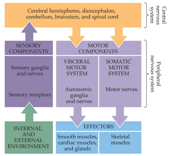
parts of CNS (general ←2) (specific ← 5)
parts of PNS (general ←2) (specific ← 2 that are described)
CNS: spinal cord & brain
cerebral hemispheres
diencephalon
cerebellum
brainstem
spinal cord
PNS: cranial nerves & spinal nerves
sensory
(internal and external environment →) sensory nerves, ganglia, and receptors
motor
visceral motor/autonomic nerves → smooth and cardiac muscle, glands
somatic motor nerves → skeletal muscles
state 3 ways biomedical research is funded
Alexander Graham Bell developed the telephone with funding from wealthy parent of his student
lots of government funding (for research) after WW2, but decreased since
NIH (national institute of health) is the largest funder
model organisms commonly used in neuroscience research (8)
c. elegans
aplysia californica
drosophila melanogaster
mouse
rat
rhesus monkey/macaque
human
iPS cells / iPSCs
(^ all are listed in order of simple to complex neural circuits in terms of # of neurons, except for the iPSCs)
__
C. elegans (302 neurons)
Aplysia californica (15,000)
monkey (50 billion)
^^ this latter part is simple to complex neural circuits
state the 2 building blocks of the NS
__
the cell is the fundamental unit of living organisms, whereas
the neuron was discovered to be the fundamental unit of the (1)
describe the 2 theories/models
which is wrong
who first used the term “synapse”
neurons & glia
__
the cell is the fundamental unit of living organisms, whereas
the neuron was discovered to be the fundamental unit of the nervous system
reticular theory (Golgi): neurons are not discrete/distinct cells, but are fused together into ONE continuous/interconnected network (“continuum of protoplasmic links”)
where axons and dendrites branch into one another
BUT is wrong b/c there are synapses/tiny gaps between neurons, so it is NOT a continuous interconnected network
neuron doctrine (Cajal): neurons are discrete entities/cells that communicate at specialized contacts/synapses (“protoplasmic kisses”)
_
Sherrington first used “synapse”
describe what Cajal discovered the __ __ of neuronal signal flow (2)
dynamic polarization of neuronal signal flow (aka directionality aka functional polarity) (aka that signals flow in ONE direction in neurons)
inferred that in sensory systems, info should generally flow from sensory organs → to the brain
found that dendrites are at the receiving end of a connection, axons deliver that info to the next neuron
ex. of neuron-to-neuron in vertebrate retina: photoR cells → bipolar cells → RG cells
name the 3 types of glia in CNS, 1 type in PNS
__
describe glia (3)
its general functions (~6)
CNS: astrocytes (support), microglia (immune), oligodendrocytes (myelin)
PNS: Schwann cells (myelin)
__________
more glia than neurons
don’t directly participate in signaling, but are still important in neuronal signaling
NOT excitable, BUT have ion channels
__
supportive roles (3): trophic factors, remove debris, regulate synapse
-
maintain extracellular ionic concentration
speed up signal propagation (by forming myelin) ← b/c glia cells like oligodendrocytes & Schwann cells
NT uptake ← NT uptake from synaptic cleft into astrocytes b/c astrocytes are part of the tripartite synapse
help with structure during development
neurons are __
includes 3 parts (not referring to dendrites, soma, or axon) (glia only have soma, usually no dendrites or axons)
function (4)
neurons are cells
membrane, cytoplasm, nucleus
__
functions (done w/in the parts mentioned above)
cellular metabolism
protein synthesis
protein modification
transport
for the functional polarity of neurons in a circuit
despite __, most neurons have a basic common structure
describe the 3 parts involved & what they do
degrees of order of magnitude of axon
__
t/f: for many neurons, axons are VERY long compared to the dendrites
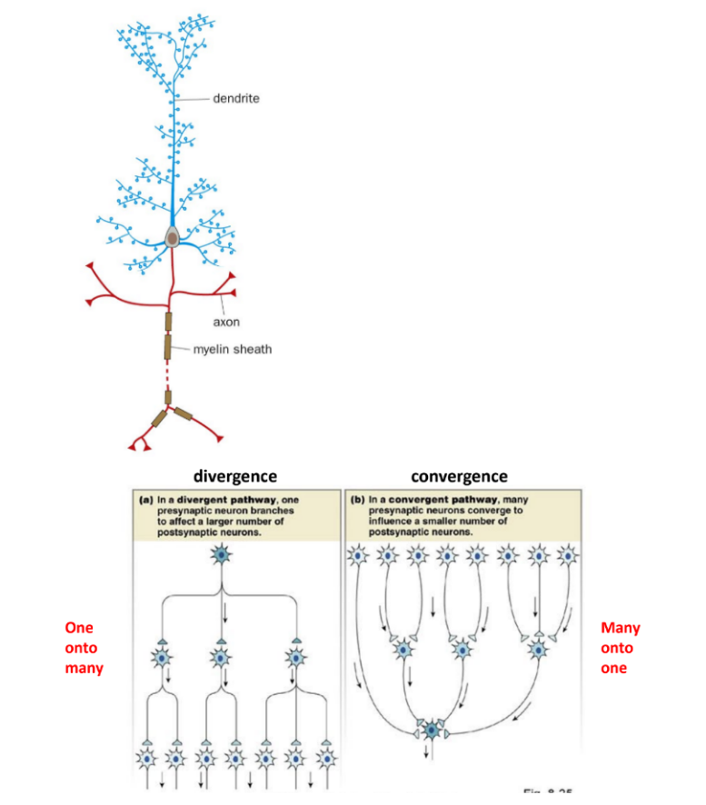
neurons have __ axon that can be highly __
state and def. 2 ways of branching of neurons
t/f: the directional flow of signals/info also occurs at the subcellular level, aka at synapses
despite heterogeneity/diversity, most neurons have a basic common structure
dendrites & soma receive inputs
soma integrate inputs
axon makes & transmits the signal/nerve impulse to its targets (some of these axons are myelinated, so some have faster transmission of signals/nerve impulses)
axon is 6 orders of magnitude (few micrometers - tens of cm)
__
true
_
neurons have ONE axon that can be HIGHLY BRANCHED
divergence: 1 axon → 10,000 different post- neurons (one to many)
1 pre- branches/diverges to affect many post-
convergence: multiple highly branched dendrites that receive multiple connections (from multiple neurons’ axons) (many to one)
many pre- converge to affect 1 post-
_
true
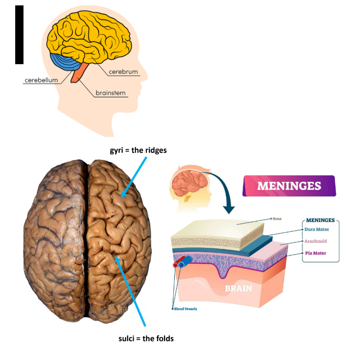
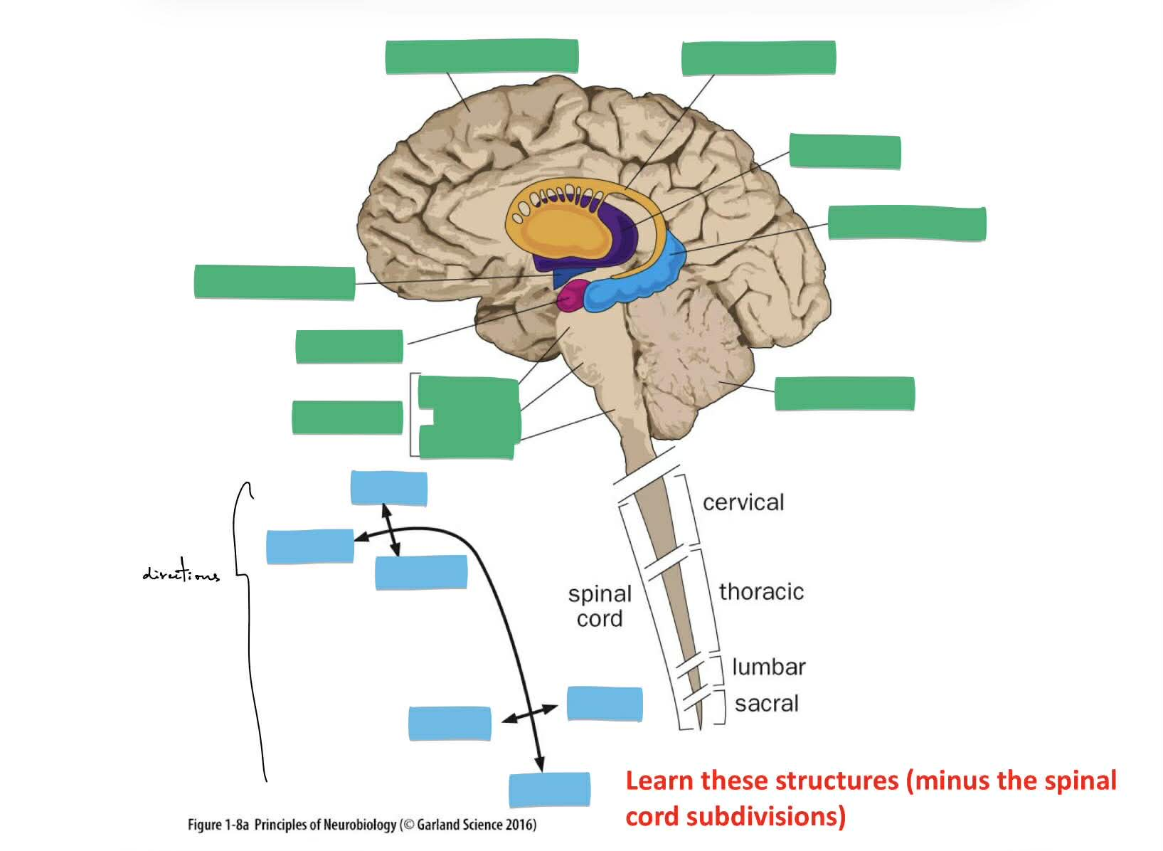
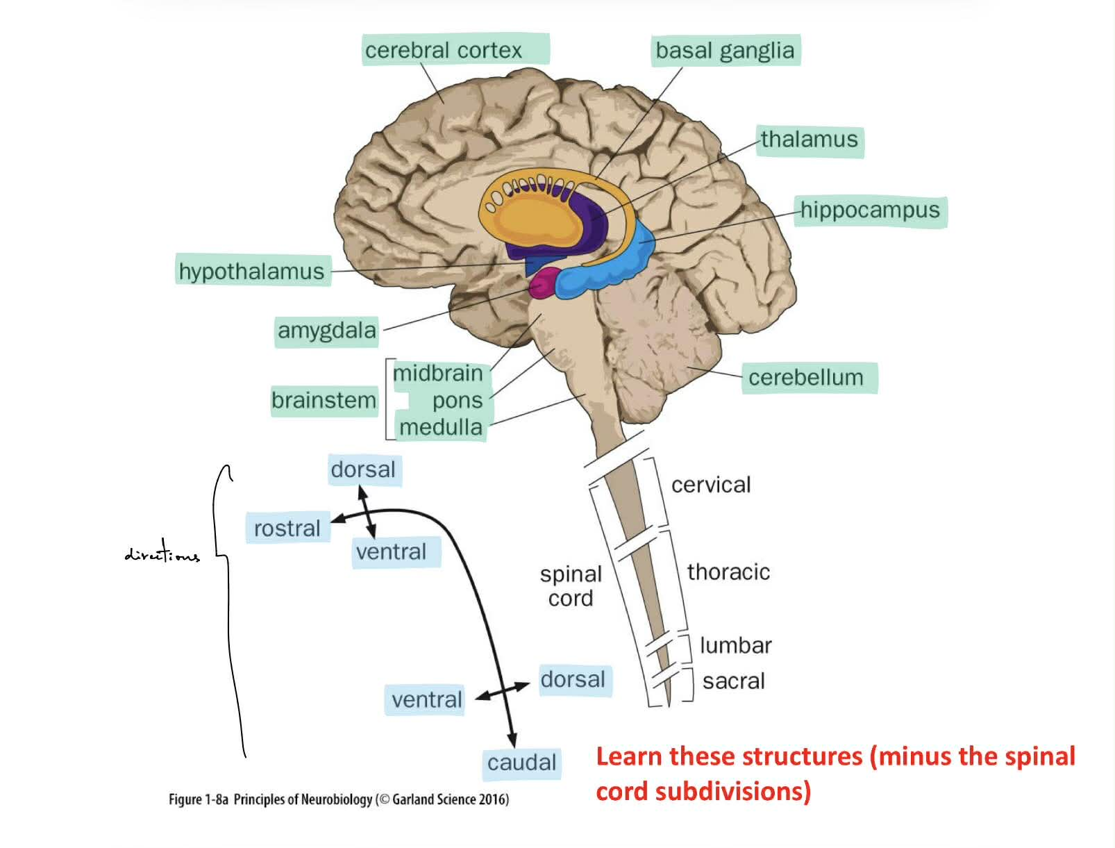
state 3 main parts of the brain (draw and label where each is)
def. meninges (name the 3 parts from outer to inner ← aka bone—3layers—brain)
def. sulci vs. gyrus
_
the brain contains a rich __ (def.)
def. phrenology
cerebrum, cerebellum, brainstem
_
meninges: 3 layers of tissue that cover & protect the brain
dura mater, arachnoid, pia mater
sulci - the folds (“suck”)
gyri - the ridges (“rise”)
__
the brain contains a rich vasculature (← arteries & veins)
phrenology: different brain regions have different functions
(old study & was proven wrong)
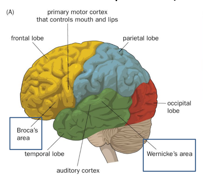
the neocortex is organized into regions that specialize in specific tasks/behavior
name the 4 lobes & location relative to e/o
what lobe are these in:
primary motor cortex (for mouth and lips)
auditory cortex
Broca’s area
Wernicke’s area
AND primary somatosensory cortex
__
state the 3 types of evidence to study/look at the specialized function and region of Broca’s and Wernicke’s area
def./describe each, where 1 of them explains Broca’s & Wernicke’s aphasia
frontal lobe — anterior
primary motor cortex
Broca’s area (“fb”) ← speech production
parietal lobe — superior to temporal
primary somatosensory cortex
temporal lobe — inferior to parietal
auditory cortex
Wernicke’s area (“tw”) ← speech comprehension
occipital lobe — posterior
__
fMRI
looks at/gives information about brain structure & brain activity, where highlighted regions show higher activity
brain stimulation during epilepsy surgery
stimulation via electrodes
brain lesions (Broca’s/Wernicke’s aphasia)
Broca’s aphasia: bad at language production, but good at language comprehension
Wernicke’s aphasia: good at language production, but is meaningless speech & bad at language comprehension
for brain stimulation during epilepsy surgery
def. topographic map
name 2 types of visualizations of this
__
parts of the body that are more densely __ occupy greater regions of the brain
_
t/f: different species organize their brain differently depending on what’s important to the organism, like the “whisker barrels” in rodents’ somatosensory cortex
_
t/f: certain sensory & motor systems have “crossed” innervation (contralateral) and also ipsilateral innervation
(^ aka crossing over or not of innervations/pathways that travel towards the primary motor cortex & primary somatosensory cortex)
topographic map:
where adjacent regions of the periphery map (body) map onto adjacent regions of the brain, visualized w/ the:
motor homunculus (primary motor cortex)
sensory homunculus (primary somatosensory cortex)
^^ homunculus is a distorted map of body parts to the cortex
__
parts of the body that are more densely innervated occupy greater regions of the brain
_
true
_
true
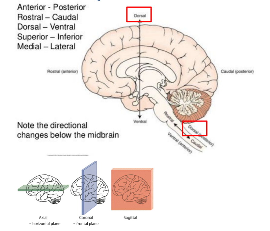
def. the directions:
lateral, medial
anterior, posterior
rostral, caudal
dorsal, ventral
superior, inferior
^^ how do some of these differ below the midbrain?
_
name and def. the 3 planes
medial (towards middle), lateral (away from middle/center line)
anterior(front), posterior (back)
rostral (nose), caudal (tail)
dorsal (back/up), ventral (belly/down) ← like for rats
superior (up), inferior (down)
^^ below the midbrain:
dorsal is posterior
ventral is anterior
__
coronal / frontal / transverse plane
into front and back
sagittal plane
into R and L
axial / horizontal plane
into up and down
white matter vs. gray matter (2 each)
_
fill in blanks
the directions are either dorsal/ventral or rostral/caudal
white matter
has the axon tracts (the communication pathways)
is mostly myelinated axons
gray matter
everything else (soma, dendrites, synapses, glia)
is mostly unmyelinated axons
(hippocampus is “C” shaped)
when neurons are excited, they create nerve impulses (← def. term)
__
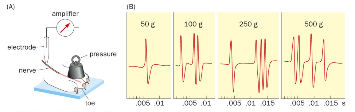
describe the 2 basic principles of somatosensory nerve responses/impulses
nerve impulses - transient/short-term changes in membrane potential that propagate down/along the nerve
__
basic principles:
nerve impulses have a uniform size & shape (“elementary unit” ←→ unitary EPP/smallest mEPP)
increasing stimulus strength will increase the frequency of nerve impulses, but does NOT change the impulses’ magnitude
aka nerve impulse frequency is encoded by stimulus strength
(stronger stim. → more APs released/higher frequency BUT has “quantal release”/same magnitude)
__ __ are the basis for membrane potential (name the 3 that determine Vm/membrane potential)
where cell’s interior/intra- is __ relative to the exterior/extra- (describe in terms of the 3 ions)
__
neurons are electrically polarized cells
name 3 types of potentials (describe each ← 1)
ion gradients ← Na+, K+, Cl- (not Ca2+)
intra- is more negative than extra-
(at RMP, more intra- K+, while more extra- Na+ and Cl-)
__
RMP
action potentials
graded potentials:
synaptic potentials
when external stimuli, like light, are transduced into electrical signals (from inside post- neuron)
receptor potentials
caused by NTs in the synapse binding to the post- cell/neuron (used by sensory neurons, from sensory receptor)
neurons exhibit 2 types of membrane potential changes: APs and graded potentials
describe each (AP ← 4, graded ← 2)
also describe RMP (4)
_
t/f: all APs are transient
explain
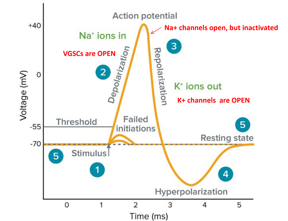
APs (aka “spike”): elementary unit of nerve impulses ← transient (b/c delayed activation of VG-K+ channels reverse the de-)
used for long-distance transmission
is “all-or-none”
first made at the axon initial segment (AIS) aka axon hillock, when Na+ influx → causes neuron to depolarize to ~ +30 mV
need to reach the minimum threshold depolarization to trigger it (~ -55 mV)
graded potentials: ← transient
used over short distance
is a gradient in the magnitude of the response (is not all-or-none)
RMP:
made by different ion concentrations across the membrane & the different permeabilities of these ions
~ -70 mV
3 major ions: Na+, K+, Cl-
is maintained at around -70 mV by the Na+/K+ ATPase (against concentration gradient: 3 Na+ out, 2 K+ in)
__
true
all APs are transient b/c VG Na+ channels will inactivate (at the peak) & VG K+ channels will open to repolarize the membrane potential back to its resting membrane potential
(picture)
VGSC / VG-Na+ channels open during de-
at peak, VG-Na+ are open & in inactivated state
VG-K+ channels open during re-/hyper-
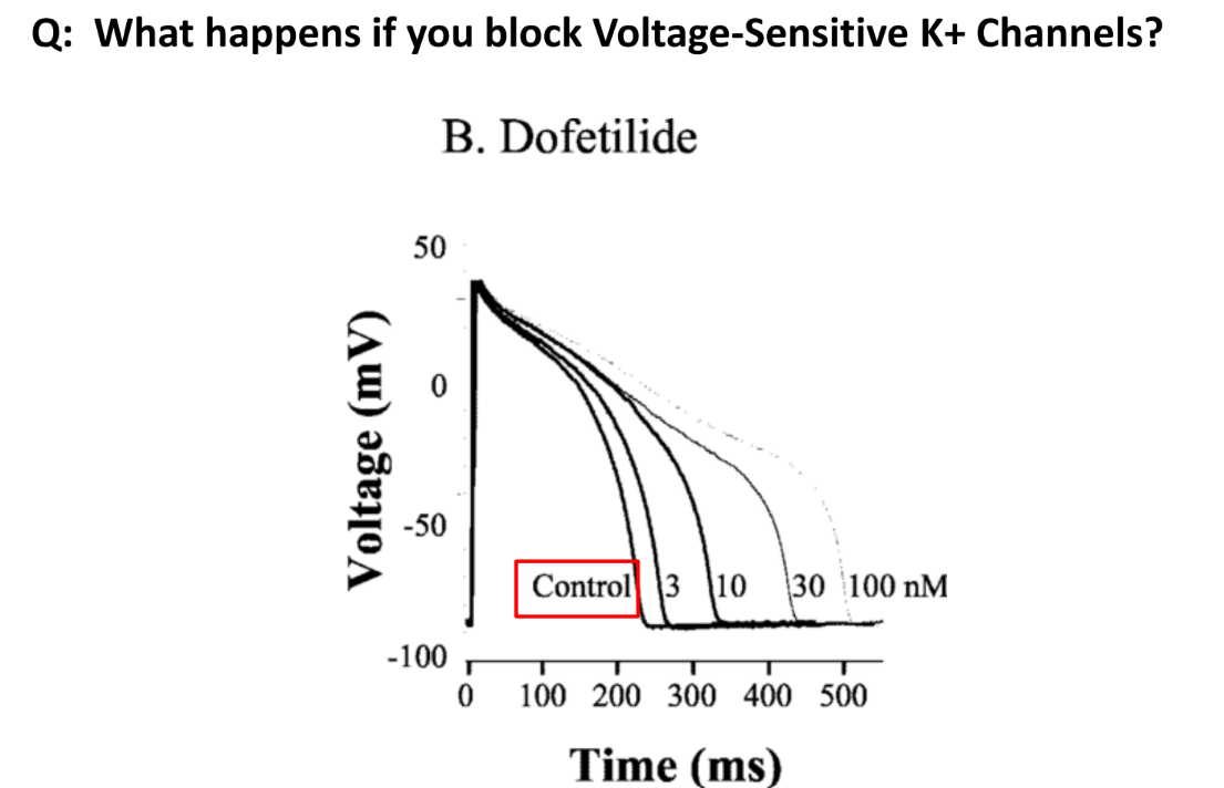
what happens if you block voltage-sensitive K+ channels?
AP is prolonged
describe 3 states of VGSCs
_
ion channels are typically multimeric transmembrane proteins (describe ← 1)
closed - inactivation gate open, activation gate closed
(resting state)
open - both gates open
(depolarization)
inactivated - inactivation gate closed, activation gate open
(hyperpolarization IF also have open VG K+ channels)
__
part of the ion channel forms a voltage sensor that causes a conformational change when opening the pore
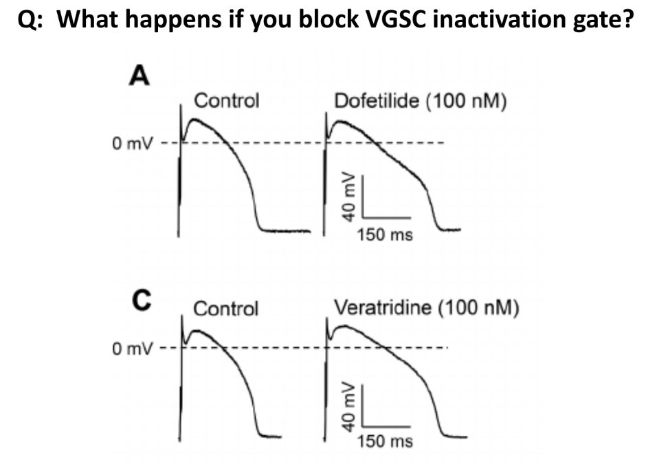
what happens if you block VGSC inactivation gate?
(blocking the VGSC inactivation gate means that it will stay open / inactivation gate is prevented from binding + closing)
AP is prolonged
(in dep-, both inactivation and activation VGSC gates are open)
the VGSC inactivation gate will stay open at & past the peak, which allows for more Na+ influx
K+ influx cannot perfectly counter this effect, so there is slower re- and hyperpolarization → which is seen as a prolonged AP
t/f: oligodendrocytes (CNS) & Schwann cells (PNS) create a myelin sheath around some axons
_
rate AP conduction velocity for unmyelinated “C” fibers (peripheral nerve fibers) vs. myelinated fibers
in PNS and/or CNS?
relevance of speed of AP conduction (1)
__
def. nodes of Ranvier
what type of conduction?
true
_
unmyelinated “C” fibers (peripheral nerve fibers) ← PNS
~ 1 m/s
myelinated fibers ←- PNS or CNS
~ 100 m/s
the speed of AP conduction/propagation determines how somatosensory signals are perceived (ex: heat vs. cold, immediate sharp pain vs. dull throbbing pain)
__
nodes of Ranvier:
gaps b/w myelin sheaths, where the current jumps from node to node which increases the speed of impulse travel along the axon
^ does saltatory conduction
what would failure of myelination (i.e. defect in oligodendrocytes or Schwann cells) do to circuit function? (1)
name and def. 1 type of disease that could result from this (w/ 4 symptoms)
is there a cure?
slowed or failed conduction (which can lead to sensory and/or motor deficits)
_
multiple sclerosis (MS): an autoimmune demyelinating disease of the CNS (spec. demyelination of oligodendrocytes)
symptoms:
motor weakness
pain
vision loss
cognitive dysfunction
(^ no cure; degree of axon and soma damage deter. the disease severity)
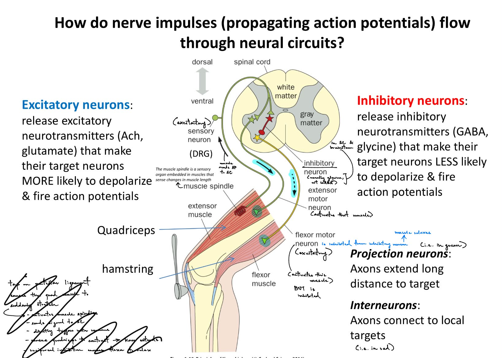
what specific neural circuit do nerve impulses (aka propagating APs) flow through? (1)
describe the pathway of these signals
__
def. excitatory vs. inhibitory neurons (& each release what 2 specific NTs)
def. projection neurons vs. interneurons
__
describe the pathway (~1/2) of how a tap on the patellar ligament simultaneously causes the quadriceps to suddenly stretch/extend AND the hamstrings to suddenly flex/bend
via the sensory-motor circuit b/w the SC and brain
from sensory neuron → DRG → spinal cord → brain stem → thalamus → primary somatosensory cortex → primary motor cortex → spinal cord → motor neuron (in muscles)
__
excitatory neurons
release excitatory NTs (glutamate, ACh) to make target neurons MORE likely to dep- and fire APs
inhibitory neurons
release inhibitory NTs (GABA, glycine) to make target neurons LESS likely to dep- and less likely to fire APs
__
projection neurons — axons extend long distance to targets
(i.e. in green)
interneurons — axons connect to local targets / axons that connect sensory neurons to motor neurons
(i.e. in red)
________
tap on patellar ligament activates muscle spindles w/in the quadricep (← the extensor muscle) (muscle spindles — sensory organ that detects change in muscle length)
the excitatory sensory neuron will send signals to SC
directly triggers/activates the extensor motor neuron
SO quads will contract→ causing knee to extend
(the sensory neuron activates the extensor motor neuron, so the quad stretches/knee extends)
___ WHILE THIS HAPPENS, there is also simultaneously-occurring reciprocal inhibition in order to have coordinated movement / to prevent opposing muscles from working against e/o ——
tap on patellar ligament activates muscle spindles w/in the quadricep (← the extensor muscle) (muscle spindles — sensory organ that detects change in muscle length)
the excitatory sensory neuron will send signals to SC
that excitatory sensory neuron will activate an inhibitory interneuron
that inhibitory interneuron will activate the excitatory flexor motor neuron
SO hamstrings will contract → causing knee to flex/bend
reciprocal inhibition causes flexor to relax
(the sensory neurons activates the inhibitory neuron, which inhibits the flexor motor neuron, so it does not stretch BUT relaxes instead)
imagine each
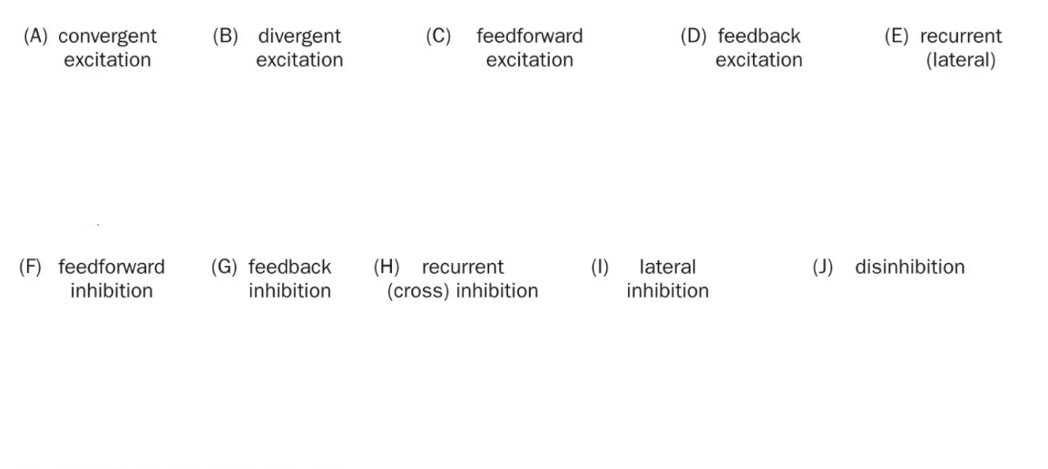
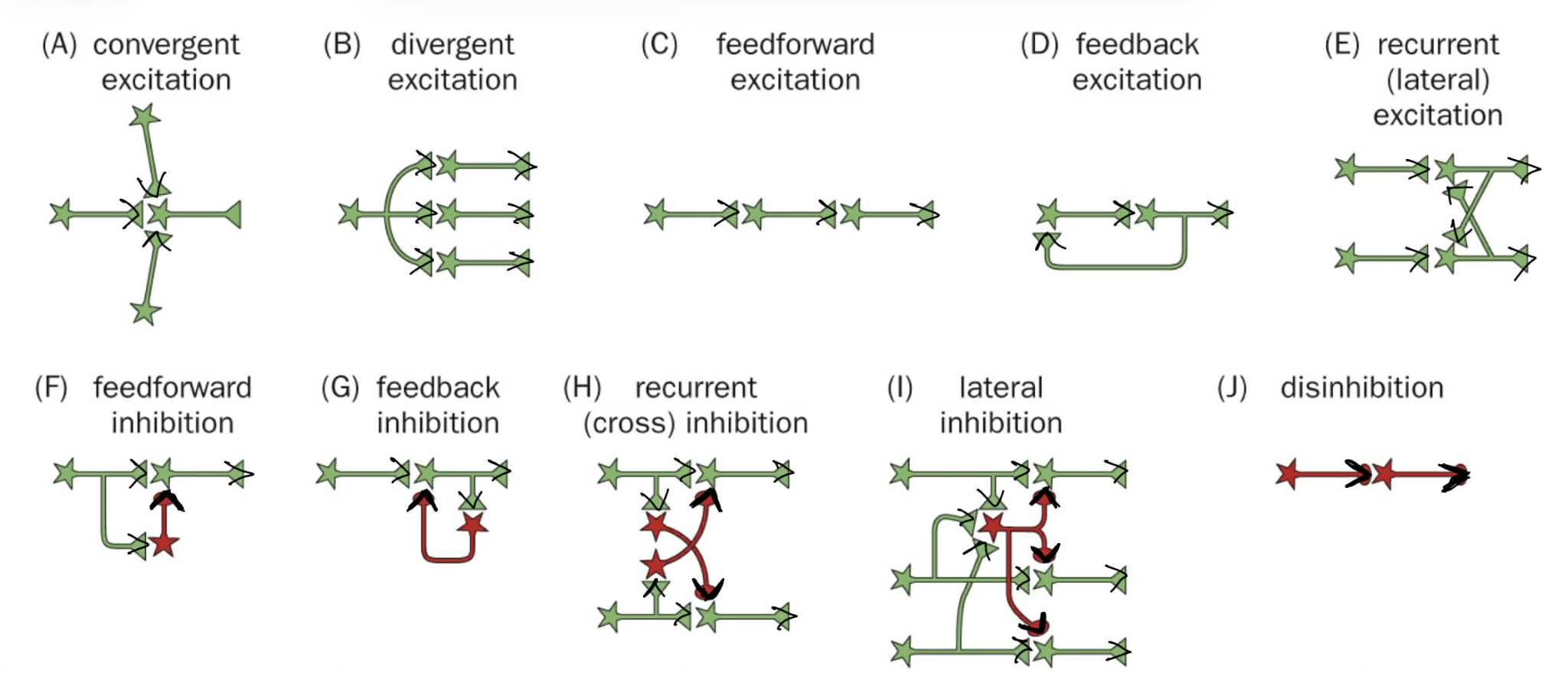
describe E, H, I, and J
recurrent lateral excitation: acts on the same neuron that just fired
_
recurrent cross inhibition AND lateral inhibition: acts on the next neuron
disinhibition: inhibits an inhibitory neuron
don’t have to memorize any of these, but just understand the basic concepts in this diagram
(MB can think of “lateral …” as the neurons in the same stage/step being equally affected)
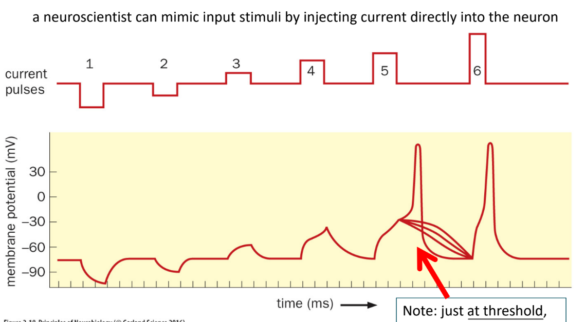
what is made if a stimulus is too weak to produce an AP? (1)
t/f: even when MP reaches the AP threshold, some pulses can elicit an AP, but some still fail to do so
graded potentials
_
true
name the 3 types of mechanosensation
_
name the specialized nerve endings for the 2 of the 3 types of mechanosensation, pain, and temp
^ they all produce what type of potential?
mechanosensation (touch, pressure, vibrations)
_
Meissner’s corpuscle — senses touch
Pacinian corpuscle — sense pressure
nociceptor — senses pain
thermoreceptor — senses temp (heat or cold)
.
^^ all located w/in the dermis AND produce receptor potentials
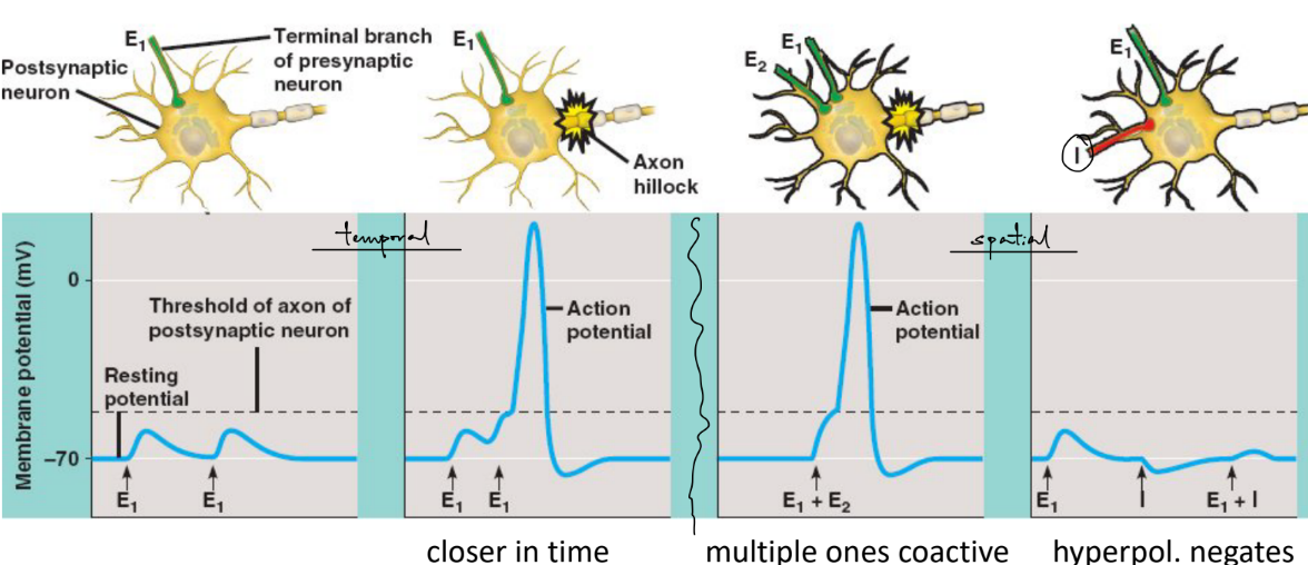
a neuron’s decision to fire or not is determined by the SPATIAL and TEMPORAL summation of __ __ potentials at the (1)
def. the 2 types of summations
_
t/f: inhibitory and excitatory pre- neurons (aka at different locations) that fire at the same time can cancel e/o out, and perhaps cause hyper-
_
t/f: AP is propelled forward, not backwards, due to (serial activation of VGSCs and) the afterhyperpolarization
a neuron’s decision to fire or not is determined by the SPATIAL and TEMPORAL summation of graded, synaptic potentials at the AIS (axon initial segment)/axon hillock (which has a high density of VSSCs aka VGSCs & other proteins)
temporal: 1 pre- neuron firing in close in time (aka same neuron at different times) → compound EPSP
spatial: multiple pre- neurons at different locations firing simultaneously (aka same time at different locations) → larger peak EPSP
__
true (IPSPs can cancel out EPSPs to prevent firing)
_
true
the neuron doctrine raises a key Q:
how does the nerve impulse cross from a neuron to its target? (1)
which is characterized by (3)
__
chemical synaptic transmission at NMJ shows that ACh mimics nerve stimulation
is this correlation or causation
by chemical synaptic transmission, characterized by:
synaptic delay
one-way signaling
depletion of vesicles/NTs
__
correlation; need more evidence to prove causation
what molecules block VG-K+ channels, VGSC inactivation gate, VGSCs (1 each)
its effect on APs
t/f: ACh works even when APs are blocked by TTX, which shows causation that ACh is needed to activate the muscle
__
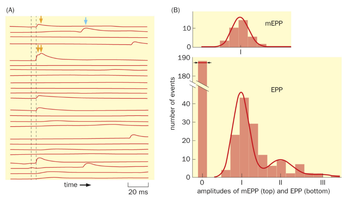
mEPPs demonstrate the quantal nature of NT release (meaning?)
dofetilide blocks VG-K+ ———→ prolonged AP
veratridine blocks VGSC inactivation gate ———→ prolonged AP
TTX (tetrodotoxin) blocks VGSC ———→ no APs
by binding to VGSC pore so that no Na+ can go into the post-, so no APs BUT there can still be depolarization
(small molecule derived from puffer fish)
__
true
b/c ACh still binds to ACh receptors on the post- and can still cause dep-
__
NTs are released in discrete packets aka quanta (shown w/ mEPPs)
(the amplitude of a mEPP determines the size of ONE quanta, in which the # of vesicles releasing NTs occur in this discrete quanta)
what is the key trigger that signals the vesicles to fuse w/ the plasma membrane? (1)
where are its channels located? why?
___
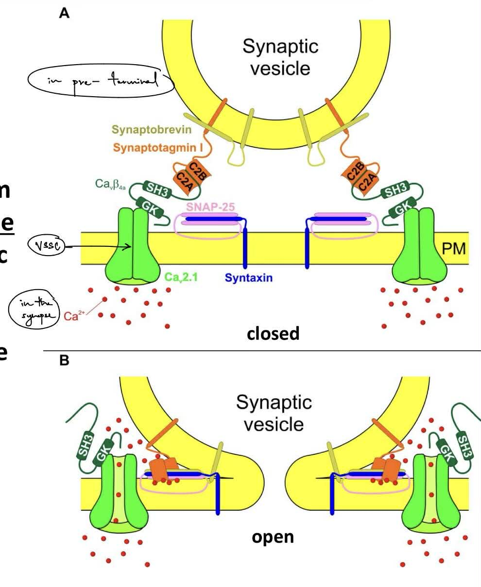
def. the SNARE complex
name SNARE proteins in vesicle membrane (~1/2) vs. plasma membrane (2)
name the 4 protein/parts of synaptic vesicles
Ca2+ influx (via VSCCs aka VG-Ca2+ channels ← which have multiple pore-forming & accessory subunits)
VSCCs are clustered next to the docked vesicles at the “active zone” (aka clustered closer to the synapse)
b/c for fast NT release by the AP (aka once the AP reaches the pre- terminal)
___
SNARE complex — regulates fusion b/w the vesicle membrane & plasma membrane, that will form a fusion pore when fused into a single, fused membrane
-vesicle membrane:
synaptobrevin (VAMP)
synaptotagmin
-plasma membrane:
SNAP-25
syntaxin
_
synaptic vesicles consist of:
synaptotagmin
synaptobrevin
V-ATPase
vesicular NT transporter
state steps of (chemical) synaptic transmission (~6/7)
AP reaches axon terminal
VSCCs open
Ca2+ influx causes vesicles to release NTs into synapse / exocytosis
NTs cross the synapse
NTs bind to neuroreceptors on post-
triggers signal in post- (aka evoked post- current / EPSC)
(summation of EPSCs can trigger a post- AP)
what would happen to synaptic transmission if proteins of the SNARE complex were disabled? (1 w/ 4)
__
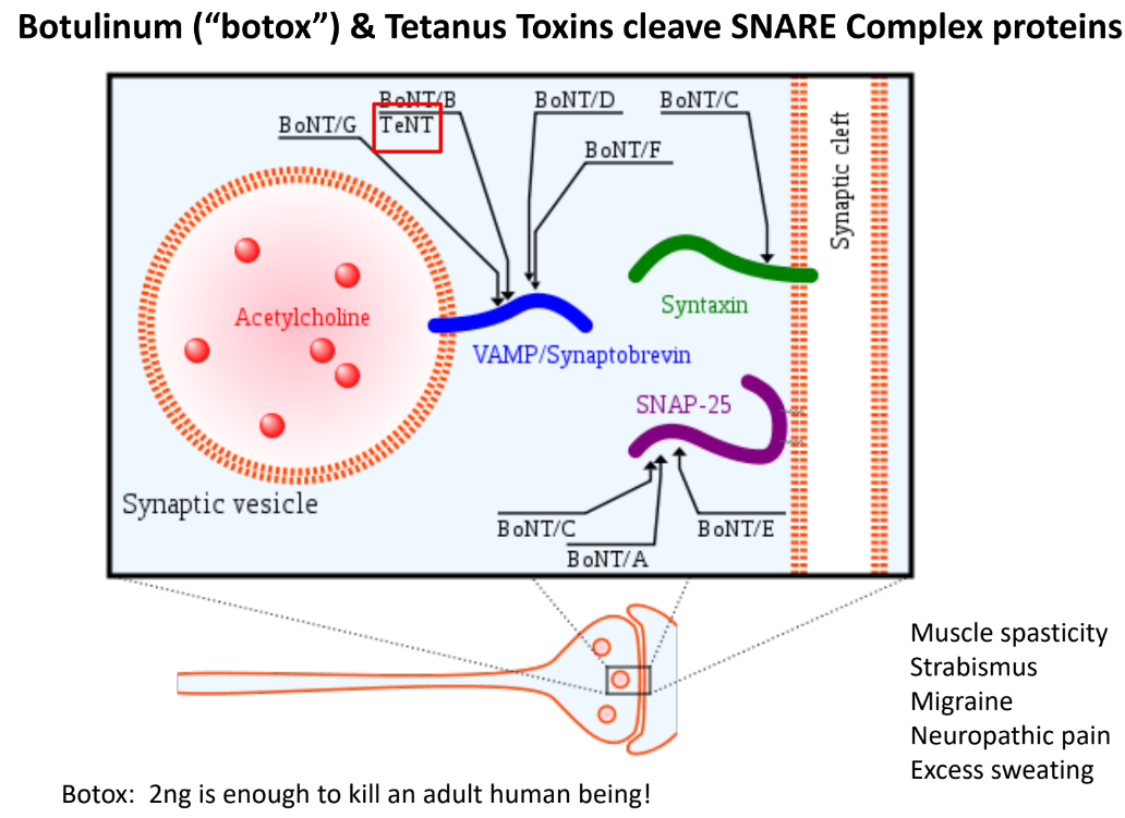
effect of botulinum (botox) and tetanus toxins on the SNARE complex?
which toxins act on which SNARE complex proteins?
NS disorders, like:
Alzheimer’s
schizophrenia
depression
ADHD
__
botulinum/botox & tetanus toxins cleave the SNARE complex proteins (synaptobrevin/VAMP, SNAP-25, syntaxin), which prevent vesicle-membrane fusion SO no NT release (like no ACh release into NMJ)
BoNT A,C,E — SNAP-25
BoNT B,D,F,G — synaptobrevin
BoNT C — syntaxin
fast neurotransmission w/ ionotropic Rs vs. slow neurotransmission w/ metabotropic Rs (2 each w/ time)
__
ligand-gated ion channels can be __ or __, depending on if they de- or hyper- the post- cell SO post- potentials are either that or that
describe EPSP vs. IPSP (← involves NTs & NT binding open “-” channels)
for EPSP, which NT is in CNS vs. PNS
_
what deter. if it is fast or slow neurotransmission?
fast neurotransmission w/ ionotropic Rs ← ~1 ms
involves ligand-gated ion channels
NT binds → channel opens → ions flow across membrane
slow neurotransmission w/ metabotropic Rs
involves G-protein-coupled Rs ← ~10-10,000 ms
NT binds to GPCR → activates G-protein → subunits / intra- messengers indirectly modulate ion channels (→ ion channel opens → ions flow across membrane)
__
either excitatory (de-, EPSPs) or inhibitory (hyper-, IPSPs)
EPSP
by glutamine (CNS) or ACh (PNS)
NT binding opens cation channels (Na+, K+, Ca2+ channels)
IPSP
by GABA or glycine
NT binding opens anion channels (Cl- channels)
__
the receptor type (not the NT) deter. if it’s fast or slow neurotransmission
name NTs that act fast (ion-) vs. slow (metabo-) (4 each)
_
t/f: some NTs can operate in both fast and slow neurotransmission
name them (5)
which NT mediates only fast neurotransmission (1)
fast:
ACh
glutamate
GABA
glycine
slow ← neuromodulators
dopamine (DA)
norepinephrine (NE)
serotonin
neuropeptides
__
true
ACh, glutamate, GABA, serotonin, ATP (← not glycine) can act in either iono- or metabo- neurotransmission
glycine mediate only fast neurotransmission
name 3 sub-classes of glutamate Rs
what do NMDARs require to bind to it? (2)
what are NMDARs blocked by? (~1)
what 2 enzymes are involved with mGluRs?
__
why does opening of glutamate R channels cause de- if both Na+ and K+ enter the cell?
__
where does glutamate neurotransmission in mammalian brain most often occur at?
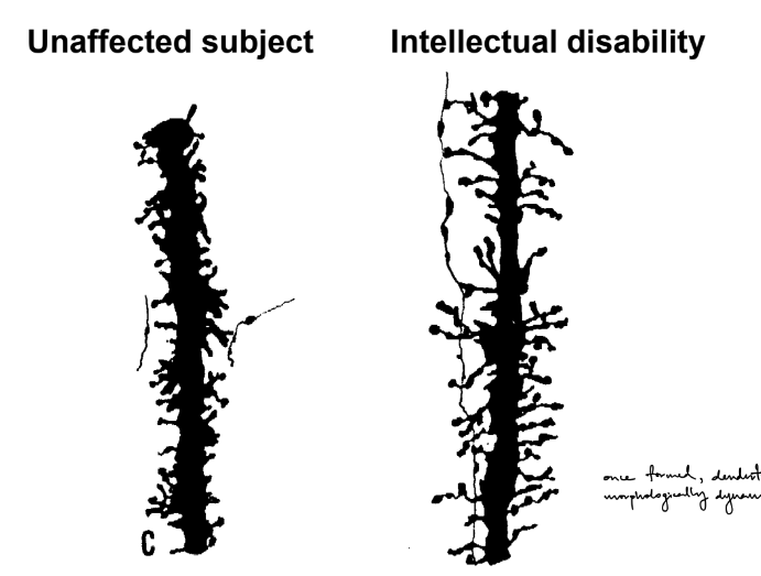
t/f: once formed, dendritic spines are morphologically dynamic
t/f: abnormal dendritic spines are observed in neurodevelopmental disorders & are lost early in many neurodegenerative diseases
AMDARs / kainate Rs (aka non-NMDA receptors)
NMDARs ← w/ Mg2+ block
require glycine co-agonist (glycine & glutamate have to bind to NMDAR to open the ligand-gated ion channel pore)
is blocked by PCP (a psychoactive drug)
mGluRs
involves AC (adenylyl cyclase) & PLC
__
b/c concentration gradient for Na+ is much greater than that for K+ (aka the greater Na+ driving force)
__
glutamate neurotransmission in mammalian brain most often occurs at dendritic spines
aka dendritic spines often receive excitatory signals from glutamate
^ dendritic spines have thin neck & bulbous head, where glutamate synapses form at
_
true
true
def. connectome
_
name & describe 3 types of dye that help visualize neurons (3/4, 4, 3)
connectome: a complete map of all the connections/synapses in a neural circuit
^ is not limited to connections b/w neurons
__
Golgi stain
the first technique to view neurons
fixed/dead tissue is submerged in potassium dichromate & then silver nitrate
seen as black
labels only 1% of neurons
_
Nissl stain
labels RNA in soma (mostly labels RNA in the rough ER)
the dye is “+” & binds to “-” charged structures, like RNA
labels most or all cells
helps view cell densities and boundaries of brain areas
_
DAPI
the dye binds to DNA
labels nuclei
helps view cell densities and boundaries of brain areas
describe direct vs. indirect immunofluorescence assay (2 each)
__
said don’t have to memorize the below
given the proteins/markers, what type of cells do they label?
parvalbumin
CamKII
GAD
GFAP
NeuN
c-fos
direct
uses 1 antibody (labeled primary)
is faster BUT more expensive & lower sensitivity
indirect
uses 2 antibodies (unlabeled primary & labeled secondary)
is more common & has higher sensitivity & is cheaper BUT slower
__
parvalbumin — PV inhibitory neurons
CamKII — pyramidal cells of sensory cortex
GAD — GABA expressing neurons (“capital G word”)
GFAP — astrocytes
NeuN — neurons
c-fos — active neurons
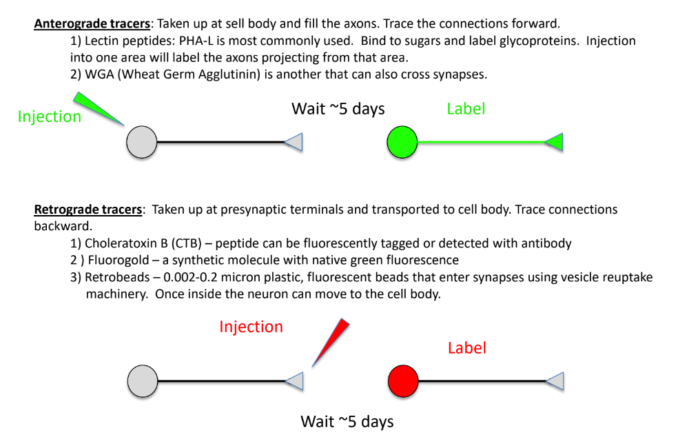
describe anterograde vs. retrograde tracing (2 each) AND 1 for both
it travels along __
just be familiar w/ the names of these:
antero- tracers
lectin peptides - uses PHA-L
(“LP” - “PL”)
WGA (wheat germ agglutinin) - can cross synapses
retro- tracers
CTB (choleratoxin B) - labels peptides
fluorogold - seen as green fluorescence
retrobeads - enter synapse via vesicle reuptake & then move to soma
anterograde tracing — identify where axons project to
(aka the post- of this neuron)
tracer into soma & moves along the axon to the distal region/terminal
labels the axon + terminal
retrograde tracing — identify the neurons that give input to this cell
(aka the pre- of this neuron)
tracer into terminal or fiber tract & moves to soma
labels the soma
^^ both require waiting ~5 days for the results
_
it travels along microtubules (used for long-distance transport)
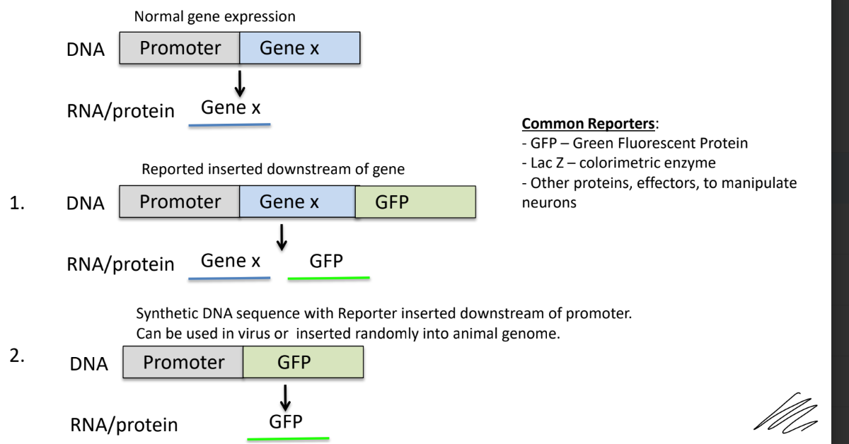
def. promoter
describe (2)
_
name & def. 2 examples of reporter genes (that track gene expression)
describe result of cases where:
reporter is inserted downstream of the gene
reporter is inserted downstream of promoter/replacing the gene
is a sequence of DNA needed to either turn a gene on or off
transcription starts at the promoter
the promoter has a binding site for the enzyme that makes mRNA
__
reporter genes
GFP (green fluorescent protein)
LacZ (identify enzymes w/ different colors)
_
if reporter is inserted downstream of the gene:
the reporter is also made whenever the gene is expressed
if reporter is inserted downstream of promoter/replacing the gene:
the promoter only expresses the reporter
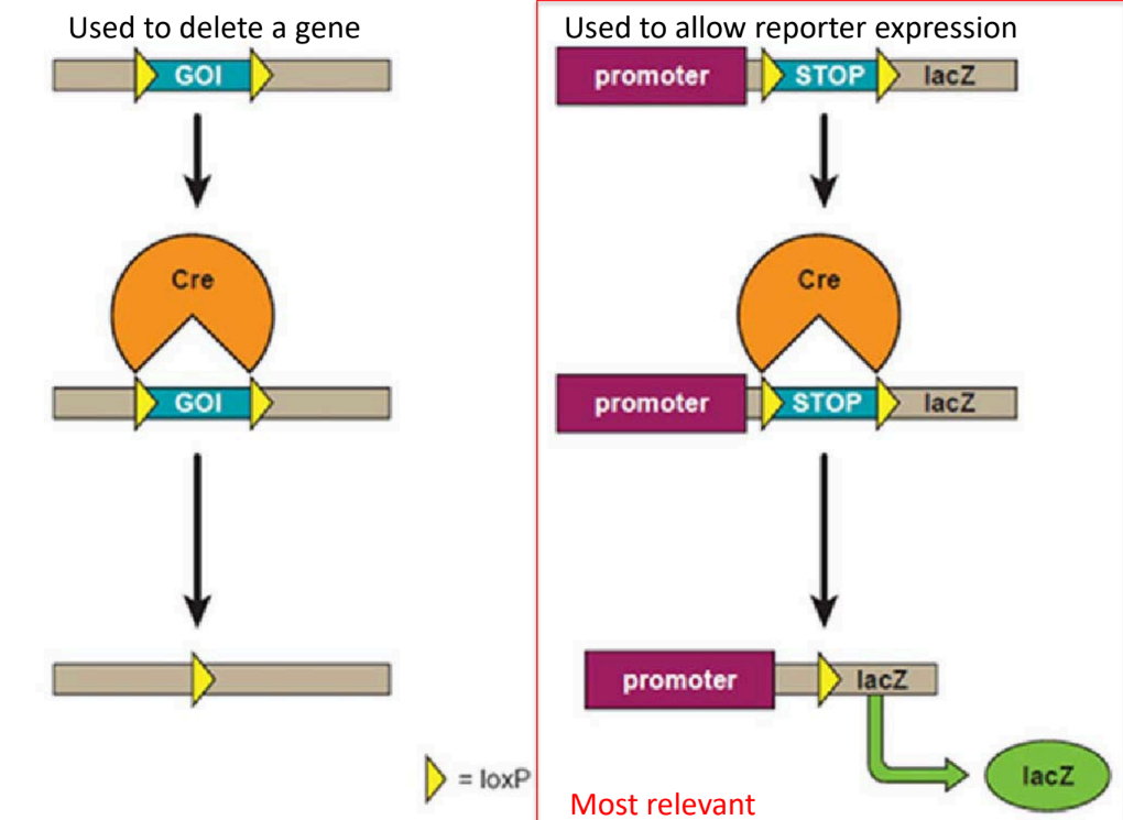
def. Cre-loxP system
def. Cre
2 functions of this system (1 of them more than the other)
is a conditional gene expression system (aka can control gene expression)
Cre — a recombinase enzyme that recombines DNA at specific LoxP sequences
_
functions:
mainly used for reporter expression (aka where the reporter gene is expressed as a result)
also used to delete a gene
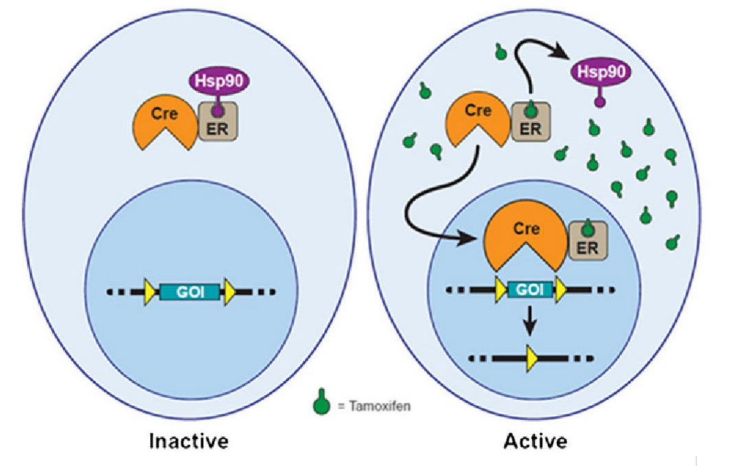
def. Cre-ER system
describe (2)
is an inducible gene expression system (aka gene expression changes depending on the conditions of place, time, type of stimulus, etc.)
Cre fused w/ an estrogen receptor (ER) (aka Cre-ER) WITHOUT tamoxifen ligand will transport the complex outside the nucleus — INACTIVE (so no gene expression)
Cre-ER with bound tamoxifen will transport the complex into the nucleus → to become ACTIVE (so induce gene expression)
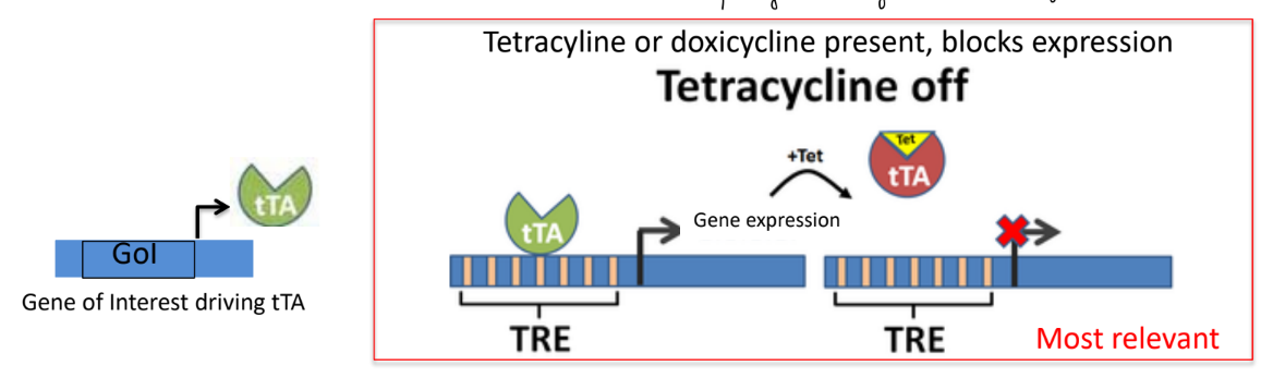
for tetracycline inducible system
def. tTA
function (1)
what happens if tetracycline or doxicycline is present
tTA — a yeast transcription factor (aka yeast TF)
binds to the TRE sequence to either turn on or off downstream gene expression
_
if tetracycline or doxicycline is present, then gene expression is blocked (SO tetracycline is NOT expressed)
(don’t think about v.v.)
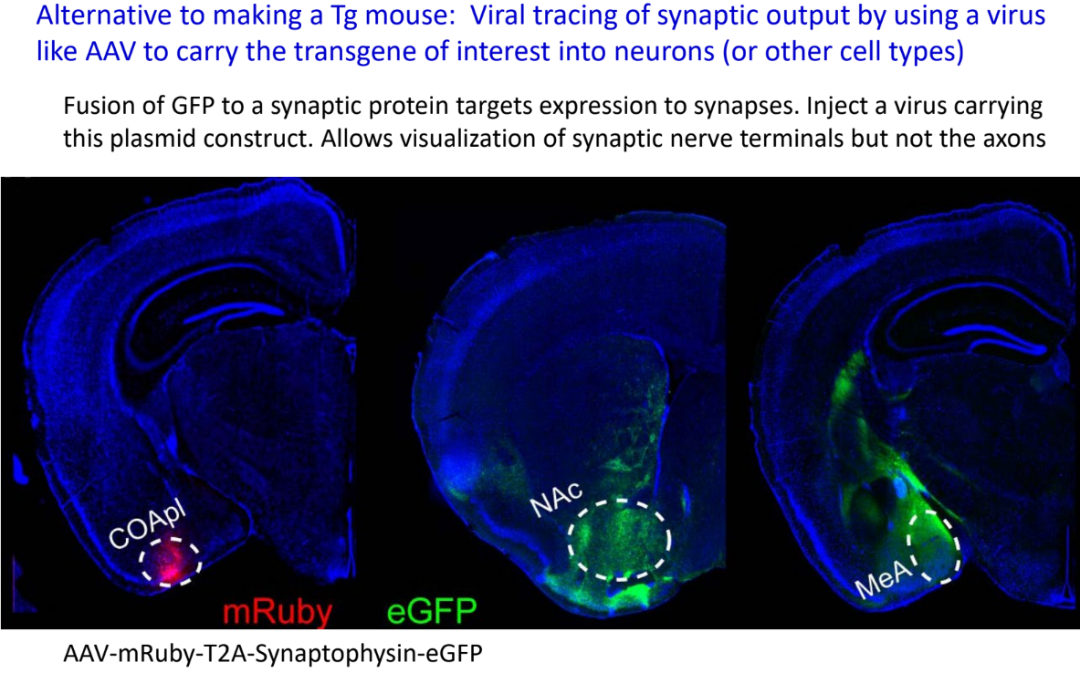
using synaptophysin (vesicle protein) & GFP together lets you view what?
is it antero- or retro- tracing
allows you to see the axon terminals, but not the axons
is anterograde tracing
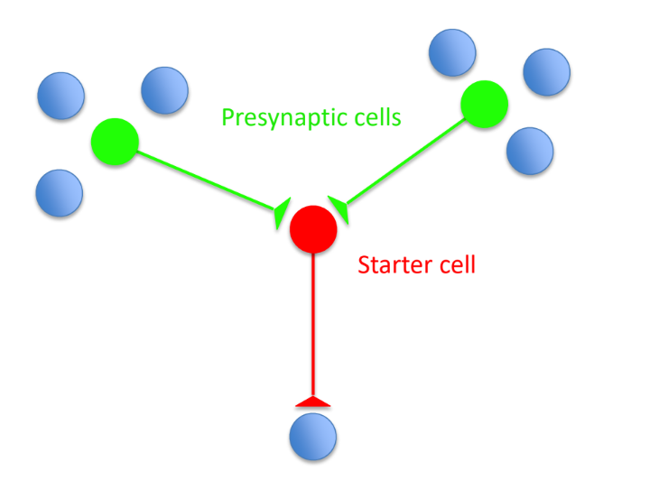
rabies travel antero- or retrogradely?
modified rabies can only jump __ synapse (aka used for __ __ grade tracing)
what does using modified rabies allow you to view?
rabies travel retrogradely
modified rabies can only jump ONE synapse (monosynaptic retrograde tracing)
used to view neurons that are synaptically connected to a specific neuron
(aka view the pre- neurons in green that are synaptically connected to the starter cell in red)
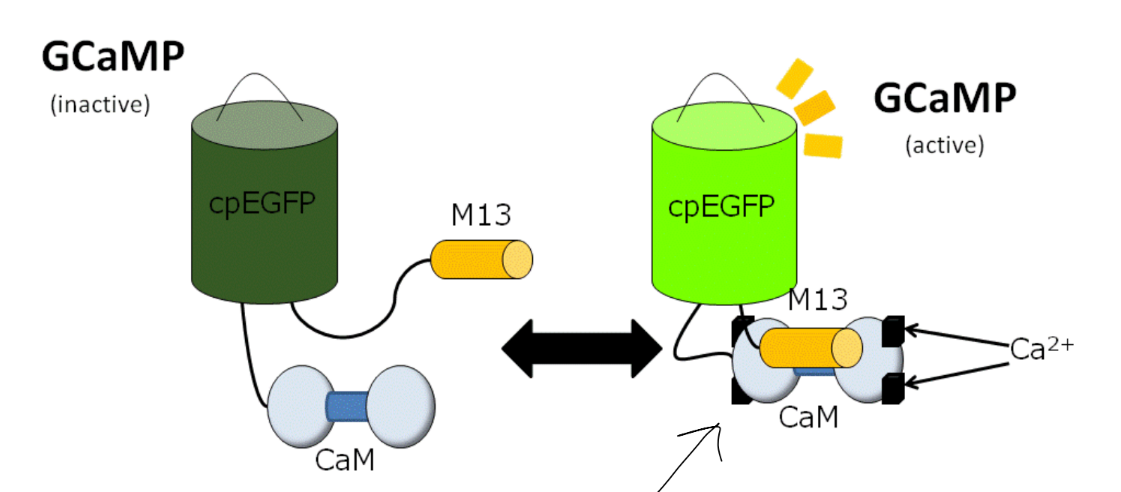
calcium imaging is a method to measure/record neuronal activity ← describe (1)
calcium imaging
when neurons fire APs, Ca2+ influx is detected by GCaMP (a fluorescent Ca2+ sensor)
2 ways to manipulate brain activity in order to test for CAUSALITY b/w brain activity and function ← name & def.
_
for classic approaches
name 2 ways to silence brain areas (LoF)
name 1 way to activate brain areas (GoF)
^ what’s the problem with classic approaches?
SO what do we use instead? (1 w/ 2)
loss of function — take something away in order to see if it’s required for something to work
deter. if it is required aka NECESSARY
gain of function — add something in to see if it causes some effect
deter. if it is able to cause an effect aka if it’s SUFFICIENT
_
classic approaches have little specificity
silence brain areas w/:
lesions (through chemical, electrical, or surgical removal)
drugs
(muscimol, GABAA agonists — inhibit neurons)
(NBQX glutamate antagonists — block excitation)
activate brain areas w/:
electrical stimulation
_
SO use modern approaches of chemogenetics and optogenetics
def. optogenetics
describe (2)
__
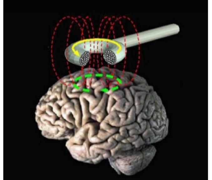
describe the TMS method (transcranial magnetic stimulation) (3) ← is neither opto- or chemogenetics
opto-: manipulate neuronal activity w/ light
by activating w/ Channel Rhodopsin (ChR2) → dep-
by silencing w/ Halo Rhodopsin (eNpHR) → hyper-
__
TMS
uses a magnetic field to cause current flow in target brain areas
either activate w/ brief pulses OR inactivate w/ high frequency pulses
lacks specific cell-type manipulation, SO instead acts broadly/over a large area of different cell types
(picture is of TMS)
summary
name & def. 3 types of experiments to test if a mechanism is BOTH “necessary” and “sufficient”
^ for 1 of the above, name 2 general ways to test if something is required/necessary OR able/sufficient through manipulation of brain area(s) (← aka 1 each)
test if BOTH necessary AND sufficient w/:
anatomy: antero- and retrograde tracers
functional activity: recording neural activity
manipulation: turning on and off activity in specific neurons to test causality via Loss of Function & Gain of Function
__
silence it to test if required/necessary for something to work
activate it to test if able/sufficient to cause a specific effect
def. neural plasticity / neuroplasticity
explains __ and __
also explains __ __ after brain damage
caused from/results from interactions b/w (2 aka 2)
__
name 4 key features that a learning mechanism must be/have
are long-lasting/long-term changes to the brain throughout a person’s life (i.e. during development and into adulthood)
explains learning & memory
explain cortical remapping after brain damage
caused from interactions b/w genes & environment (aka nature & nurture)
__
a learning mechanism must be/show:
long-lasting
caused by brief events
(aka able to encode something very quickly)
(brief events mean short/brief, high frequency bursts) ← tetanic stimulation
have synapse specificity
have associativity
(aka able to associate b/w 2 or more different events)
name the 2 different types of learning (1 of them has 2) & def. each
also name 2 types of non-associative learning
give ex. of associative learning
^ what 1 is hippocampus-dependent, what 1 isn’t
declarative (memory for facts)
procedural (memory for habits, skills, motor responses)
non-associative (change in response that occurs over time)
habituation — decreased response w/ repetitive stim.
sensitization — increased response w/ repetitive stim.
associative (associations form b/w 2 events)
ex: classical conditioning
_
declarative is hippocampus-dependent
procedural is NOT hippocampal-dependent
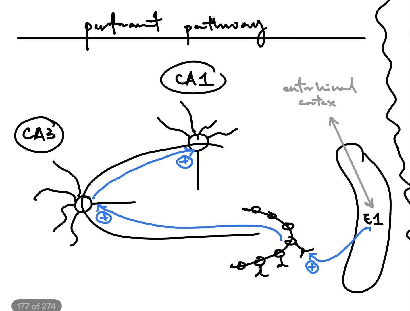
name & describe the pathway of this major hippocampal circuit
all the synapses use __ as their NT
^ this is a type of __ excitation
^^ DRAW THIS MAJOR HIPPOCAMPAL CIRCUIT
__
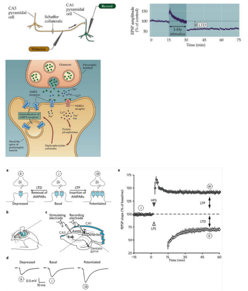
electrophysiological studies of rodent hippocampus led to discovery of __ (def. term)
we observed extra- _PSP
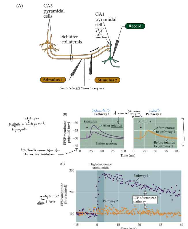
describe the simple vs. complex setup used in hippocampal recording to study LTP
trisynaptic prime circuit (all 3 synapses use glutamate as their NT)
perforant path (from the entorhinal cortex) → dentate gyrus → CA3 pyramidal cell → CA1 pyramidal cell
^ is a type of feedforward excitation (can refer to a previous flashcard if needed for picture)
__
discovery of LTP (long-term potentiation) — is enhancement/increase of the post- response ← cellular and molecular mechanism for learning & memory
observed extra- EPSP (b/c all 3 synapses release & use glutamate)
simple setup: 1 stimulating & 1 recording electrode
complex setup: 2 stimulators/stim. electrodes AND take both intra- & extra- recordings
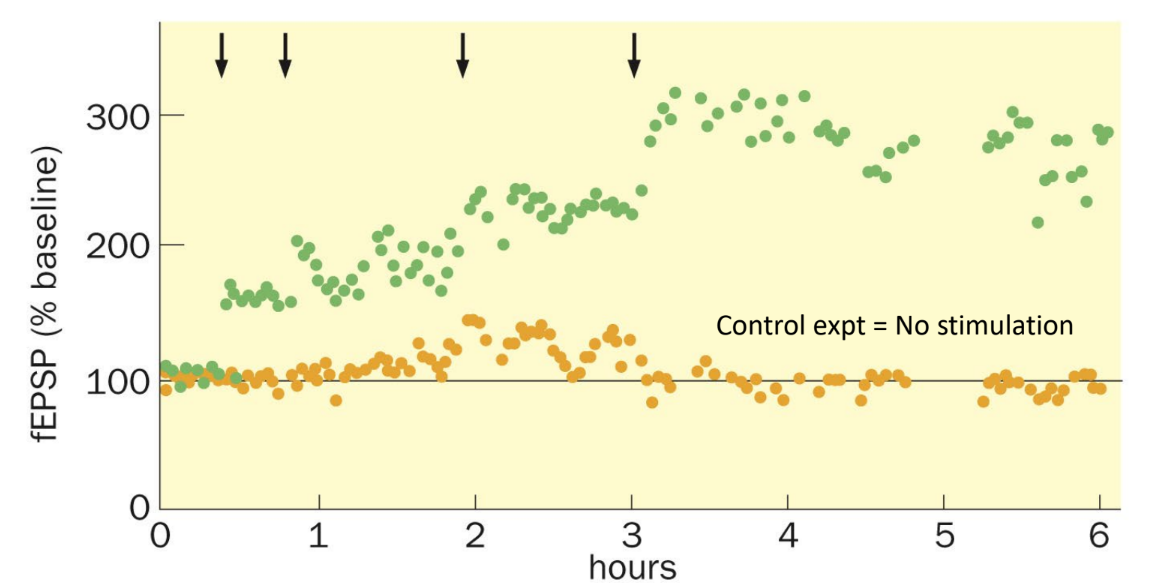
def. tetanus
can a tetanus induce a LTP/enhancement of the post- response?
(what is fEPSP?)
tetanus — a brief, high frequency (tetanic) stimulation
yes, a tetanus can induce a LTP/enhancement of the post- response
_
fEPSP is a field EPSP (aka collective synaptic strength of multiple neurons)
(arrows point to where tetanus occurs)
diff. b/w stable & unstable potentiation
_
brief sleep deprivation will impair the maintenance/stability of (1) in the hippocampal __ region
stable potentiation is long-lasting (LTP)
unstable potentiation is short-term/lasting potentiation
_
brief sleep deprivation will impair the maintenance/stability of cAMP-PKA signaling-depending LTP in the hippocampal CA1 region
for LTP at the CA3-CA1 synapse via (two) Schaeffer collateral axons
describe the experiment that observes LTP in CA1 hippocampal neurons if one stimulus on each of the Schaeffer collateral axons (← 1 w/ 2)
_
what is the EPSP amplitude?
you apply a stimulus to both axons, but one has tetanic stimulation (brief, high frequency stim.) while the other is the control
want to see if the pathway w/ tetanic stim. on CA3 induces a LTP on CA1 (seen if that pathway’s response increases)
if the control pathway doesn’t change, then the LTP is input-specific
(aka ONLY the tetanized pathway shows enhancement/increased response)
(aka strong stimulation is specific to that synapse/pathway)
__
EPSP amplitude is usually the slope of the EPSP
(picture shows pathway 1 w/ tetanic stim. & pathway 2 as control)
tetanized of ~100 Hz (aka activation pulses per sec.)
pathway 1 has time to recover b/w stimuli SO no habituation occurs
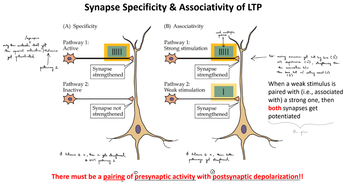
def. synapse specificity/input-specificity & associativity of LTP
_
what are the coincidence detection conditions needed for LTP?
what is the coincidence detectors?
specificity:
only the synapses that get the tetanized activation get potentiated/strengthened
(independent from e/o)
associativity:
when a weak stimulus is paired w/ a strong one (tetanic stim.), then BOTH synapses gets potentiated/strengthened
^^ doesn’t matter which pathway has which type of stim. b/c the concept still applies
__
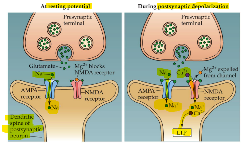
LTP only occurs if there is simultaneous pairing/coincidence of pre- glutamate release & post- depolarization
coincidence detectors are NMDARs (meaning that NMDARs only open when both conditions are met simultaneously)
def. Hebb’s postulate
what is Hebbian’s rule of plasticity
__
pre- release of glutamate binds to what? post- dep- is driven by what?
_
explain the voltage “gate” of NMDARs
__
what happens if:
only glutamate binds to NMDAR
there’s both post- dep- from Na+ influx through AMPARs & glutamate bound to NMDARs
learning occurs when pre- activity is paired with post- activity
rule: neurons that fire together, wire together
“wire together” means strengthened association/synapse OR grows more connections/synapses
^ ALSO “out of sync, fail to link”)
__
pre- glutamate release, where glutamate binds to the NMDAR ligand binding site
AND
post- dep- caused by AMPARs’ Na+ influx
_
voltage “gate” is due to the voltage-dependent Mg2+ block of NMDARs
__
if only glutamate binds to NMDAR:
then only Na+ influx (monovalent cations)
if both post- dep- from Na+ influx through AMPARs & glutamate bound to NMDARs:
then Mg2+ block is removed → allow for BOTH Na+ and Ca2+ influx (divalent cations)
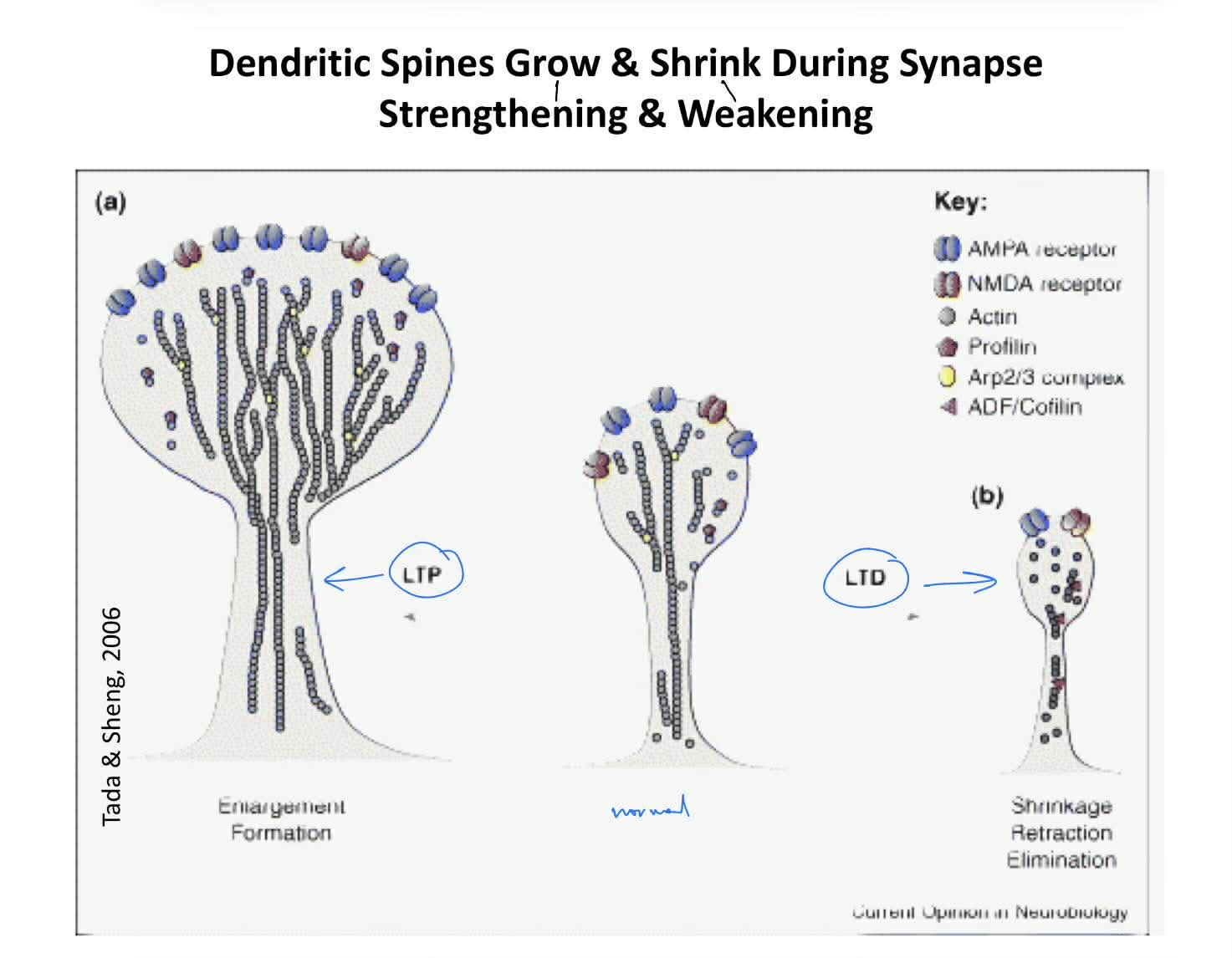
do dendritic spines grow or shrink when there is synapse strengthening vs. weakening? (← aka match them)
__
synapse strengthening during LTP (and morphological plasticity of spines) depends on __ __ cytoskeleton
what do substances that block this "- -” cytoskeleton do?
dendritic spines:
grow during synapse strengthening (LTP)
shrink during synapse weakening (LTD)
^ identify by comparing the size (expansion/shrinking) and/or # of dendritic spines (new or no new spines)
__
synapse strengthening during LTP (and morphological plasticity of spines) depends on dynamic actin cytoskeleton
pharmalogically blocking actin filament dynamics cause LTP to be unstable
(aka still have LTP, but it in unstable / fades off more quickly ← b/c unstable is short-term potentiation)
name the 2 key features of NMDARs
for NMDARs, what do you need to induce LTP? (1)
blocked by Mg2+ ion, unless post- membrane dep-
is Ca2+ (and Na+ and K+) permeable
_
CANNOT cause LTP w/o post- Ca2+ influx (aka NMDARs need Ca2+ influx in order to induce LTP
(picture shows that the post- neuron is affected specifically at its dendritic spines)
describe the steps/cascade of molecular mechanisms that induce & maintain synapse strengthening (5)
NMDARs allow Ca2+ influx
activates calcium-dependent protein kinases: PKC & CaMKII
inserts more AMPARs into the post- membrane
aka exocytosis of AMPARs from endosome’s reserve pool
subsequent pre- glutamate release induces a stronger post- response (LTP)
b/c have stronger post- dep- due to more AMPARs in post- membrane
potentiation of synapse strength is converted to a long-lasting form (days or more) via changes in gene expression (which is mediated by TFs, like CREB)
describe the 1 piece of evidence that support the idea that NMDARs are necessary for LTP
__
explain this:
initial activation of AMPAR can provide enough dep- to allow NMDAR to become activated quickly
_
what happens if the synapse has AMPAR, but no NMDAR?
silencing (aka removal) of NMDARs prevents LTP induction
__
it means that AMPAR provide small depolarization (from Na+ influx) that is strong enough to remove the NMDAR Mg2+ block, SO if there’s pre- glutamate release, then NMDAR can immediately activate
_
if synapse has AMPAR but no NMDAR, then post- still depolarizes BUT no Ca2+ influx → so no LTP is induced
LTD (long-term depression) in hippocampal CA1 is caused from activation of __ __ AND removal of __ (__)
LTD is caused by __ frequency stimulation
LTD (long-term depression) in hippocampal CA1 is caused from activation of protein phosphatases AND removal of AMPARs (endocytosis)
LTD is caused by LOW FREQUENCY STIMULATION
for CORRELATION studies b/w LTP and memory
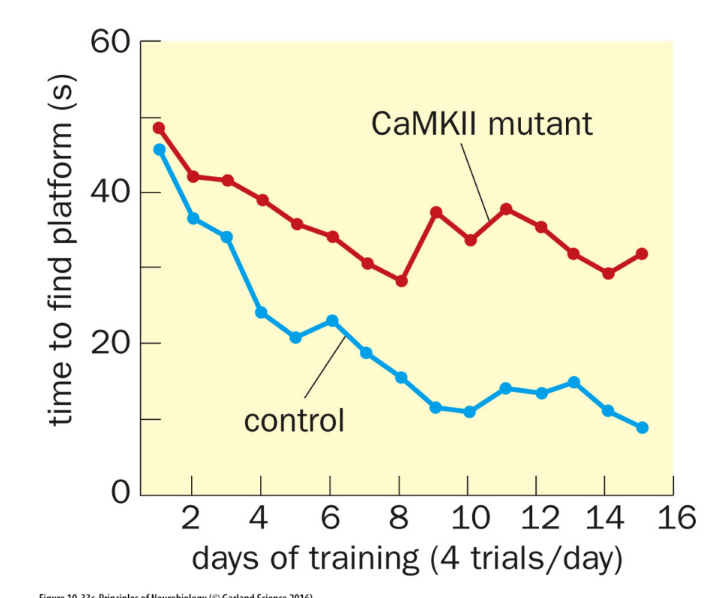
explain the Morris water maze experiment
conclusion?
used to test the memory of rats on where the hidden platform is (doesn’t like being wet)
control rat: finds the hidden platform
hippocampal-lesioned rat: has difficulty in finding the platform
also tests if control w/ NMDAR vs. AP5 that blocks NMDARs (which are important for LTP learning and memory) will affect performance
control spends most time in the quadrant w/ the platform
AP5 spends about the same amount of time in each quadrant (AP5 is a NMDA antagonist)
_
since NMDAR are necessary for LTP, then memory should be impaired if NMDAR are not functional
so control group should take less time to find the platform compared to the NMDAR knockout group
is the fact that “activated calcium-dependent protein kinases are necessary for LTP” correlational or causation? why?
give example relating to the Morris water maze experiment
more correlational than causal
b/c protein kinases are active when LTP is induced, BUT this fact alone does NOT prove that active kinases induce LTP
ex: CaMKII mutant group spends more time to find the platform than the control group