lec 12 - limb development
1/72
There's no tags or description
Looks like no tags are added yet.
Name | Mastery | Learn | Test | Matching | Spaced | Call with Kai |
|---|
No analytics yet
Send a link to your students to track their progress
73 Terms
3 principle axes of the limb
proximo-distal (PD)
AP
DV
PD axis runs from
humerus → digits
parts of the PD axis
stylopod, zeugopod, autopod
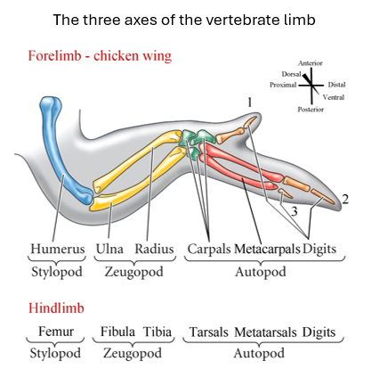
the AP axis runs from
thumb → pinkie
DV axis runs from
back of hand → palm of hand
the early limb bud consists of _____ surrounded by _____, with a ___
mesenchyme cells
an ectodermal (epithelial) layer,
thickened distal ridge called the AER
what are the 3 key signaling centers and which axes do they control
ZPA → AP
AER → PD
Dorsal ectoderm → DV
ZPA stands for
Zone of polarizing margin
the ZPA expresses ___ and specifies _____ via ____
Shh
AP digit identity
morphogen gradient
AER =
apical ectodermic ridge
the AER expresses ___ and specifies _____
FGF8
PD outgrowth and patterning
the Dorsal Ectoderm expresses ____ and specifies ____
the Ventral Ectoderm expresses ____ and specifies ____
Wnt7a; dorsal identity
BMP/Eg1; ventral identity
together pattern the DV axis
limbuds first appear as
bumps at defined positions along the AP axis
Limb bud positioning is determined by
Hox gene products in the lateral plate mesoderm
hox genes - forelimb position determed by
Hox4 and Hox5
hox genes - hindlimb position specfied by
Hox9-100
____ provide positional information interpreted by Hox proteins
signaling gradients like RA or FGF
Hox gene products activate
limb type-specific TFs
limb type-specific TF function
determine forelimb vs hindlimb identity
limb type-specific TFs - what specifies the forelimb
Tbx5
T-box TF
limb type-specific TFs - what specifies the hindlimb
Tbx4 + Pitx1
T-box TFs
Describe the takeuchi experiment
Ectopic Tbx5 expression in the leg → feathers normally restricted to wing appear.
Ectopic Tbx4 in the wing → four digits (like leg).
Demonstrates these TFs can re-specify some limb identity features.
describe the mingullion experiment
Forced expression of Pitx1 transgene in forelimb → elbow develops a knee-like articulation. Confirms Pitx1 contributes to hindlimb-specific morphology.
describe the limb bud initiation loop
Tbx4/5 induces Fgf10 in the lateral plate mesoderm/early limb bud mesenchyme via Wnt signaling
Fgf10 signals to the overlying ectoderm via Wnt 3a to induce Fgf8 in the AER (PD axis)
Fgf8 signals back to the limb bud mesenchyme to maintain Fgf10 expression, creating a self-sustaining feedback loop
this loop drives PD outgrowth
Fgf10 KO in mouse →
complete absence of limbs
descirbe the Cohn and Tickle experiments
an Fgf-soaked bead grafted into the interlimb flank is sufficient to induce an entire ectopic limb
cohn and tickle experiments - what was the identity of the limb they grew in the interlimb flank
identity of the limb (wing-like vs leg-like vs chimeric) depended on how close the bead was to the Tbx5/Tbx4 expression domains, which expanded into the new bud.
conclusions of the cohn/tickle experiments
Fgf induction is sufficient in inducing limb growth
Fgf does not directly induce Tbx 4/5
The AER is essential for ____ outgrowth
PD
what did Saunders test for
what would happen if you surgically removed the AER
what happens if you surgically remove the AER
timing-dependent
remove early → only humerus forms
the later the removal, the more distal the elements that are lost become
the later the removal, the more complete the skeleton
Saunders AER removal experiment - conclusion
AER is required for PD outgrowth
skeletal elements are laid down in a proximal-to-distal order
three models of PD patterning
progress zone model
two-signal model
signal-progress zone model
progress zone model was found by
summerbell and wolpert
describe the progress zone model
Limb patterning depends on timing/how long you stay in the progress zone
Undifferentiated cells at the distal tip (progress zone) are maintained by AER-Fgfs
Cells leaving early → proximal fate (stylopod)
Cells leaving late → distal fate (zeugopod, then autopod)
Timing relates to progressive Hox gene activation
describe the two-signal model
pattern depends on opposing gradients
high RA from the flank specifies stylopod (proximal fate)
high FGF from AER specifies autopod (distal fate)
as the limb grows, both drop → zeugopod (intermediate fate)
what is the issue with the two-signal model
This implies that the proximal and distal structures are developed first, and then intermediate structures are formed.
However this contradicts the AER removal experiment and the Hox gene layout (which patterns the same way as the body forms, humerus → digits), as both show that limb patterning occurs from proximal to distal.
what is the current consensus on PD limb patterning
signal-progress zone model - limb patterning depend on both timing and morphogen concentration
describe the signal-progress zone model
high RA early
prevents progress zone from forming
specifies proximal stylopod before progress zone is established
FGFs antagonize RA from AER, allowing a distal progress zone to form
once the progress zone exists, timing of the cells leaving the progress zone specifies zeugopod and autopod
_____ are colinearly expressed along the PD axis
HoxA and HoxD gene paralogs
Hox genes in PD patterning
____ → stylopod
____ → zeugopod
____ → autopod
Hox10
Hox11
Hox12/13
in what order do HoxA and HoxD genes express along the PD axis
10 → 13, 3’ → 5’, proximal → distal
what are hox paralogs
each Hox gene has a number and a letter. two genes with the same number but a different letter are paralogs
HoxA11 + HoxD11 + HoxC11 triple KO
zeugopod fails to form (both fore- and hindlimb)
HoxA13 + HoxD13 double KO
autopod cartilage fails to form (soft tissues remain but no phalanges)
Hox gene paralog KO to cause zeugopod to fail to form
HoxA11/D11/C11 triple KO
Hox gene paralog KO to remove phalanges and cartilage
HoxA13/D13 double KO
ZPA
zone of polarizing activity (aka polarizing region)
ZPA is important for ___ patterning
AP
who discovered the ZPA and how
Saunders and Gasseling
grafted tissue from posterior margin to anterior margin of host wing bud
result: mirror image duplications of digit pattern (321123)
grafts of only a few cells → only digit 1 induced
conclusion: the region must produce a morphogenic signaling molecules in the posterior margin - ZPA
ZPA morphogen hypothesis
predicted the ZPA produced a diffusible morphogen forming a posterior → anterior gradient
high ZPA concentration = posterior digit identity (pinkie)
intermediate = digit 2
low = digit 1/thumb
what is the ZPA morphogen
Shh
How do we know that Shh is the ZPA morphogen + who found out
Riddle et al
Shh-expressing cells/Shh beads grafted to the anterior margin produced concentration-dependent digit duplication - same as ZPA grafts
Shh specifies digit identity in tissue adjacent to ZPA
non-cell-autonomous
caused by external factors
what did Towers’ GFP fate mapping find
ZPA cells do not contribute to the duplicated digit skeleton.
Shh acts on host tissue in a non-cell-autonomous manner.
Shh takes 12 hours to pattern the limbs
how long does it take for Shh to specify digits 1→3 + how do we know + who did it
12 hours
towers et al
Cyclopamine (blocks Shh pathway) applied at 4hr intervals.
3 administartions of cyclopamine (12 hrs) for all 3 chick wing digit identities to be specified, from digit 1→3
wolley et al
made a mathematical model of Shh gradient, found the same timeframe
how are more than 3 digits patterned
timing - length of Shh exposure
ZPA cells are specified as digit 4 and 5 because they were exposed to Shh the longest
Shh signaling operates through
Gli TFs
Describe the Gli3A/R gradient + what it depends on
Gli3 activator posteriorly, Gli3 repressor anteriorly, forms a gradient
depends on Shh concentration (high posterior, low anterior)
Shh/Gli3 targets
Ptch1
Gremlin1
Tbx2
Tbx3
What are the current strongest downstream Shh signaling candidates for specifying digit identity
Tbx2 - digit 4
Tbx3 - digit 3
what is AERMF
BMP antagonist secreted by Gremlin (Shh target)
explain how AP and PD patterning are coordinated
context
AER-FGF4/8 are required for Shh expression
BMP inhibits AER-FGF4/8
Gremlin1 encodes BMP antagonist AERMF
gremlin feedback loop
Shh → gremlin
gremlin —| BMP, which → AER-FGF4/8
AER-FGF4/8 → Shh
how is Shh only expressed in posterior limb?
ZRS (ZPA regulatory sequence) controls it
TFs like Hand2 and HoxA/D bind to ZRS
chromosome looping → ZRS contacts Shh promoter → TFs swithc on Shh transcription posteriorly
transcriptional repressors also bind ZRS to prevent Shh from being expressed anteriorly
ZRS mutation + example
Shh expressed ectopically at anterior margin → polydactyly
ex: hemingway cats wihth polydactyly on anterior margins of paw
____ controls DV polarity
the ectoderm
how do we know that the ectoderm instructs DV identity
classic chick experiment - ectoderm was removed from the mesoderm of an early chick wing bud, rotated 180 degrees (flipped DV), then replaced.
result: DV polarity of mesoderm was reversed, even though only the ectoderm was rotated - showing that the ectoderm instructs mesoderm DV identity
what is the default state - dorsal or ventral
ventral
which signaling molecules are important for DV patterning
Wnt (Wnt7a)
BMPs
explain how DV patterning is set up
dorsal ectoderm: Wnt7a → TF Lmx1b → dorsal fates
ventral ectoderm: BMPs → Engrailed1 —| Wnt7a ventrally → ventral fates
Wnt7a mutant mouse
Lmx1b is not induced, whole limb becomes ventral (double ventral pattern
Lmx1b mutant
same double ventral outcome as Wnt7a mutant - Wnt7a can express but cannot act without Lmx1b
Engrailed1 mutant
Wnt7a expressed in the entire ectoderm → Lmx1b induce everywhere → whole limb becomes dorsal (double dorsal)