5.2.1 Nervous system
1/44
There's no tags or description
Looks like no tags are added yet.
Name | Mastery | Learn | Test | Matching | Spaced | Call with Kai |
|---|
No analytics yet
Send a link to your students to track their progress
45 Terms
Overview of the nervous system
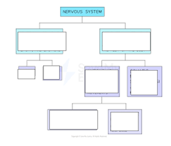
CNS = brain + spinal cord
PNS = neurones that connect CNS to rest of body
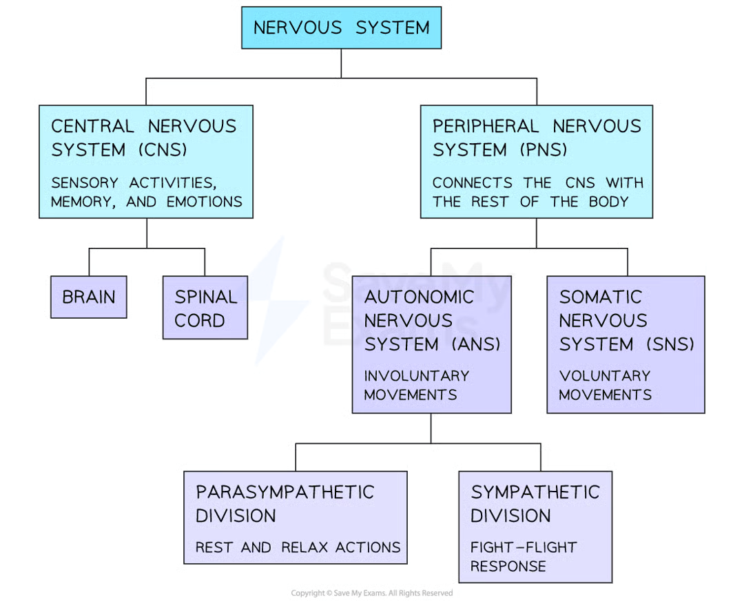
Difference between somatic and autonomic nervous system
Somatic NS | Autonomic NS |
Voluntary actions | Involuntary actions |
Carries impulses to skeletal muscles | Carries impulses to endocrine glands, smooth muscle + SAN in RA of heart |
SNS neurones fully myelinated | ANS only myelinated from ganglion to CNS |
1 neurotransmitter released – Acetyl Choline | 2 NTs released – Ach + noradrenaline |
Doesn’t divide any further | Divides into sympathetic + parasympathetic |
differences between parasympathetic + sympathetic NS
structure of human brain
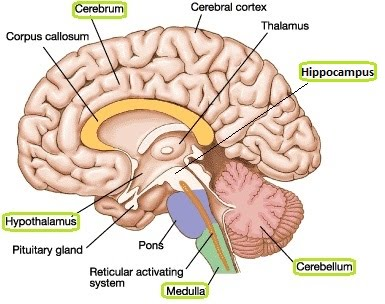
function of cerebrum/cerebral cortex in the brain
cerebrum
has 2 hemispheres —> Left hemi receives sensory inputs from receptors on RHS of body
surface covered by cell bodies
extensively folded
controls conscious thought, language, memory, emotional responses + intelligence
function of cerebellum
controls non-voluntary movements + muscle coordination
function of medulla oblongata
controls autonomic (involuntary) activities e.g heart rate, blood pressure, peristalsis (relaxing/constriction of gut muscle)
function of hypothalamus + pituitary gland in brain
hypothalamus:
controls thermoregulation, osmoregulation, homeostasis, secretion of hormones
pituitary gland:
posterior lobe: stores + secretes hormones made by hypothalamus
anterior lobe = makes + secretes hormones
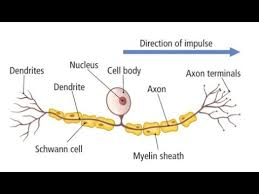
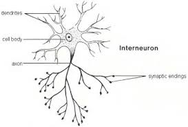
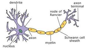
what are the 3 general structures of neurones
cell body
contains nucleus
has lots of ER + mitochondria for production of neurotransmitters
Dendrons
short extensions of cell + cytoplasm from cell body —> increases SA to receive nerve impulses from other neurones
divides into smaller branches = dendrites —> transmit electrical impulses TOWARDS cell body
Axon
single, elongated nerve fibre that extends from cell body
transmits electrical impulses AWAY from cell body
surrounded by plasma membrane + can be myelinated
sensory neurone
carries impulse from receptor to relay neurone
relay neurone
carries impulse from relay (in CNS) to motor neurone
motor neurone
carries impulse from motor to effector (muscle to contract, gland to secrete hormones)
what’s the difference between an axon and a dendron
an axon always goes AWAY from the cell body + to the effector
dendron always goes TO cell body from a receptor
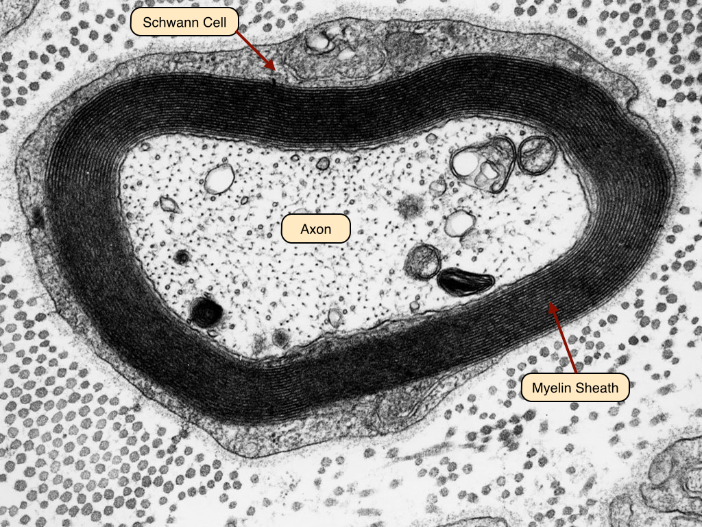
structure + function of myelin sheath
made from many layers of plasma membrane synthesised by Schwann cells
these layers wrap around axons/dendrons
they insulate the axon, making the myelinated parts impermeable to Na+/K+
small gaps between Schwann cells = Nodes of Ranvier —> sites of depolarisation that allows movement of Na+/K+
enables salutatory conduction
what’s the importance of the myelin sheath
increase lengths of local currents —> when Na+ enter the axon during depolarisation, they generate local electrical currents that spread along the axon and trigger the next node of ranvier to be depolarised —> if local currents can travel further, fewer action potentials need to be generated + depolarisation only occurs at the nodes of ranvier
significance: faster conduction of nerve impulses + less energy is used as fewer Na+'/K+ pumps needed to restore resting potential
what happens if there’s no myelin sheath
no electrical insulation of axon
axon becomes permeable to Na+/K+ ions
no saltatory conduction
shorter local circuits - depolarisation happens along whole membrane, more APs need to be generated along the axon
slower transmission of electrical impulses
what is an action potential
the change in electrical potential associated with the passage of an impulse along the membrane of a neurone
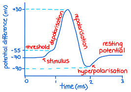
what stages are in an action potential
Ressting potential
depolarisation
repolarisation
hyperpolarisation / refractory period
what happens in depolarisation
occurs when a neurone is stimulated
Na+ channels in axon membrane open
Na+ diffuse into the membrane, down the electrochemical gradient
inside of axon becomes LESS negative
if membrane potential reaches -50mV (threshold level), this will stimulate more voltage gated Na+ channels to open —> more Na+ diffuse into cell
what happens after depolarisation (propagation along axon)
the Na+ ions diffuse sideways along axon
this triggers Na+ channels in the next region (next node of ranvier) of axon to open
Na+ diffuse into axon + membrane becomes LESS negative
this causes wave of depolarisation to travel along the axon

what happens in repolarisation (peak of AP)
all voltage gated Na+ channels close
voltage gated K+ channels open
K+ diffuse out of the axon, down the electrochemical gradient
inside of the membrane becomes MORE negative
Na+/K+ pumps restores ionic balance
what happens during hyperpolarisation
K+ channels are slow to close, so diffusion of K+ out of the axon continues until the inside of the membrane becomes more negative than the resting potential (-70mV)
voltage gated K+ ion channels close + action of Na+/K+ pumps restores membrane to resting potential
What is resting potential
when the neurone is not transmitting an impulse but it’s still active
what happens in a resting potential
active transport of 3 Na+ OUT of axon, and 2K+ IN via proton pumps
inside of membrane is MORE negative than outside
so Na+ slowly diffuses back in to membrane and K+ diffuses out via non-voltage gated channels
what is the refractory period
delay between one AP and the next to prevent the overlapping of impulses
importance of the refractory period
ensures action potential travels in only 1 direction - if more depolarisation occurs during 1 AP, this can cause the AP to travel backwards - never reaching target cell
ensures impulses aren’t sent too quickly - this limits frequency of APs
prevents axon becoming depolarised after an AP so no impulses overlap
how does a larger stimulus affect the AP
larger stimulus increases the frequency of APs
but it does NOT change the size of the AP
what is the all or nothing response
an AP can only occur if the threshold level is reached
structure of a synapse
synaptic knob = lots of mitochondria + SER to produce NT
synaptic vesicles = contain NT - fuses with presynaptic membrane, allowing NT to be released into synaptic cleft via exocytosis
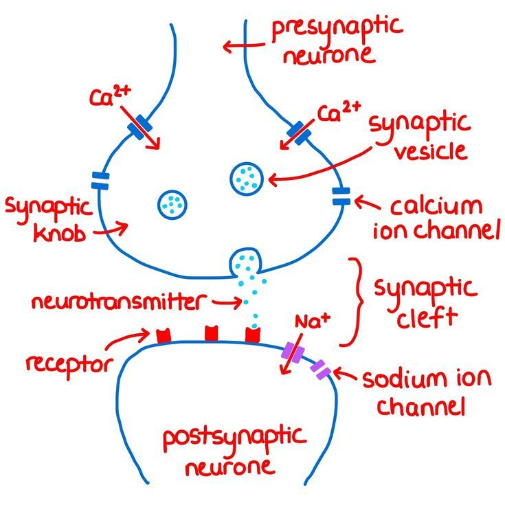
How does a nerve impulse cross a synapse
axon potential reaches end of presynaptic neurone
causes depolarisation of presynaptic membrane
Ca2+ ion channels open + C2+ diffuses into presynaptic knob
influx of Ca2+ activates the synaptic vesicles to fuse with the presynaptic membrane
causes NT to be released via exocytosis into synaptic cleft
NT diffuses across cleft, down the conc gradient
NT binds to specific, complementary receptors on post synaptic membrane
stimulates Na+ channels (on post sm) to open —> Na+ diffuse in to Post neurone
if sufficient Na+ enters that overcomes the threshold value, an AP is generated
impulse is propagated along post-synp neurone
what are the 2 types of neurotransmitters
excitatory - causes depolarisation of postsyn neurone
if threshold is reached, AP is generated
AcH, glutamate
Inhibitory - causes hyperpolarisation of postyn membrane
prevents AP being generated
GABA, glutamate (rod cells only)
What happens to the neurotransmitter once an AP has been generated in post synaptic membrane
must be broken down to prevent stimulus being maintained
AcH is hydrolysed by acetylcholinesterase into choline + acetate (ethanoic acid)
Ach is released from receptors on postsyn membrane
Na+ ion channels close
choline + acetate reabsorbed back into synaptic knob by endocytosis
Ach is reformed by aerobic respiration using ATP
Ach repackaged into vesicles —>NT are recycled
types of postsynaptic potentials
roles of synapses
ensure impulses travel in 1 direction - NT receptors only found on post syn membranes so diffusion of impulses can only occur from pre - post
synaptic divergence -
what’s the nature of a reflex arc and how does this compare to a normal reaction
rapid, involuntary responses to stimuli to prevent harm to body —> not taught
different to normal reaction as it’s INVOLUNTARY
what types of reflex arcs are there
blink reflex - rapid closing of the eye in response to stimulation of the cornea e.g bright lights/dust in eye
plantar reflex - used as a diagnostic tool to assess NS damage/consciousness —> sole of foot stimulated with blunt object —> NORMAL response = foot flex down, ABNORMAL = foot flexes up
iris reflex - involuntary changing of pupil size in response to different light intensities (protects retina) —> NORMAL = pupils constrict to same degree, ABNORMAL = constrict differently
what are the 2 types of brain damage
traumatic - severe head injury e.g car crash, fall
non-traumatic - not caused by head injury e.g stroke, infection
what techniques are used to assess brain + spinal cord damage
MRI
functional MRI
CT scans
PET scans
EEGs
why are brain scans important
detects location and extent of injury
necessary treatments can be carried out quickly
needed to diagnose brain damage
effect of brain + spinal cord damage
what drugs can be used to treat brain damage
effect of recreational drugs
drug dependency
how can CT scans be used to detect the location/extent of injury in nervous system
uses X-rays to build up a detailed 3D image of brain/spinal cord—> shows areas with bleeding/poor blood supply
how can MRIs/fMRIs be used to detect the location/extent of injury in nervous system
MRIs
uses magnetic fields to detect swelling/inflammation/areas of demyelination
shows difference between healthy and damaged areas
RISKS: ionising radiation, not suitable if pt has metal implants, pt must be still
fMRIs:
shows difference between healthy and damaged areas
detects changes in blood flow
no ionising radiation used, non invasive
RISKS: pt must be still