Basics. Exam 3.
1/45
Earn XP
Description and Tags
Joint Type, Articulating Surfaces, Ligaments, Movements, Innervation, Blood Supply. [Lec 7, 8,9] done as of 4/13/26]
Name | Mastery | Learn | Test | Matching | Spaced | Call with Kai |
|---|
No analytics yet
Send a link to your students to track their progress
46 Terms
Classification of Sacroiliac Joint (2x)
Anteriorly - synovial
Posterior - syndesmosis
Articulating surfaces of the Sacroiliac Joint
auricular surfaces of sacrum and ilium
Ligaments of the Sacroiliac Joint (8x)
anterior sacroiliac lig
sacrospinous lig
iliolumbar lig
lateral lumbosacral lig
posterior sacroiliac lig
interosseous sacroiliac lig
sacrotuberous lig
longitudinal posterior sacroiliac lig
Innervation of the Sacroiliac Joint
Branches from:
anterior and posterior rami of 1st, 2nd sacral spinal nerves
superior gluteal nerves
May Contribute:
obturator nerve
lumbrosacral trunk
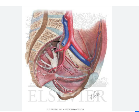
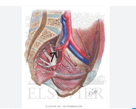
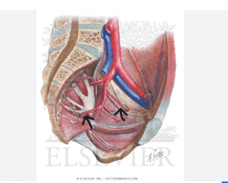
Blood Supply Pathway of Sacroiliac Joint
left common iliac → internal iliac → posterior division → iliolumbar, lateral sacral, superior gluteal a.
Main Function of the Sacroiliac Joint (2)
relieves stress on pelvic girdle during movement of trunk and lower appendage during muscle contraction
Handles body weight and ground reaction forces
Classification of Hip Joint
Synovial Ball and Socket
Articulating surfaces of the Hip Joint
acetabulum - lunate surface, fossa, fibrocartilage fat
acetabular labrum
head of femur
Capsule Attachments of Hip Joints (4)
neck of femur
greater trochanter and intertrochanteric crest
intertrochanteric line
acetabulum
Movements at Hip Joint
Flexion, extension, abduction, adduction, internal rotation, external rotation, circumduction
Ligaments of Hip Joint (6x)
transverse acetabular lig
lig of head of femur (round lig)
iliofemoral lig
pubofemoral lig
ischiofemoral lig
zona orbicularis
Innervation of Hip Joint (4)
femoral n
obturator n
nerve to quadratus femoris
superior gluteal n
First Blood supply pathway of Hip Joint
left common iliac → internal iliac → posterior division → superior gluteal a.
second blood supply pathway of the hip
left common iliac → internal iliac → anterior division → obturator a., interior gluteal a.
Third Blood supply pathway to Hip Joint
(left) common iliac → external iliac → femoral → profunda femoris → medial circumflex femoral, lateral circumflex femoral → ascending branch
Function of Hip Joint
support weight and movements of upper body
supports lower body movements
The knee joint complex is composed of what two joints?
Tibiofemoral, Patellofemoral
Classification of Joint Type Knee (include both)
Tibiofemoral - double condyloid
Patellofemoral - synovial
The knee complex as a whole is classified as
modified hinge joint
Ligaments of Knee Complex (10x)
Transverse lig of knee
Coronary lig
Anterior cruciate lig
Posterior cruciate lig
Posterior menisco-femoral lig
Tibial collateral lig
Fibular collateral lig
Patellar lig
Oblique popliteal lig
Arcuate popliteal lig
Movements of knee
Flex/Ext
Med rot/Lat rot
Abd/Add
Innervation of Knee Complex (4x)
Branches from
Femoral n
Obturator n
Common peroneal n
Tibial n
First blood supply pathway of Knee joint
descending branch of lateral circumflex femoral a.
Second blood supply pathway of Knee joint complex
articular branch of Descending genicular a
Third blood supply pathway of knee
popliteal a. → superior and inferior; medial and lateral genicular aa. (4x)
Fourth blood supply pathway of knee
anterior tibial a → anterior and posterior tibial recurrent aa.
Proximal Tibiofibular Joint Classification Type
Synovial Plane
Articulating surfaces of Proximal Tibiofibular Joint
Head of fibula
posteroinferior aspect of lateral tibial condyle
Ligaments of Proximal Tibiofibular Joint
anterior lig of fibular head
posterior lig of fibular head
Movements at the Proximal Tibiofibular Joint
Slide/Glide
Innervation of the Proximal Tibiofibular Joint
Branches from:
common fibular n.
tibial n.
Blood supply of the Proximal Tibiofibular Joint
Anterior and posterior tibial recurrent aa.
Distal Tibiofibular Joint Type Classification
Syndesmosis
Articulating Surfaces of the Distal Tibiofibular Joint
medial convex surface on distal fibula
fibular notch of tibia
Ligaments of Distal Tibiofibular Joint
Anterior tibiofibular lig
Posterior tibiofibular lig
Interosseous lig (of Distal Tibiofibular Joint)
Movements at the Distal Tibiofibular Joint
no movement
Innervation of the Distal Tibiofibular Joint
Branches from:
Deep fibular n.
Sural nn.
First blood supply pathway of Distal Tibiofibular Joint
Perforating branch of fibular a
Second blood supply pathway of Distal Tibiofibular Joint
Anterior and posterior tibial arteries → auricular, malleolar, tarsal branches
Classification of Ankle Joint Type
Synovial Hinge
Articulating surfaces of ankle joint
lateral (fibular) and medial (tibial) malleolus
trochlea of talus
Medial ligament and parts thereof of Ankle Joint
Medial Collateral lig
posterior tibiotalar
tibiocalcaneal
tibionavicular
anterior tibiotalar
Lateral Ligaments of ankle joitn
Lateral Collateral Ligs
posterior talofibular lig
calcaneofibular lig
anterior talofibular lig
Movements at Ankle Joint
dorsiflexion/plantarflexion. inversion/eversion. add/abd
First blood supply pathway of ankle joint
perforating and communicating branches of the fibular a.
Second blood supply pathway of the ankle joint
Branches of anterior and posterior tibial aa. → tarsal, malleolar branches