BIOSCI 107- module 5 neurons
1/113
Earn XP
Description and Tags
1) Describe and recognise the structure of a neuron 2) Explain and discuss how the resting membrane potential is generated. 3) Explain and discuss how action potentials are generated and transmitted. 4) Explain and discuss mechanisms and features of synaptic communication.
Name | Mastery | Learn | Test | Matching | Spaced | Call with Kai |
|---|
No analytics yet
Send a link to your students to track their progress
114 Terms
Nervous system is made up of?
CNS and PNS
cells of the nervous system
Neurons and glia
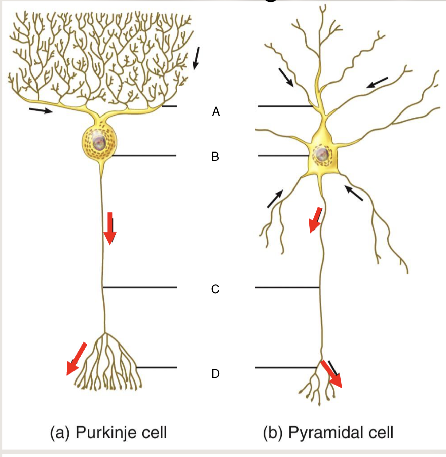
A?
Dendrites

B?
Cell body

C?
Axon

D?
Axon terminal
Synaptic potentials flow?
Into the cell body through the dendrites (passive electrical signals)
Action potentials flow?
Out of the cell body towards the axon terminal (non passive)
Are action potentials passive or not?
Not passive
Are Synaptic potentials passive or not?
Passive electrical signals
Function of dendrite?
Receives input
Function of Cell body?
Passively conduct electrical signals
Function of axon initial segment?
Initiate action potentials (AP)
Function of Axon?
Propagate AP
Function of Axon terminals?
Releases chemical signals
Examples of electrical signals
Dendrites, cell body, axon
Example of chemical signals
Synapses
The RMP experiment
2x electrodes, one inside cell (recording electrode), one outside, measure voltage difference (-50 to -70mV), when both outside mV=0
In neurons and their processes, the ———- has a potential that is 50 to 70mV lower (ie. more negative) than the potential of the extracellular fluid.
Cytosol
In neurons and their processes, how much lower is the potetial of the cytosol compared to the extracellular fluid?
50-70mV
What is it called when cytosol of a neuron has a lower mV (potential) than the extracellular fluid?
Seperation of charges
What is seperation of charges?
a difference in potential, causing electrical potential and therefore current, therefore voltage.
How many cells in the body have RMP?
Almost all except red blood cells or dead / dying cells
What can RMP affect in the body?
circadian rytheym and wound healing
What are excitable tissues?
Tissues that can suddenly respond with a transient change of this potential (ie. an action potential) in response to a stimulus (neurons and muscle fibres)
What are the two kinds of excitable tissues?
Muscle fibers and neurons
Two ways of recording intracellular potentials?
Microelectrodes and patch clamp technique
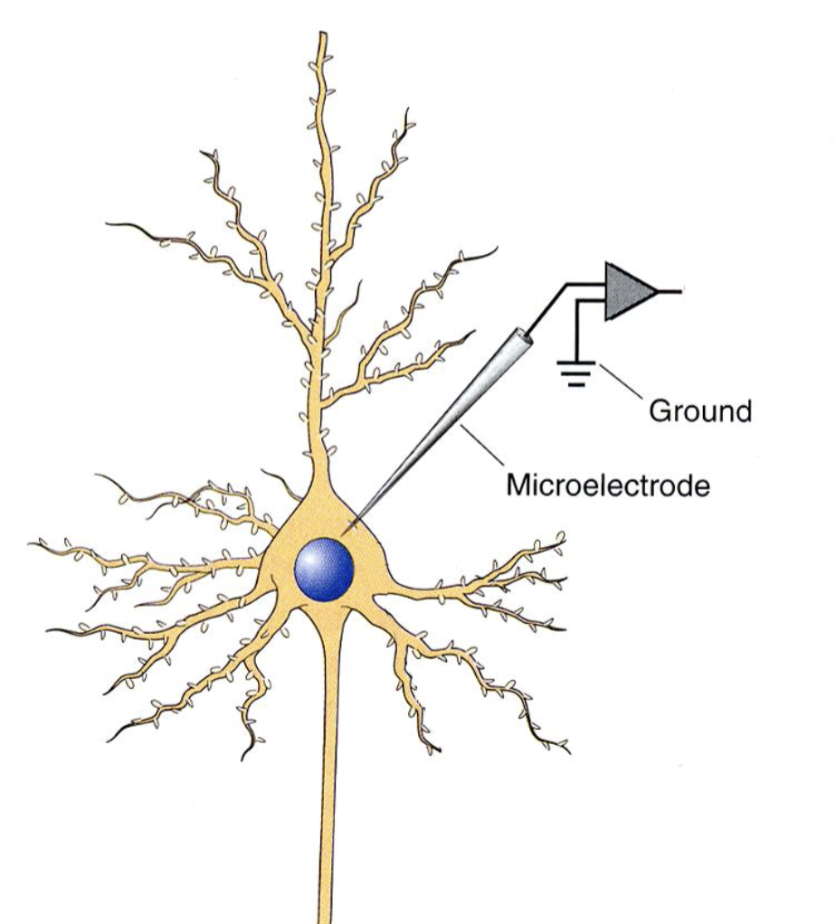
Microelectrode recording technique
Same as the RMP experiment but electrodes are much smaller and made of glass
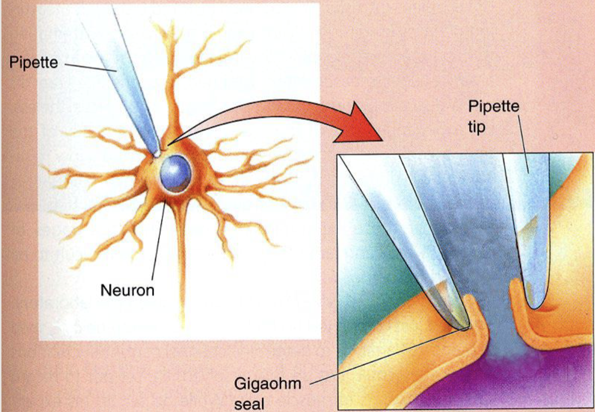
Patch-clamp technique
Clamp and rupture membrane, intracellular fluid fills pippette
Two kinds of signals
Electric and chemical
Two kinds of electric signals
Synaptic potentials (passive) and action potentials
What are synaptic potentials?
Movement of charges
What is an action potential?
When the cell actively regenerates, allowing signal to propagate a longer distance
Equilibrium potentials tell you what?
The potential for the ion (not cell)
What equation do we use to calculate equilibrium potential?
Nernst Equation
What does the Nernst equation include?
intracellular and extracellular ion conc, R, F, T, Z constants (61.5)
Does the Nernst equation calculate for 1 ion type or 2?
1
What equation calculates RMP?
Goldmans equation
Does goldmans equation calculate for 1 ion type or 2?
2
What factors does Goldmans equation have?
intracellular and extracellular ion conc, R, F, T, Z constants (61.5), permeability
Ek+
-80mV
ENa+
60mV
Pk+ / PNa+
40/1
RMP
~ -65mV
Receiving neurotransmitters is an ____ signal
Chemical
Propagating neurotransmitters is an _______ signal
Electrical
Potential inside neurons is not constant. When does it change?
membrane permeability or ion concentrations change
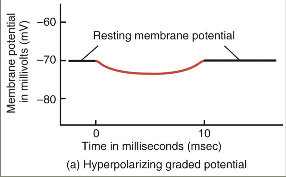
Hyperpolarisation
When the cell membrane potential becomes more negative than the resting membrane potential because the cell moves closer to the equilibrium potential of potassium
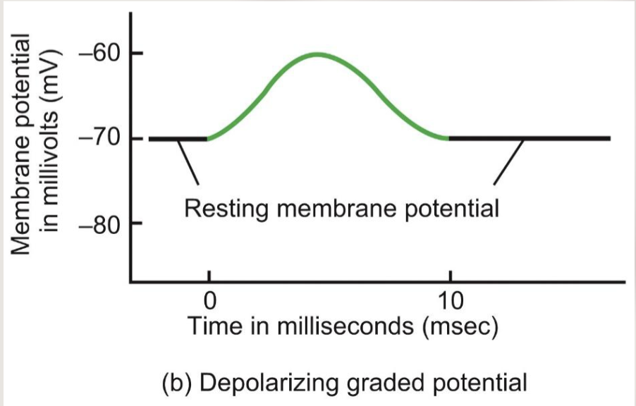
Depolarisation
When cell membrane potential moves positively (away from RMP) because something has changed that moves it closer to the equilibrium potential of sodium
Action potential is
A brief fluctuation in membrane potential caused by a transient opening of voltage-gated ion channels which spreads, like a wave, along an axon.
Action potential occurs
after the membrane potential reaches a certain voltage called the threshold (~ -55 mV).
When talking about action potentials, we are only talking about______________, because they can suddenly change their membrane potentials
Excitable tissues (muscles and neurons)
Action potentials are a key element of _________ along (often very long) axons.
signal transmission
The _________ of action potentials encodes information (a language by which neurons communicate)
frequency
Why are action potentials a key part in signalling along long axons?
They communicate with minimum decay to signals, (wont get degraded when sent far away)
Stimulus
can be physical (eg electric current or mechanical stretch) or chemical (eg a drug or synaptic excitation), pain
What do all action potentials start with?
RMP
1st step of action potentials
Membrane potential reaches threshold, followed by fast depolarisation to ~ +30 mV (‘overshoot’)
Fast depolarisation
voltage gated sodium channels open allowing large amounts of sodium to enter the cell, going towards the equilibrium potential of Na
Where does graded depolarisation occur?
After RMP but before threshold
What is graded depolarisation?
When the depolarisation it shows is relative to the amount of stimulus you have
What happens in repolarisation?
Na channels shut, K+ channels open
When does repolarisation occur?
After fast depolarisation
What is the last step of Action potentials?
After-hyperpolarisation
Example of graded depolarisation
The more you stretch a receptor, the more it gets depolarised
2nd step of action potentials
Repolarisation
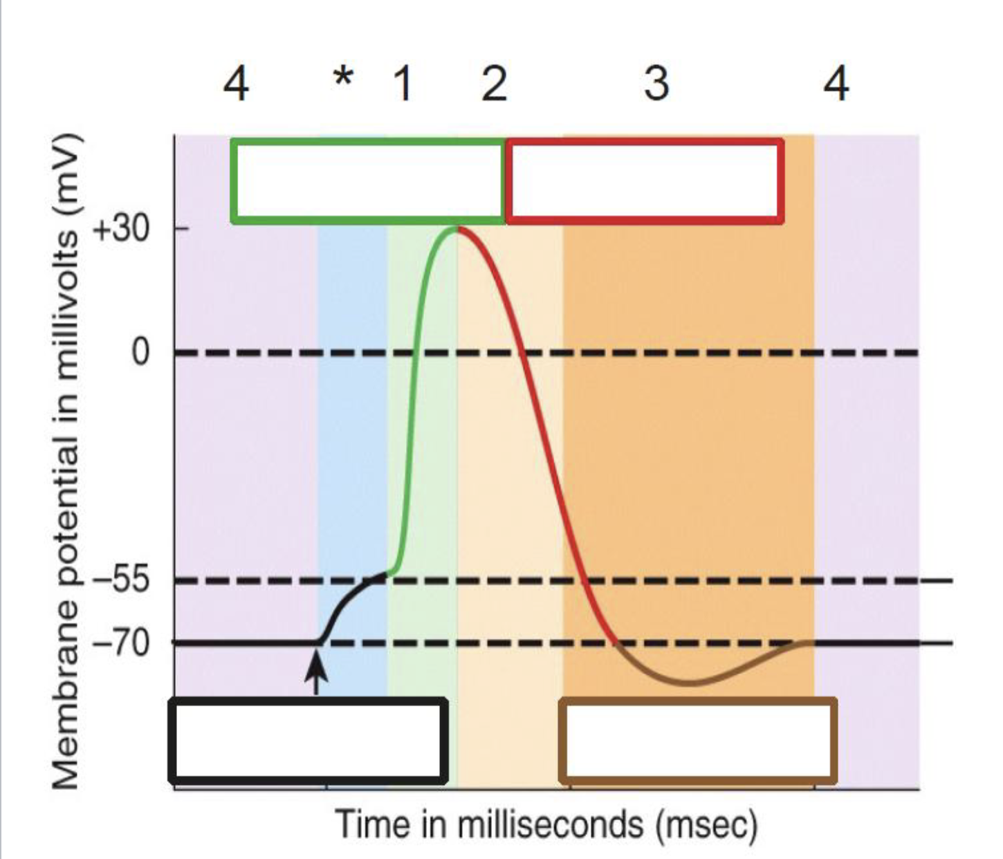
Area 1 and 2 are the
absolute refractory period

Area 3 is the
relative refractory period
If you used a second stimulus to try and stimulate the same neuron during this period, it wont do anything.
Absolute refractory period
Why will a neuron not be stimulated in the absolute refractory period?
Because all voltage gated ion channels already open after threshold, so there is no more to open
During this period the same neuron can be stimulated if a larger stimulus is used
Relative refractory period
during Relative refractory period, what is key to stimulating the neuron again?
A larger stimulus for action potential
After-hyperpolarisation AHP. Membrane potential shifts towards EK+ since PK/Pna becomes
~100:1
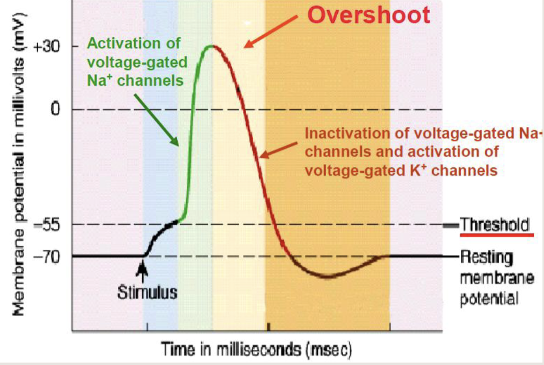
When MP reaches the ————- there is a sudden activation (opening) of voltage-gated —— channels (PNa increase). At this moment PK/PNa is 1: 20 (before it was 40:1), therefore MP shifts towards the ENa+ towards +60 mV = overshoot.
threshold, Na+
Opening of voltage-gated —- channels is short-lasting, as these channels inactivate quickly. This is followed by transient opening of voltage-gated —- channels, leading to repolarisation
Na+, K+
What happens when membrane potential reaches the threshold?
Sudden opening of voltage-gated Na+ channels (PNa increase)
When the voltage-gated Na+ channels open, they inactivate quickly. What happens next?
transient opening of voltage-gated K+ channels, leading to repolarisation

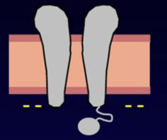
What is happening during purple period?
Resting membrane potential. Na+ channels are in the resting state and K+ channels are closed

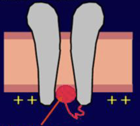
What is happening during the blue period?
Stimulus causes depolarisation until threshold is reached
What is happening during the green period?
Voltage gated Na+ channel activation gates opened

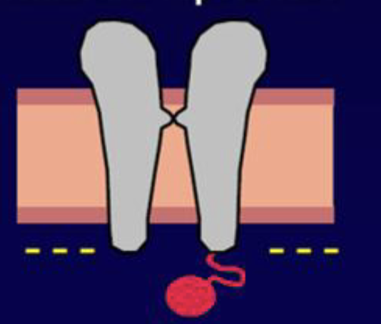
What is happening during the yellow period?
Voltage gated K+ channels are open, Na+ channels are inactivating

What is happening during the orange period?
Also known as the relative refractory period, Voltage gated K+ channels are still open, Na+ chanels in resting state

What two periods make up the absolute refractory period?
Green and yellow
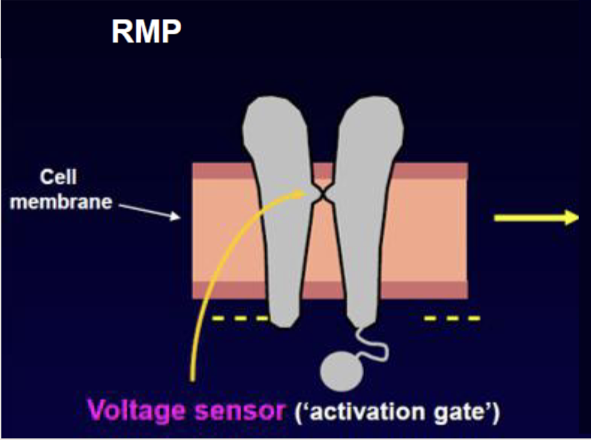
Activation gate closed, inactivation gate open- RMP
Voltage-gated Na+ channel in resting state
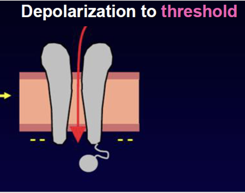
Depolarisation to threshold
When the voltage threshold is reached, sodium channels open and Na+ ions move into the cell along both the concentration and electrical gradient.
Influx of Na+ slows down and stops when:
The inside potential becomes positive (moves towards ENa) and thus attracts Na+ less, Na+ channels inactivate
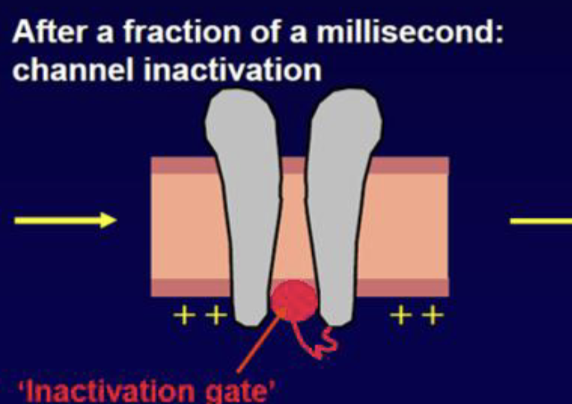
Peak of arc, after a fraction of a millisecond the inactivation gate closes
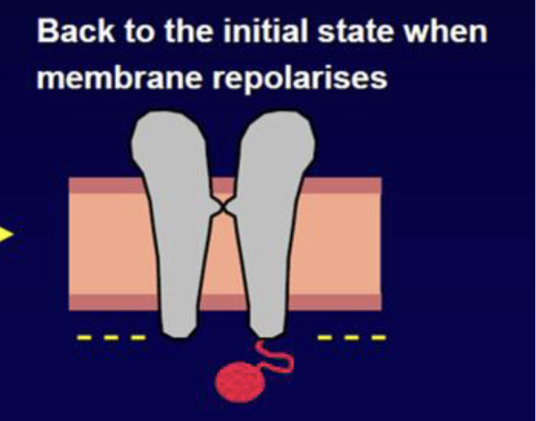
Back to initial resting state when membrane repolarises (when it goes under threshold)
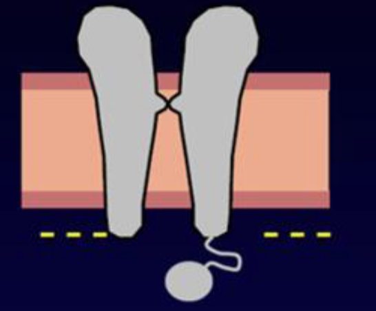
RMP, first step
Depolarisation to threshold, step 2
Inactivation gate, peak of arc, step 3
back to initial state after repolarising, under threshold, last step
When the cell stimulus allows the cell to reach threshold, your activation gate will now open, Na rushes in contributing to depolarisation phase. After being open for a fraction of a millisecond, Na+ channels inactivate, ball and chain shut channel.
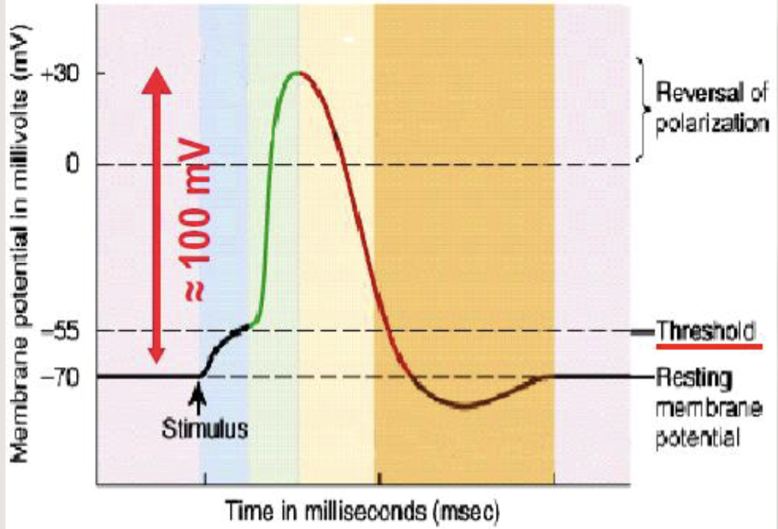
Name each step of the line
RMP, depolarisation to threshold, fast depolarisation, repolarisation, after-hyperpolarisation
Why is each action potential is an all- or-none event?
Once voltage gated ion channels are open, they are ALL open. Unlike graded depolarisation or hyperpolarisations.
Frequency ______ with more stimulus
Increases
If a large signal needs recognising, what gives information about it?
the change in frequency of action potential, not amplitude of ap. This is because a large stimulus may invoke several APs closer together, but all APs are same.
Normals amplitude of an Action potential?
~ 100 mV
amplitude of action potentials does not depend on the ‘stimulus’ intensity as long as the stimulus is ————
above the threshold