ANS 123 Lecture 1-6 QUIZ 1
1/109
There's no tags or description
Looks like no tags are added yet.
Name | Mastery | Learn | Test | Matching | Spaced | Call with Kai |
|---|
No analytics yet
Send a link to your students to track their progress
110 Terms
Development
Cellular changes over time that enable tissues and subsequently organs to take on different and increasingly more complex roles & functions
Process through which a single, totipotent cell (e.g., fertilized egg) gains complexity to become a complete organism
• Increasing complexity ⇄ Decrease in number/range of genes being expressed ⇄ Loss of potency[expression]
Growth
General and normal expansion of size through accretion of tissues similar in composition to the original tissue or organ
Accretion
Gradual accumulation/buildup through adhesion of external parts & particles
“-blast”:
To sprout
o Used to indicate an immature cell that can undergo mitosis and retains potency
• The prefix indicates the cell lineage of the immature cell
Ex. Chondroblast
= Immature cartilage cell
“-clast”:
To destroy
o Used to indicate a differentiated cell that destroys tissue
• The prefix indicates what type of tissue
Ex. Osteoclast
= Bone destroying cell
“-cyte”:
Cell
o Used to refer to a mature, completely differentiated cell
Ex. Myocyte
= Mature muscle cell
Fertilization
Fusion of sperm & egg
Cytokinesis
Cell division
Cell determination
When an immature cell “shuts down” gene expression of portions of its genome and reduces its cellular potency to become a more specialized cell type
o Still cannot function as the specialized cell type
Cell differentiation
When a precursor cell undergoes morphological changes through differential, specialized, and limited gene expression so it can perform its assigned function
o Genes that express products for that specific function get “switched on”
o Results in cellular diversity
Apoptosis
Programmed cell death
o Critical process throughout a living organism’s lifetime
Hypertrophy
Growth that occurs when cells increase in size
Hyperplasia
Growth that occurs when cells increase in number
o Can occur in combination or separately
Average daily gain
Typically measured in mass (e.g., pounds/ kilograms)
o Generic measure that includes fat, muscle, and bone
What types of tissues are there?
• Nervous
• Epithelial
• Muscle
• Connective
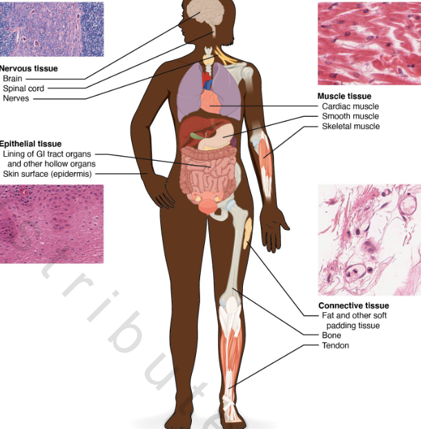
What are tissues?
Aggregate of cells of a particular kind + Intercellular substance (i.e., “cellular packaging”)
• Act synergistically [come together and work together] to fulfill specific function
• Intercellular substance typically made up of/produced by connective tissue cells
Ex. Muscle tissue
• Aggregate of muscle tissue cells + Connective tissue sheaths
• Responsible for movement
Nervous Tissues: Brain, Spinal Cord, Nerves
• Aggregate of:
1. Neurons/Nerve cells
2. Neuroglia/Glial cells
• Responsible for coordinating the various functions of the body
Nervous tissue: 1. Neurons/Nerve cells
Communicate/carry messages throughout the body using electrical pulse
Nervous tissue: 2. Neuroglia/Glial cells
• Support, bind, & defend nervous tissue cells
o Insulation
o Structure
Nervous system
Collection of nervous tissue that interact
1. Central nervous system (CNS) – Consists of brain and spinal cord
2. Peripheral nervous system (PNS) – Consists of bundles of nervous
1. Central nervous system (CNS) – Consists of brain and spinal cord
Integrates and coordinates all bodily functions
2. Peripheral nervous system (PNS) – Consists of bundles of nervous
o Sensory component: Detects stimuli (both internal & external) and delivers information to CNS→ receive info through neurons
o Motor component: Carries response messages from CNS to appropriate tissues throughout body→ have info effect something via motor system
Soma
Body of the neuron
o Contains nucleus + other typical cell organelles (mitochondria)
Processes
Extensions from soma
o Dendrites: Receive signals from other neurons/cells & carry TO soma
o Axons: Carry signals FROM soma & transmit to other neurons/cells
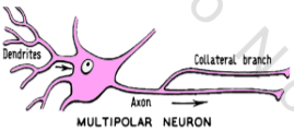
Multipolar Neuron [Most common]
Description: Multiple dendrites from soma in opposite direction of axon
Location: Brain and spinal cord (e.g., CNS)
Function: Integrates information from multiple inputs
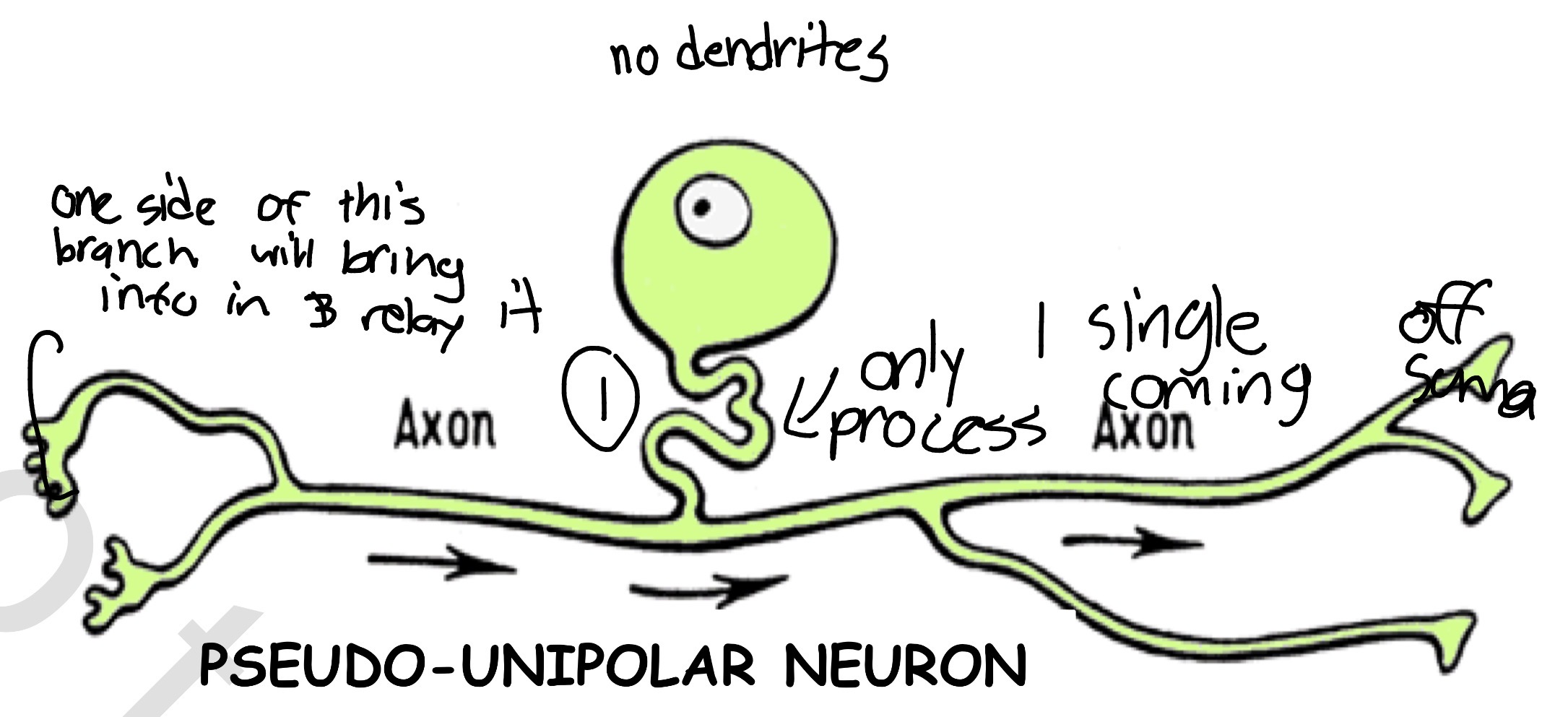
Pseudo-unipolar Neuron: Pseudo” because axon branches split from a single one
Description: Single axon split into 2 branches; each branch goes in different direction
Location: Throughout body, closely associated with spinal cord
Function: Receives sensory information & transmits to spinal cord
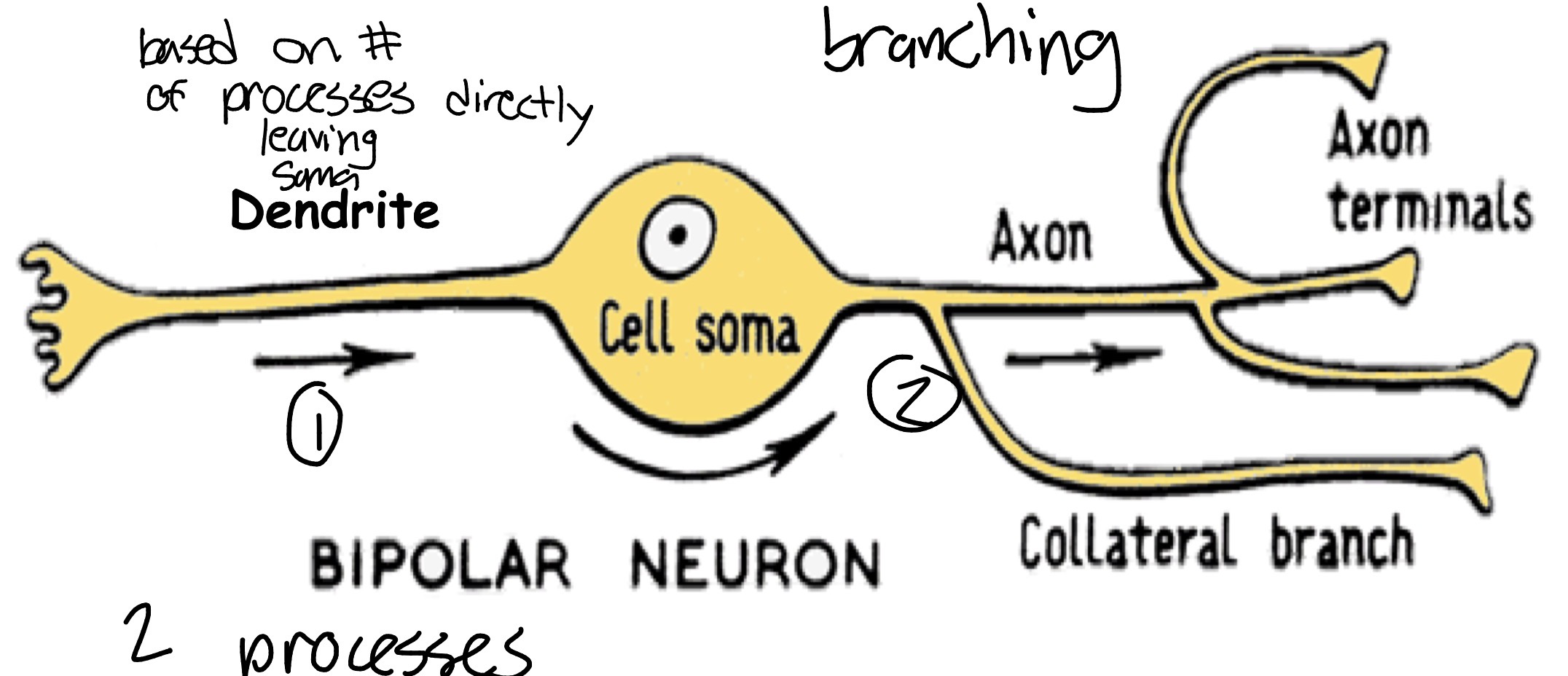
Bipolar Neuron
Description: Single dendrite extends from soma in opposite direction of axon
Location: Eye (e.g., retina), nose (e.g., olfactory tissue), and ear (e.g., vestibular-cochlear nerve)
Function: Receives highly specialized signals & transmits to nerve bundles that link to the brain
• Plays crucial role during neurogenesis
Glial cells: Supporting cells
• Constitute ~ ½ total mass of nervous tissue
• Do not participate in electrical signaling
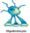
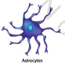
CNS: Oligodendrocytes, Astrocytes, microglia, Ependymal Cells
PNS: Schwann cell, Satellite cells
Glial Cells: Oligodendrocytes
Central Nervous System
Produce myelin sheaths that wrap around some axons; gives white appearance; single cell → multiple sheaths
Glial Cells: Astrocytes
Central Nervous System
Control levels of neuro- transmitters & ions at synapses; participate in blood-brain barrier
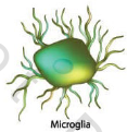
Glial Cells: Microglia
Central Nervous System
Immune cells of nervous tissue - behave as macrophages
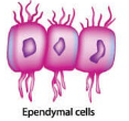
Glial Cells: Ependymal Cells
Central Nervous System
Line Ventricles & produce cerebral-spinal fluid [aqueous cushion for protecting brain]; participate in blood-brain barrier
Glial Cells: Schwann Cells
Peripheral Nervous System
Produce myelin sheaths around some axons; single cell → single sheath
[NOTE: Text considers these to be specialized oligodendrocytes]
![<p>Peripheral Nervous System</p><p>Produce myelin sheaths around some axons; single cell → single sheath</p><p>[NOTE: Text considers these to be specialized oligodendrocytes]</p>](https://assets.knowt.com/user-attachments/2922d544-97fa-4ac4-a6c3-a3ad3a157f74.png)
Glial Cells: Satellite Cells
Peripheral Nervous System
Control levels of neuro- transmitters & ions at synapses
[NOTE: Same term used for cells in muscle tissue]
![<p>Peripheral Nervous System</p><p>Control levels of neuro- transmitters & ions at synapses</p><p>[NOTE: Same term used for cells in muscle tissue]</p>](https://assets.knowt.com/user-attachments/da81bee9-1fd7-496f-94ef-488ff4f5d443.png)
Epithelial Tissues: Lining of GI tract organs and other hollow organs, Skin surface [epidermis]
Cover exposed surfaces of body
o Protect underlying tissue from its environment (both internal and external)
Control nutrient flow to underlying tissues
House secretory cells
Aid in sensory communication
Ex. Cells in taste buds, retinas, and nasal cavity linings
Typically associated with basal lamina/basement membrane
o Basal lamina/Basement membrane : Attaches & anchors epithelial cells to underlying tissue
Extracellular, connective tissue matrix
Integument: Largest organ = Skin
Composite membrane
o Epithelial tissue = Epidermis
• Appendages: Hair follicles, sebaceous & sweat glands, hooves/nails, horns
o Connective tissue = Dermis (houses secretory cells)
• (Hypodermis: Not part of integument – anchors integument to rest of body)
Integument: Associated with commercially important products
o Wool/hair
o Feathers
o Leather
• Also produces: Hooves/nails, Horns
Muscle Tissue: Cardiac Muscle, Smooth Muscle, Skeletal muscle
1. Enables movement: Muscle cells contract & relax
2. Participates in thermoregulation: Contracting muscle cells generate heat
• 30-40% of total body mass: Major drain on energy efficiency
Muscle cells→ Muscle fiber, myocyte, myofiber, muscle cell
Unique characteristic: Physiological & Functional plasticity ⇒ Retain ability to change physiology and thus, function throughout life
o Embryonic, fetal/perinatal, α-cardiac, Types 1, 2A, 2B, 2X*
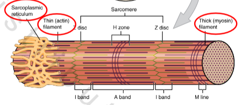
Sarcolemma
Muscle fiber cell membrane
o Responsive to electrical stimulation[ion changes] by motor neurons
o Multiple pores enable communication throughout cell
![<p>Muscle fiber cell membrane</p><p>o Responsive to electrical stimulation[ion changes] by motor neurons</p><p>o Multiple pores enable communication throughout cell</p>](https://assets.knowt.com/user-attachments/398473b1-d766-4f70-b832-1224cf297be1.png)
Sarcoplasm
Muscle fiber cytoplasm
o High concentrations of proteins & mitochondria
o Sarcoplasmic reticulum: Muscle fiber endoplasmic reticulum
• Repositories of Ca2+: controls calcium/release or suck back to reticulum
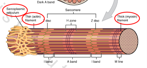
Cytoskeletal elements
o Actin filaments (microfilaments)
o Myosin
Types of Muscle Tissue
1. Smooth
2. Cardiac
3. Skeletal*
Muscle tissue: Characterized based on:
Function
Cell structure
Shape
# of nuclei
Presence or absence of striations: Organization of striations, if present
Contractions
Type - Voluntary or Involuntary
speed, direction, coordination
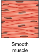
Smooth muscle
Function: Move substances within body: Surrounds many internal organs & vessels of circulatory system
Cell structure
o Shape: Spindle
o Nuclei #: 1/cell
o Striations?: No
Contractions
o Type: Involuntary
o Speed: Slow
o Direction: Multi-directional
o Coordination: Loose
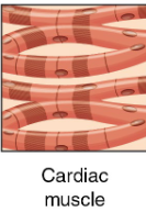
Cardiac muscle
Function: Rhythmic contractions of heart; creates tight jxns, more opportunities to grab each other
Cell structure
o Shape: Short & branched
• Interdigitated: branching allows this, intercalated disks allow end to end of fiber holding
o Nuclei #: 1-2/cell
o Striations?: Yes
Contractions
o Type: Involuntary
o Speed: Fast & slow
o Direction: Unidirectional
o Coordination: High
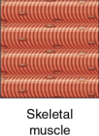
Skeletal muscle
Function: Movement of body; Attached to bones in skeleton
Cell structure
o Shape: Elongated & cylindrical
o Nuclei #: Multi-nucleated (hundreds/cell)/ Actual # directly proportional to length
o Striations?: Yes/ Aligned
Contractions
o Type: Voluntary
o Speed: Fast & slow
o Direction: Unidirectional
o Coordination: High
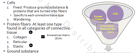
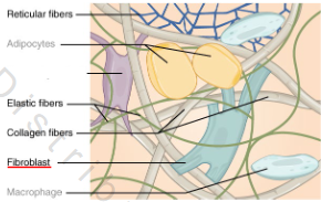
Connective Tissue: Fat and other soft padding tissue, Bone, Tendon
Cells
1. Fixed: Produce ground substance & proteins that are turned into fibers
• Specific to each connective tissue type
2. Wandering
Protein fibers: At least one type found in all categories of connective tissue
1. Collagen
2. Reticular
3. Elastic
Ground substance
1. Chondroitin sulfates
2. Hyaluronic acid
Extracellular Matrix:
Protein fibers: At least one type found in all categories of connective tissue
Ground substance:
1. Chondroitin sulfates
2. Hyaluronic acid
Connective tissue Fxns
• Provides structure to body
• Supports body
• Most diverse tissue type: Function dictates structure
3 main categories
1. Proper
2. Supportive
3. Specialized
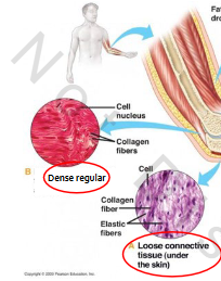
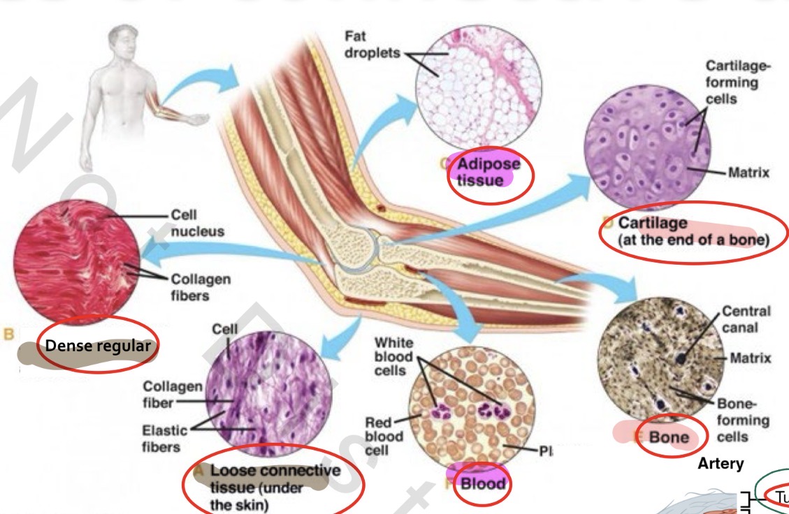
Specific types of connective tissue: Proper
A. Loose/Areolar
B. Dense
i. Regular
ii. Irregular (not pictured)
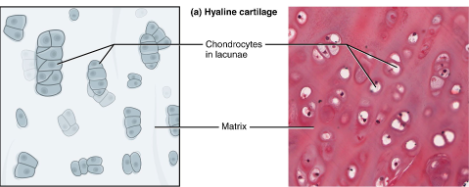
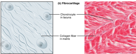
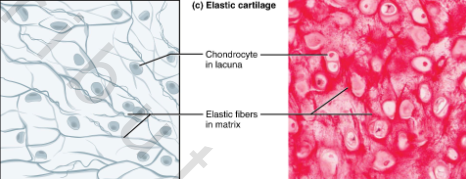
Specific types of connective tissue: Supportive
A. Cartilage
i. Hyaline
ii. Elastic
iii. Fibrocartilage
B. Bone
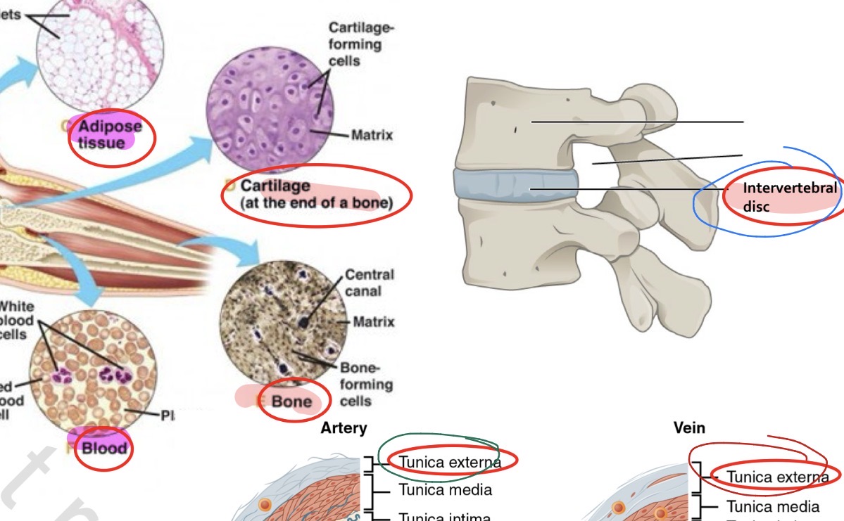
Specific types of connective tissue: Specialized
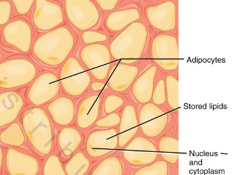
A. Adipose
B. Blood
Connective Cells Fixed: Produce ECM (fibers + ground substance)
Proper = Fibroblasts
Supportive
o Cartilage = Chondrocytes
o Bone = Osteocytes
Specialized
o Adipose = Fibroblasts + Adipocytes
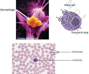
Connective Cells Wandering: Defend & clean
• Proper, Supportive & Specialized = Macrophages & Mast cells
• Specialized only: Blood & Lymph = Erythrocytes, Lymphocytes, Platelets
Connective tissue fibers: Collagen
o Protein: Fibrillar collagen
o Structure & support, esp in skin & muscles
Connective tissue fibers: Reticular
Protein: Collagen III
o Support within organs, glands, etc., and/or fine structural support
Connective tissue fibers: Elastic
o Protein: Elastin
o Stretchiness
Fibroblasts (Proper)
Mostly fibers; some ground substance
• Fibers:
o Collagen
o Reticular
o Elastic
Chondrocytes (Supportive)
Cartilage fibers
Hyaline, Fibrous, Elastic
Contained in small chambers (e.g., lacunae [grows that way/holding space) in extracellular matrix
Cartilage fibers: Hyaline
Collagen fibers; slightly more ground substance firm/rubbery
Cartilage fibers: Fibrous
Collagen fibers; little ground substance
Less Squishy
Cartilage fibers: Elastic
Mostly elastic fibers + some collagen fibers; more ground substance
Stretchy
Osteocytes (Supportive)
Bone fibers
o Collagen fibers; calcium-rich ground substance [hard as cement]
Contained in lacunae
all supportive cells are in lacunae [space in substance]
![<p>Bone fibers</p><p>o Collagen fibers; calcium-rich ground substance [hard as cement]</p><p>Contained in lacunae</p><ul><li><p>all supportive cells are in lacunae [space in substance]</p></li></ul><p></p>](https://assets.knowt.com/user-attachments/1dd8a9eb-bb17-4b75-871b-3032615d8341.png)
Adipocytes (Specialized)→ Fat cells
Triglyceride storage
• Mostly storage; very little ground substance
Wandering cells
Proper, Supportive & Specialized
o Macrophages: Seek out & destroy foreign bodies & damaged cells
o Mast cells: Release histamine
Specialized only
o Erythrocytes: Transport O 2 & CO 2
o Lymphocytes: Immune response
o Platelets: Clotting
EARLY development
Cellular changes over time that enable tissues and subsequently organs to take on different and increasingly more complex roles & functions
Process through which a single, totipotent cell (e.g., fertilized egg) gains complexity to become a complete organism
Karyogamy
Fusion of ovum & sperm pro-nuclei
Germ layers
Ectoderm, Mesoderm, Endoderm
o Form the embryo
“Natal”/“Birth” and “Hatch” will often be used inter-changeably
Pre-natal: Before birth
• Peri-natal: Around birth
• Post-natal: After birth
• Neo-natal: Newly born
Cellular/Cell potency
How much of the organism’s genome a cell can express
Stem cells
1. Totipotency
2. Pluripotency
3. Multipotency
4. Unipotency
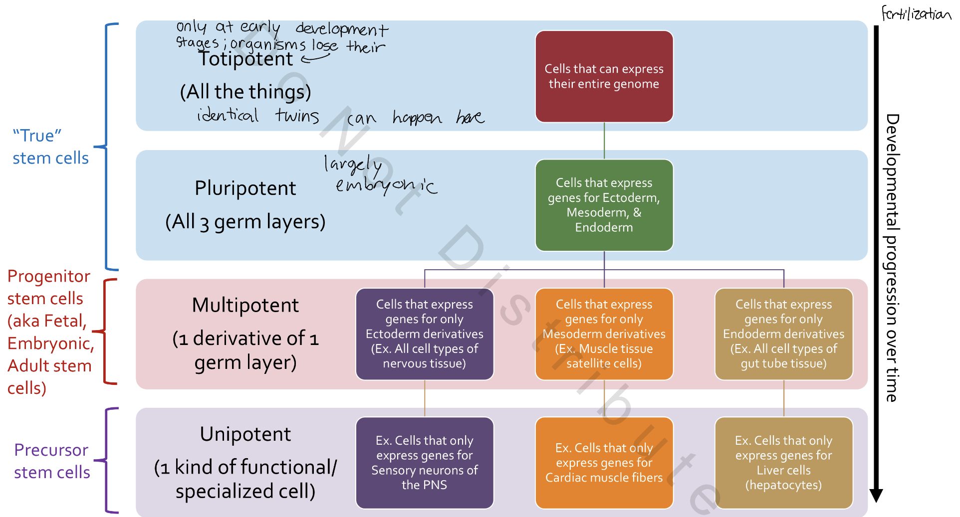
Totipotency
ability to express ALL the genes in the organism’s genome
• Differentiate into ANY type of cell; both embryonic AND extraembryonic
Pluripotency
Ability to express genes that code for all germ layers (e.g., endoderm, mesoderm, & ectoderm)
Multipotency [Progenitor Cells]
ability to express genes that code for closely-related family of cells
one type of layer
Delamination
Splitting single sheet of cells through loss of adhesion
Ex. Hypoblast formation
Ingression
Division & migration of cells into internal area to form a separate layer
Ex. Internal cell mass & trophoblast separation
Invagination
Depression in cell layer deepens due to cell division – sides of depression come together and pinch off from existing layer
Ex. Neural tube formation
Epiboly
Increase in # of cells on outer surface
Ex. Primitive streak formation
Involution
Inward rolling of cells along an existing membrane to create layers
Ex. Myelin sheath formation
Morphogenesis
Generation, differentiation & growth of tissues & organs during development (i.e., morphology)
General term that covers
o Cleavage, Layer formation, Gastrulation, Neurulation, Organogenesis, Etc.
• Commences post-fertilization
Morphogenesis: Associated with increasing cellular complexity and loss of cellular potency
During early development achieved by creating 3 “new” germ layers
⇒Loss of totipotency
Obvious differences: The ovum→ Birds
Yolk→ Marcolecithal [large]
Yolk distribution→ Telolecithal [non-uniform]
Cleave pattern→ Meroblastic [incomplete discoidal] each time cells divide, you don’t get yolk by by bit remains intact entire time
only dividing cells divide little dish shape floating on top of yolk
![<p>Yolk→ Marcolecithal [large]</p><p>Yolk distribution→ Telolecithal [non-uniform]</p><p>Cleave pattern→ Meroblastic [incomplete discoidal] each time cells divide, you don’t get yolk by by bit remains intact entire time </p><ul><li><p>only dividing cells divide little dish shape floating on top of yolk</p></li></ul><p></p>](https://assets.knowt.com/user-attachments/2678927a-b0f9-4851-8148-7f651e8a1973.png)
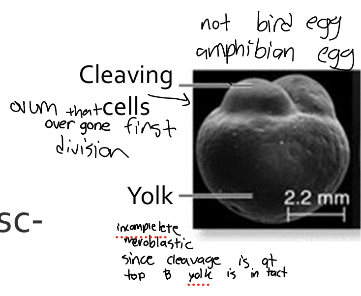
Obvious differences: The ovum→ Mammals
Yolk→ Alecithal [absent]
Yolk distribution→ Isolecithal [uniform]
Cleave pattern→ holoblastic [complete], rotational → yolk involved in same division
![<p>Yolk→ Alecithal [absent]</p><p>Yolk distribution→ Isolecithal [uniform]</p><p>Cleave pattern→ holoblastic [complete], rotational → yolk involved in same division</p>](https://assets.knowt.com/user-attachments/1809aaea-acf2-4f81-95cf-32990a8cc12f.png)
Surprising similarities in development for avian/ungulates/rodents
At some points during development, ungulate processes are more similar to avian processes than they are to rodents.
similarities between avian and rodents
cleavage, organogenesis, gastrulation
Cleavage: Functions
1. Increase # of cells via mitosis without growth
o Cell number ⇌ Cell size [daughter cells get smaller]↴
2. Establish embryonic vs extraembryonic cell lines
• Results in:
o Avian blastoderm
o Mammalian blastocyst
• Cleavage ends when blastocyst makes contact with uterine wall
Avian cleavage: Discoidal
Discoidal: Cleaving cells clump together into a disc- shape
Avian cleavage: Meroblastic
Cleavage plane is Incomplete; yolk remains intact
Avian cleavage: Functions: 1. Increase number of cells thru cell division
Meroblastic [incomplete] cleavage
about the same as moving through next division size of cell and # relatively same
![<p></p><p>Meroblastic [incomplete] cleavage</p><p>about the same as moving through next division size of cell and # relatively same</p>](https://assets.knowt.com/user-attachments/9cda7edb-266a-46e5-8cf4-999db402ab95.png)
Avian cleavage: Meroblastic 1-16 divisions
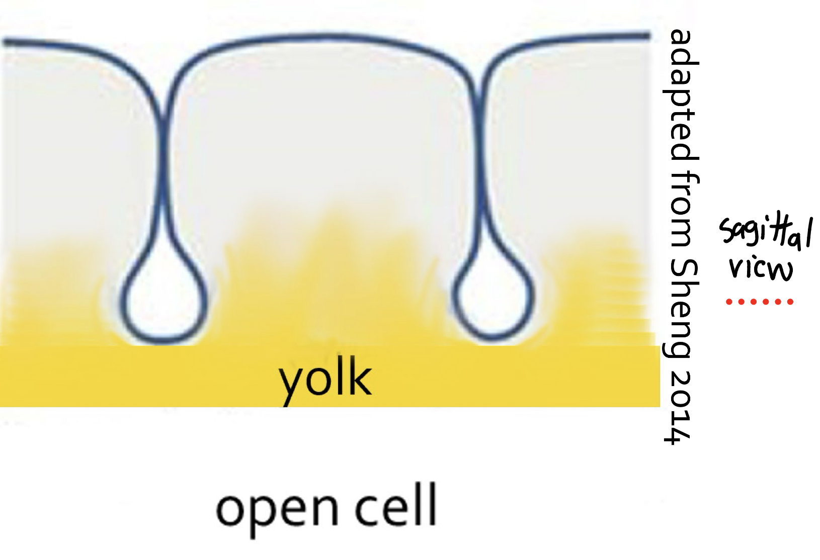
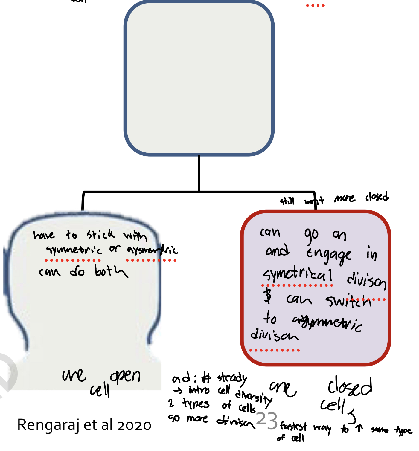
Symmetric division ONLY =
Daughter cells are identical
o BOTH daughter cells form INcomplete cell membrane = Open cell
• Cytoplasm mixes with yolk
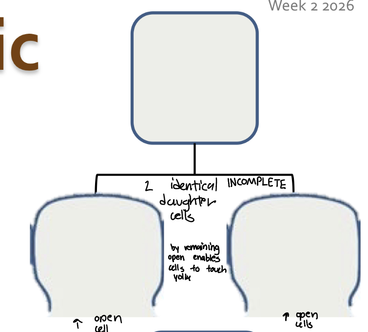
Avian cleavage: Meroblastic After 16
Asymmetric division begins =
Daughter cells not identicAsymmetric division begins =
Daughter cells not identical!
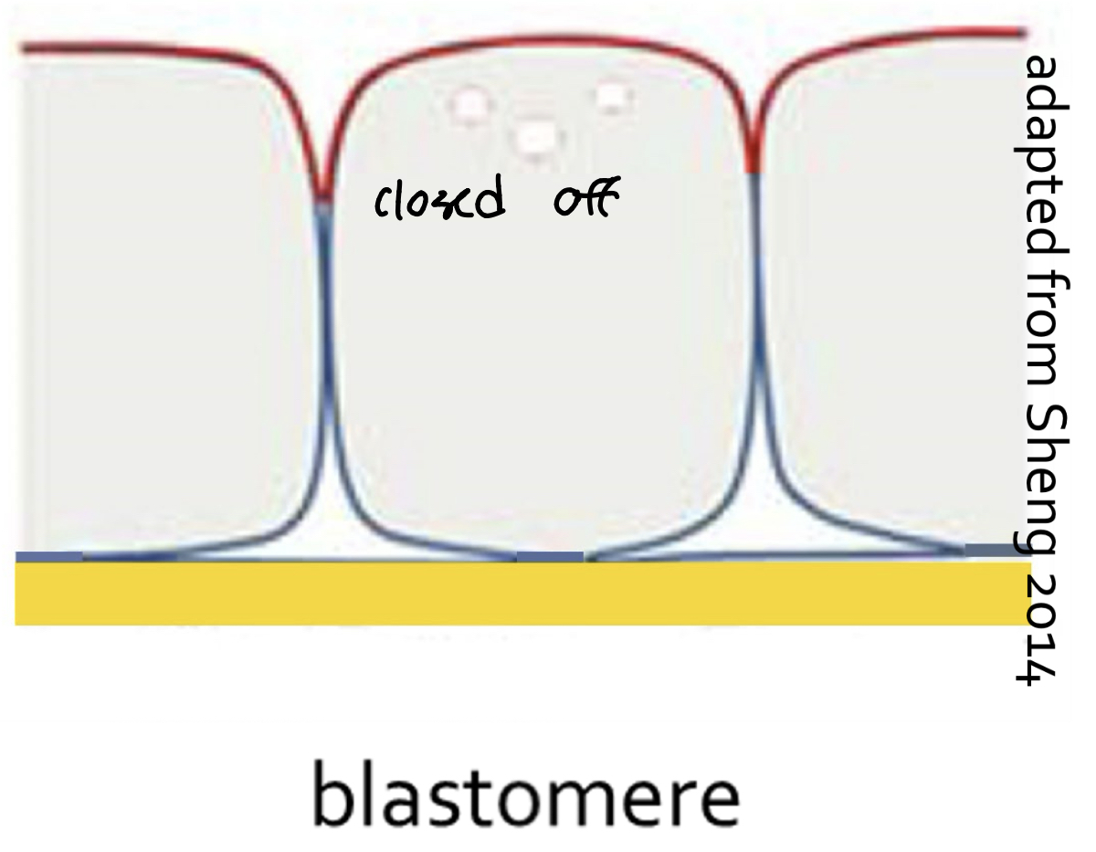
o One forms complete (“closed”) cell membrane
= Blastomere
o One is an open cel!
o One forms complete (“closed”) cell membrane = Blastomere
o One is an open cell
Avian cleavage: Functions 2. Establish embryonic vs extra- embryonic cell lines
embryonic cell lines : form of embryo
extra-embryonic cell lines: fully defer to extra embryonic tissue
Establish embryonic vs extra- embryonic cell lines→ Blastoderm (from 64-cell stage):
Most central cells = Closed
o 5-6 cell layers thick
o Embryo only
Regions of open cells beneath & around embryo begin to form
Mostly extra-embryonic [open daughter cells to do embryonic tissue in the periphery] ; some embr Most central cells = Closed
o 5-6 cell layers thick
o Embryo only
• Regions of open cells beneath &
around embryo begin to form
o Mostly extra-embryonic; some
embryo
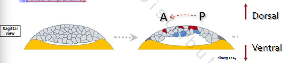
Avian cleavage: One more function→ 3. Establish axes of polarity
o Dorsal-ventral axis [HEAD]
o Anterior-posterior axis [BUTT]
cluster of cells need direction to develop
Interaction of these axes creates a left & right side of embryo
3. Establish axes of polarity
Dorsal-ventral axis→ talking about cells that are going to become embryonic
• Dorsal = Egg shell side
• Ventral = Yolk side
Anterior-posterior axis
• More complex…[slowly rotating]
![<p>Dorsal-ventral axis→ talking about cells that are going to become embryonic</p><p>• Dorsal = Egg shell side</p><p>• Ventral = Yolk side</p><p>Anterior-posterior axis</p><p>• More complex…[slowly rotating]</p>](https://assets.knowt.com/user-attachments/25b2743a-d3b7-4645-bd1b-fc2150301969.jpg)
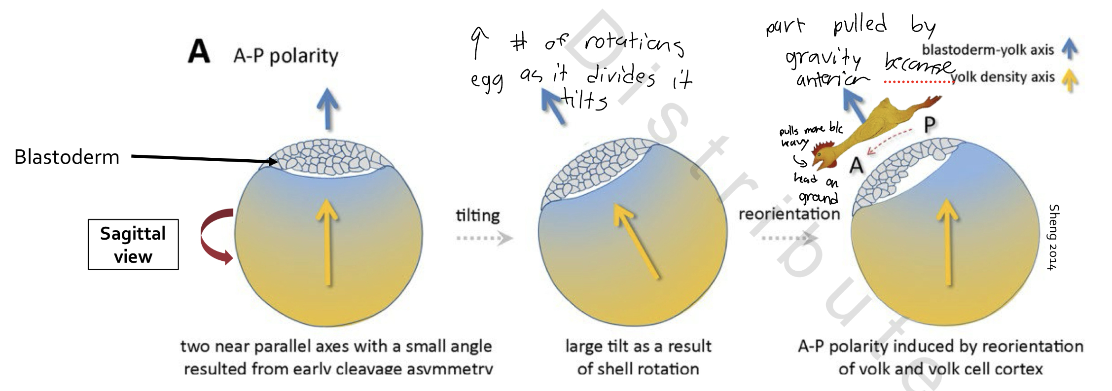
Avian cleavage: Anterior-Posterior Axis
Egg rotates 10-12 turns/hr in shell gland
• Gravity causes “heavy” blastoderm to tilt away from “top” of yolk
o Lowest end of blastoderm becomes anterior of embryo
Avian cleavage: Conclusion
The blastoderm
• Two cell lineages
1. Closed cells
2. Open cells
• Axes of polarity