Urinary System
1/41
There's no tags or description
Looks like no tags are added yet.
Name | Mastery | Learn | Test | Matching | Spaced | Call with Kai |
|---|
No analytics yet
Send a link to your students to track their progress
42 Terms
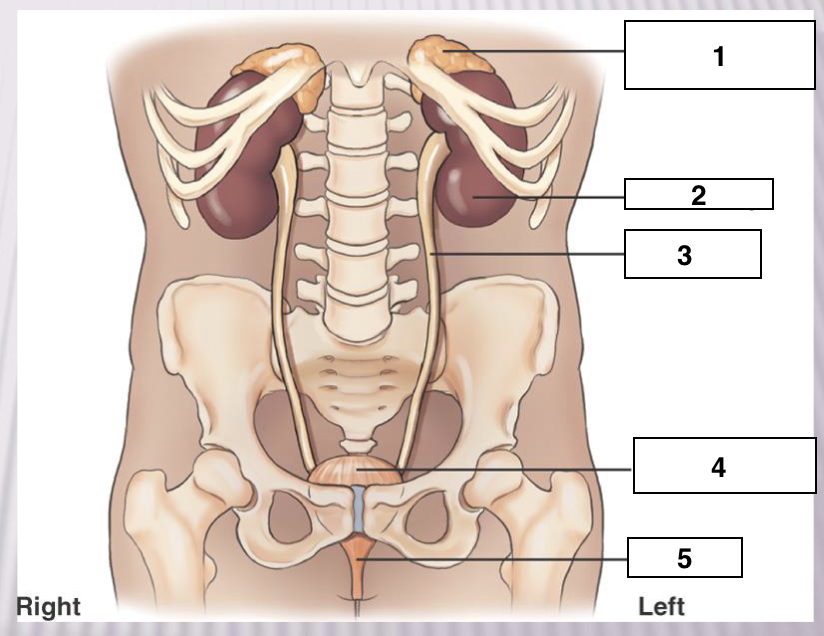
Adrenal gland
Left Kidney
Left ureter
Urinary bladder
Urethra
Functions of Urinary system
-Production of urine and it’s elimination from the body
-Remove nitrogenous wastes
-Maintain fluid and electrolyte levels
-Secreting substances that affect blood pressure
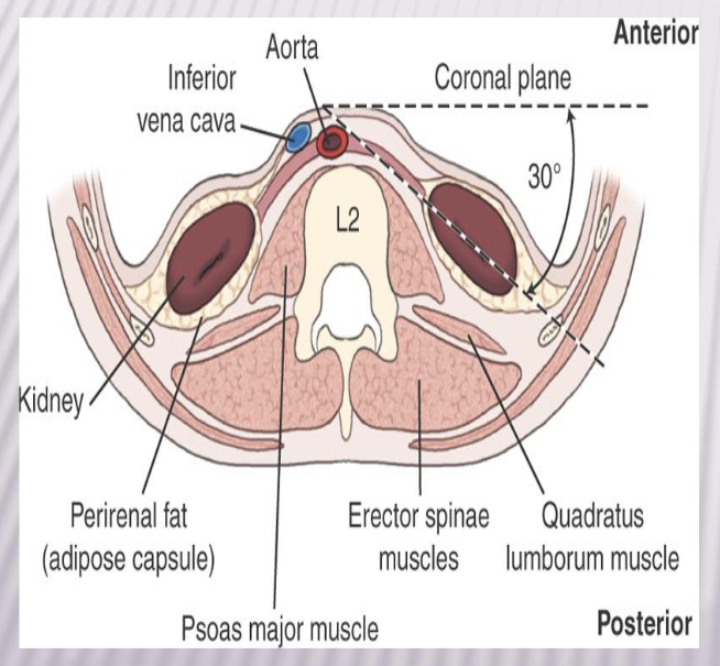
Retroperitoneal structures
-Kidneys
-Ureters
Infraperitoneal structures
Distal ureters
Urinary bladder
Urethra
Kidney orientation
30 degree angle from coronal plane
Right kidney is lower = bc of liver
Kidney Location
Halfway between xipoid process and iliac crest
Between T11-T12
Right kidney more inferior than left
Nephroptosis
a condition where the kidney descends more than 5 cm (or two vertebral bodies) when moving from a supine to an upright position
-Kidney drops down
Kidney transplant
Low in the pelvis near bladder bc you have to attach the ureter
-near the pelvis
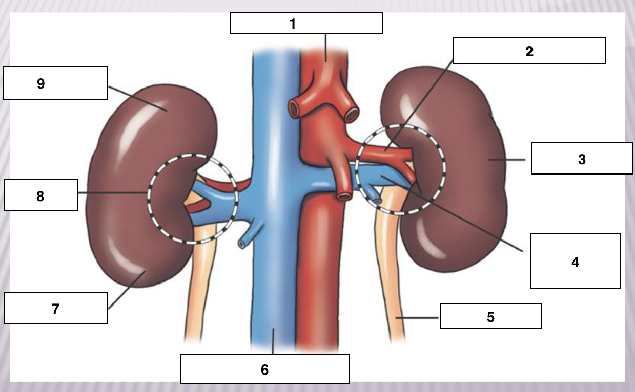
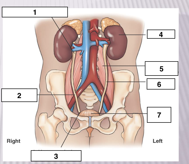
Abdominal aorta
Left renal artery
Left kidney
Left renal vein
Left ureter
Inferior vena cava
Lower pole
Hilum - mainstream, where stuff goes in and out
Upper pole
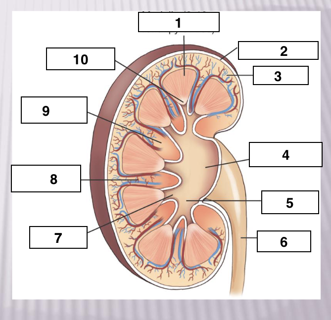
Macroscopic structure
Cortex
Fibrous capsule
Nephrons
Medulla
Renal pyramids - 8-18
Renal papilla
Mior calyces - 4-13
Major calyces - 2-3
Renal pelvis
Ureter
Medulla
Fibrous capsule'
Cortex
Renal pelvis
Major calyx
Ureter
Minor calyx
Renal sinuses
Renal column
Renal papilla
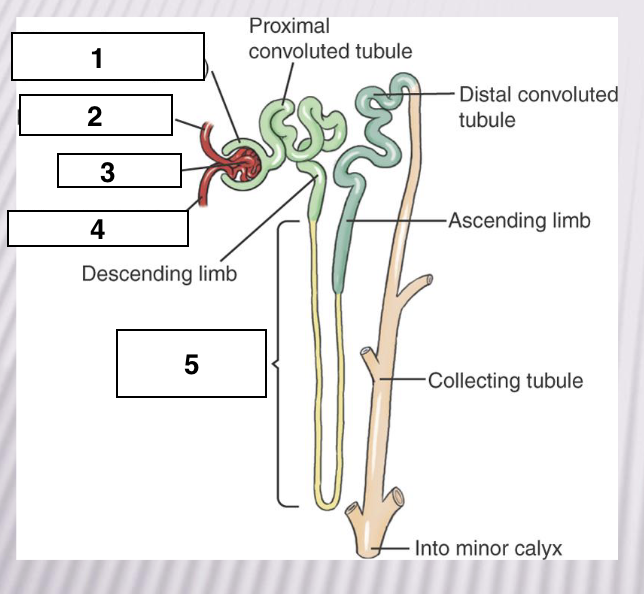
Glomerular capsule or bowmans capsule
Efferent arteriole
Glomerulus
Afferent Arteriole
Loop of henle in medulla portion
-Structural and functional unit
-Over 1 million per kidney
-Blood filtered
URINE PRODUCTION SUMMARY
H2O intake (2.5 L/day)
Bloodstream
Filtrate 99% reabsorbed
Urine (1.5 L/day)
Ureters
10-12 inches long, 1mm to 1 cm in diameter
Lie on psoas muscles
Enter posterolateral bladder
Peristaltic contractions move urine towards bladder
Points of constriction
Ureteropelvic junction (UPJ)
Pelvic brim
Ureterovesical junction (UVJ)
Ureteropelvic junction (UPJ)
Pelvic Brim
Ureterovesical junction (UVJ)
Left kidney
Left psoas major muscle
Left ureter
Urinary bladder
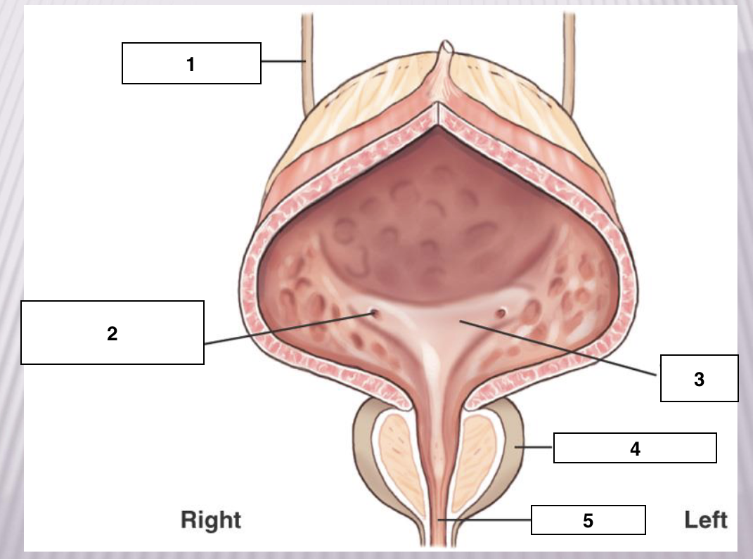
Right ureter
Ureteral opening (UV junction)
Trigone
Prostate gland
Urethra
Urinary bladder
-Musculomembranous sac
-Serves as a reservoir for urine
-Located immediately posterior and superior to pubic symphysis
Anterior to rectum in males
Anterior to vaginal canal in females
-Apex is anterosuperior aspect
-Neck is lowest part
-Trigone = triangular area of bladder base between
three openings
Micturition
process of expelling urine from the bladder
-Urination
Incontinence
Not able to hold urine, leaking urine
Retention
Holding onto the urine, not emptying the bladder all the way
Voiding
another word for urination
Urethra
-Conveys urine out of the body
-Approximately 1½ inches (3.8 cm) long in females
-Approximately 7 to 8 inches (17.8 to 20 cm) long in males
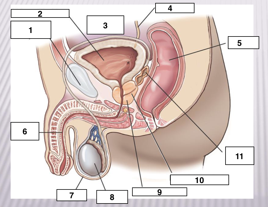
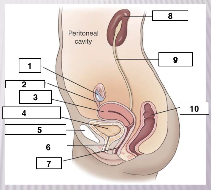
Symphysis Pubis
Urinary Bladder
Peritoneal cavity
Ureter
Rectum
Urethra
Scrotum
Testis
Prostate gland
Ejaculatory ducts
Seminal vesicles
Prostate
-Small glandular body surrounding the proximal part of the male urethra
-Considered part of the male reproductive system, but because of location, is often described with the urinary system
-Measures approximately 1½ inches (3.8 cm) transversely, ¾ inch (1.9 cm) at its base, and 1 inch (2.5 cm) vertically
-Benign Prostatic hyperplasia (BPH)
Gland gets enlarged
Bumps and lumps on the gland
Ovary
Uterine tube
Uterus
Urinary bladder
Symphysis Pubis
Urethra
Vagina
Kidney
Ureter
Rectum
IVU
Demonstrates kidneys, ureters and bladder
To demonstrate the function of the urinary system contrast media is injected into a vein and then, followed by imaging by either x-ray or computed tomography (CT)
Two filling techniques
Antegrade- Blous or IV drip
Retrograde - backward flow through the urethra, catheter
Clincal indications for IVU
Abdominal or pelvic
massRenal or urethral calculi, kidney stones
Kidney trauma
Flank pain
Hematuria- Blood in urine
Hypertension
Renal failure
Urinary tract infection (UTI) (pyelonephritis)
Lithotripsy - larger stones
Cystitis - inflammation of bladder
Hydronephrosis - the swelling of one or both kidneys caused by urine buildup
Functional procedures
-IVU/EXU (Intravenous/Excretory Urography)
-Nephrogram/Nephrotomogram
-CT IVU
-Hypertensive IVU
-Voiding Cystourethrogram (VCUG)= retrograde study
*Can watch the system
Basic routine of images for IVU
-Scout radiograph (KUB)
-Injection : Note time at beginning of injection.
-1 min nephrogram or nephrotomography
- 3-20 min intervals AP supine
- 5-10 min interval 30 degree posterior obliques
-Postvoid (AP bladder shot, KUB or erect)
-* special view- ureteric compression
Intravenous Urograhy Procedure
-Before procedure, patient must empty bladder,
remove clothing, and put on a gown
-Check blood chemistry (BUN, Creatinine)
-Ask if patient followed preparation : NP after midnight
-Informed Consent signed
-Make scout radiograph
-Perform venipuncture
Administer 50 to 100 mL of contrast for adult patient of average size (usually half of body weight)
Dosage for infants and children is adjusted according to age and weight
-Produce radiographs at specified time intervals
-When images are completed patient voids (empties bladder)
-Post Void image taken
-Patient instructed to drink plenty of fluids
-No metformin for 48 hours if applicable
CT IVU
Benefits → Minimal bowel prep: Water only at least 1 hour prior to procedure
2. Noncontrast images to evaluate for presence and location of renal calculi
3. Option to use contrast media provides a structural and functional study
4. Fast procedure with helical CT scanner
5. 3D reconstruction
Nephrogram
Radiographs taken in the first few minutes after injection to study to demonstrate the renal parenchyma or the functional portion of the kidney
- starting at 1 min
-Kidneys only- enter between xiphoid and iliac crest– one radiograph
AP Obliques
30 degrees
CR → iliac crest
2 inches lateral towards upside from the midline
LPO→ makes right kidney parallel to IR, moves left ureter off the spine
RPO→ makes left kidney parallel to IR, moves right ureter off spine
Expiration breathing
Postvoid
Can be done erect or recumbent
CR→ iliac crest
Bladder shot
CR→ 2 inches superior to symphysis pubis
10×12 IR or collimate
Used to visualize residual urine in bladder
AP with ureteric compression
CR→ iliac crest
Compression device places medial to ASIS at pelvic brim
Retrograde studies
Functional → Voiding cystourethrogram
Non functional→ Retrograde urogram, cystogram
VOIDING CYSTOURETHROGRAPHY/URETHROGRAPHY
VCUG
Purpose: Functional study of the bladder and urethra, ask to urinated and fluoro
-Performed after routine cystogram
-Catheter removed and imaged while voiding
Female= AP
Male = 30 degree RPO
Retrograde urography
Requires catheterization of ureters
-Scout radiograph taken
-Series of radiographs taken as requested
-Ureterogram taken once catheter has been removed
AP Axial bladder - cystogram
CR→ angled 10-15 degrees caudal, 2 inches abover upper border of pubic symphysis
10×12 collimation
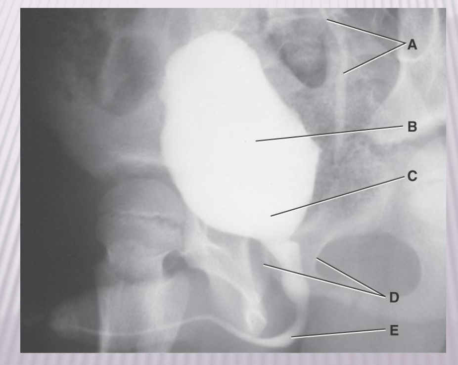
Voiding Cystourethrogram
A. Ureter
B. Bladder
C. Neck of bladder
D. prostate gland
E. Urethra
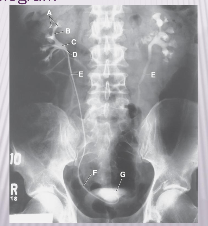
A. Minor caylces
B. Major Caylces
C. Renal pelvis
D. UPJ
E. Ureter