8: eye movements
1/66
There's no tags or description
Looks like no tags are added yet.
Name | Mastery | Learn | Test | Matching | Spaced | Call with Kai |
|---|
No analytics yet
Send a link to your students to track their progress
67 Terms
how many muscles are attached to the eye
6 muscles:
4 rectus muscles ( superior, inferior, medial and lateral)
2 oblique ( superior and inferior)
what are the 3 axis of rotation in which the orientation of the eye is defined by
horizontal
vertical
torsional
what is adduction
rotates the eye towards the nose
abduction
rotates the eye away from the nose
elevation
rotates the eye vertically up
depression
rotates the eye down
torsional movements
do not change the line of sight but rotate the eye around it
intorsion: rotates the top oc cornea towards nose
extorsion: rotates the top of the cornea away from the nose
medial rectus adducts the eye the lateral rectus abducts it
superior rectus
adduction: intorsion
abduction: elevation
inferior rectus
adduction: extorsion
abduction: depression
superior oblique
adduction: depression
abduction: intorsion
inferior oblique
adduction: elevation
abduction: extorsion
what 3 cranial nerves control the extraocular muscles
the abducens nerve CN VI
occulomotor nerve CN III
trochlear nerve CN IV
patients with lesions of extraocular musces or their nerves complain about double vision as the image of an object of gaze no longer falls in the same location of both retinae
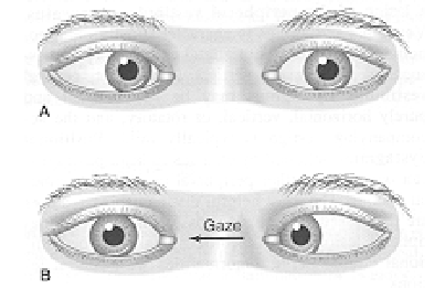
what does an isolated lesion of the abducens nerve cause
results in a loss of abduction beyond the midline, causing diplopia when patients attempt to look in the direction of the paralysed lateral rectus muscle
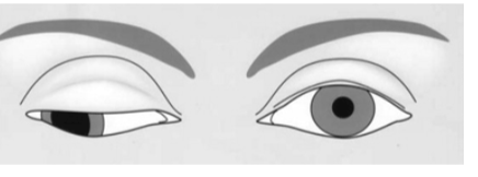
what does an isolated region of the occulomotor nerve result in
results in a loss of eye movements medially or upward from the mid position
also leeds to a drooping eye lid (ptosis) and a downward lateral gaze
what does an isolated lesion of trochlear nerve cause
causes deficits in intorsion/extorsion and elevation/depression which in turn leads to a skew deviation and a torsional deficit
what are the 2 reasons we move our eyes
to acquire targets ( gaze shifting)
to maintain gaze on targets ( gaze holding)
gaze shifting
involves 2 forms of eye movement
saccades- that shift the fovea rapidly to a peripheral target
vergence- moves the eye in opposite directions so that the image is positioned on both fovea
gaze holding
involves 3 forms of eye movements
smooth pursuit- movements keep image of a moving target on fovea
vestibulo-ocular- keep images on retina during bried head movements. driven by vestibular system
optokinetic- hold images during sustained head movements and driven byv siaul stimulation
saccades
very fast
shift the line of sight from one object to anoher
short duration
eg allow us to read text
can control saccades and use them to explore environment
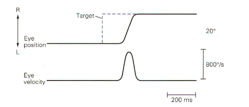
waveform of saccades
saccades have a stereotypical waveform with a single smooth increase and decrease of eye velocity
only the distance of the target from fovea determines the velocity of a saccade
movement of saccades
can voluntarily change the direction and amplitude of saccades
cannot change the velocity
can saccade to visual stimuli, auditory, tactile, memorised locations and verbal commands
what do extraocular motor neurons signal
they signal eye position and velocity
the discharge frequency of extraocularmotor neurons is directly proportional to the position and velocity of the eye
as eye velocityincreases, firing rate of motor neurones increase in a pulse of activity
what happens once the eyes have reached their final position after movement
they are held in place by contraction of extraocular muscles and this leads to a change in the baseline dishcarge rate known as a step in intensity
the saccadic signal of an ocular motor neurone has the form of a step pulse
step vs pulse signal
during a saccade, eye movements control often involves 2 signals
pulse → brief burst of neural activity that moves eye quickly
step → sustained signal that keeps the eye in the new position so the eye doesnt drift back
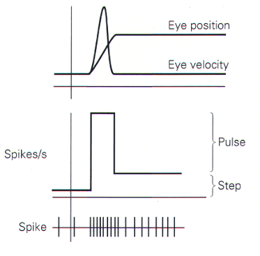
the saccadic signal
this is the nueral command sent from the brain to extraocular muscles to generate a saccade
height of the step indicates the extent of the saccade
the height of thw pulse indicates the speed of saccade
where do the motor circuits for saccades lie
they lie in the brain stem
the horizontal component of a saccade is organised in the pons and medulla
vertical component organised in mesencephalic reticular formation- in midbrain
in each cirucit lies neurones that are responsible for the step and pulse
what are burst cells
the neurons that give rise to the pulse components
motor circuits for saccades
the burst cells fire before and during ipsilateral saccades
what are medium lead burst cells
cells that make direct connections to the motor neurones
what are long lead burst cells
cells that drive the medium lead burst cells and recieve inputs from highher centres
what are inhibitory burst neurons
they suppress contralateral abducens neurons and are driven by medium lead burst cells
help coordinate the correct muscles during a saccade , ensuring the opposing muscle is switched off
what are omnipause cells
also involved in motor circuit for saccades
fire continously except around the time of a saccade
located in the nucleus of dorsal raphe
theyre GABA-ergic and inhibit the burst neurons
by pausing their firing, it allows burst cells to initiate a saccade
what helps maintain a stable saccade system
contribution of excitation of burst cells and inhibitoion of omnipause cells
tonic activity in neurons in midbrain and nucleus maintain steady signal related to eye positon
lesions in these regions doesnt affect saccades but after saccade the eyes drift back to starting positoin
what are the lateral rectus motor neurons in the pons driven by
driven by medium lead burst neurons which supply the burst and the vestibular and pepositus nuclei neurons , which supply tonic signal
vertical saccades generated in mesencephalic retincular formation
px with brain stem lesoins have characteristc deficits in eye movements
what does lesion in pontine gaze centres result in
results in deficits in horizontal saccades
what do lesions in midbrain gaze centres cause
deficits in vertical saccades
what are saccades controlle dby
controlled by the cerebral cortex
the pontine and mesencephalic burst cells provide motor signals for saccades but their output is controlled by superior colliculus
the SC is a major visuomotor integration region
what is smooth pursuit
keeps the image of a moving target on the fovea by calculating how fast the target is moving and moving the eye accordingly
neurons that signal eye velocity recieves major inputs from MT/MST and from the FEF
smooth pursuit movements have a max velocity of about 100 degrees/ sec which is slower than saccades
tracking an object
smooth pursuit occurs prior to a saccade and then smooth pursuit eye movements allow one to track the subsequent movement
what are microsaccades
very small rapid eye movements that occur while a person is trying to fixate on a single point
small and involuntary
what is the superior colliculus SC
a visuomotor integration region
main visual input comes from retina
helps the brain orient the eyes, head and attention towards objects in the environment
recieved input from V1, frontal eye fields and the retina
SC- during smooth pursuit
neurons a small distance from the front edge are activated, leading to small eye movements
for saccades, neurons are activated in a region that represents the point to which the saccade will be directed
just prior to a saccade activity builds up in cells at the target location and decreases in other parts of sc
what 2 regions is the SC divided into
superficial layers
intermediate and deep layers
the 3 superficial layers recieve direct input from retina as well as input from primary visual cortex and neurons in these layers respond to visual stimuli
what is the activity of intermediate and deep layers related to
occulomotor actions
SC indiv neurons
indv neurons within the SC fire before saccades of specific amplitdues and directions
while sc recieves most of its input from the retina its output is controlled by cerebral cortex
what is the movement field of neurone
the region of the visual field that contains the targets for the saccades
most movement related cells have large movement fields and so actual eye movements are encoded by a large number of neurones
frontal eye fields
involved in triggering intentional saccades
the supplementary eye fields, are involved in organising groups of saccades into sequences
parietal eye fields
further back in the bran
involved mainly in reflexive saccades, made in response to changes in the view
vergence movements
smooth pursuit and saccadic systems produce conjugate movements of both eyes
vergence movements produce disconjugate movements of the eyes
when looking at near objects, eyes rotate towards each other; converging
when lookinng at objects in the distance- eyes rotate away from each other; diverging
what do the disconjugate movements ensure
make sure that the object of interest is on the same place in both retinas
vergence is a function of the horizontal rectus muscles and is controlled by neurons in te occulomotor nucleus of the midbrain
what is the vestibular system
part of the body responsible for detecting head movement, maintaining balance and stabilising vision through the vestibulo-ocular reflex
done by measuring the linear and angular acceleration of the head through an ensemble of 5 sensoy organs in the inner ear
how many receptor organs does vestibular labyrinth house
5 receptor organs
linear accelerations are detected by the utricle and saccule
angular accelerations measured by semicircle canals
the 2 vestibular systems can measure linear acceleration along any axis and angular accelerations about any axis
vestibular reflexes: VORs
vest system is reposnible for vaerious reflexes that the body yses to compensate for head movements and the perception of motion in space
the VORs keep the eyes fixed on an object when the head moves
vestib apparatus signals how fast the head is rotating and the occulomotor system uses this to compensate for these movements keeping the visual images motionless on retina
VORs
visual processing is much slower and less efficient than vestibular processing for image stabilisation
if you lose these reflexes it would be hard to fixate on something whilst moving
what are the 3 different VORs that arise from 3 vestibular sense organs
rotational vestibulo ocular reflex compensates for head rotation
translational vestibulo ocular reflex compensates for linear head movements
ocular counter-rolling compensates for head tilt in the vertical
rotational vor
causes eyes to slowly rotate in opposite direction to any rotational head mocement detected by vestivular system
sustained rotation doesnt cause the eyes to be driven past the edge of their orbit instead the eyes make a rapid resetting movement back across the centre of gaze
combination of slow and quick eye movements results in a pattern called nystagmus - vestib signals drives the slow phase of nystagmus while brain stem circuits generate the quick phase
nystagmus
where the eyes make repetitive, uncontrolled movements
consist of a slow drift in one direction followed by a fast corrective movement in opposite
translational VOR
to compensate for linear head movements, the translational vor must take into account the distance to the object being viewed
keeps visin stable when moving head linearly
eg when leaning forard to look at something close, the eyes move slightly backwards to keep the object in focus
ocular counter rolling
since gravity exerts a constant acceleration on the head the vestibular system also senses the orientation of the head relative to gravity
vest system used to estimate the tilting of the head in vertical place and initiate counter rolling response of the eyes to compensate
gaze
coordination of hea and eye movements to direct the fovea
because the head has much higher inertia, during small gaze movements the fovea reaches its target before the head begins to move
small gaze movements trabslates into saccade, followed by head movement with vor
larger gaze movement
the eyes and head move simultaneously
vor temporarily suppressed in order for the eyes and head to move at the same time
problems with vor
it habituates- eg nystagmus in dark slows down and eventually stops
the canals do not respond well to very slow head movements
to compensate, the optokinetic system provides central vest system with visual information that is used to stabilise the visual scene in the eyes
optokinetic reflex
uses the fact that as we move our eyes in spaced fixed objects appear to move in the opposite direction
the reflex drives the eyes in the direction of the observed image motion, which if it were perfect it would stabilise the image o the retina
optokinetic reflex properties to VORs
it builds slowly so as to provide a motion signal that can take over as vestibular signal decays
it responds to very slow visual image motion
circular vection
type of optokinetic stimulation
illusion of self rotation, when body is stationary
visual system sees rotating environment and vest system detects no actual rotation so brain combines conflicting signals
after short period the subject percieves that rotation to have slowed down
perception of self motion in opposite direction builds up
integration of optokinetic and vestibular inputs
vest nuclei recives peripheral input when stimuli rotates at constant speed
but continued rotation means vest input fall to 0
meanhwile optokinetic pathway feeds a rising signal of similar time constant into tsame vestibular brain stem nuclei
hence in normal circumstances the summed output of these 2 signals results in a constant response