Contagious state - bacteria + mycosis (q1-12)

0.0(0)
Studied by 1 person
call kaiCall Kai
learnLearn
examPractice Test
spaced repetitionSpaced Repetition
heart puzzleMatch
flashcardsFlashcards
GameKnowt Play
full-widthPodcast
1
Card Sorting

1/11

encourage image

There's no tags or description

Looks like no tags are added yet.

Last updated 9:47 AM on 5/25/26
Name
Mastery
Learn
Test
Matching
Spaced
Call with Kai

No analytics yet

Send a link to your students to track their progress

12 Terms

1
New cards

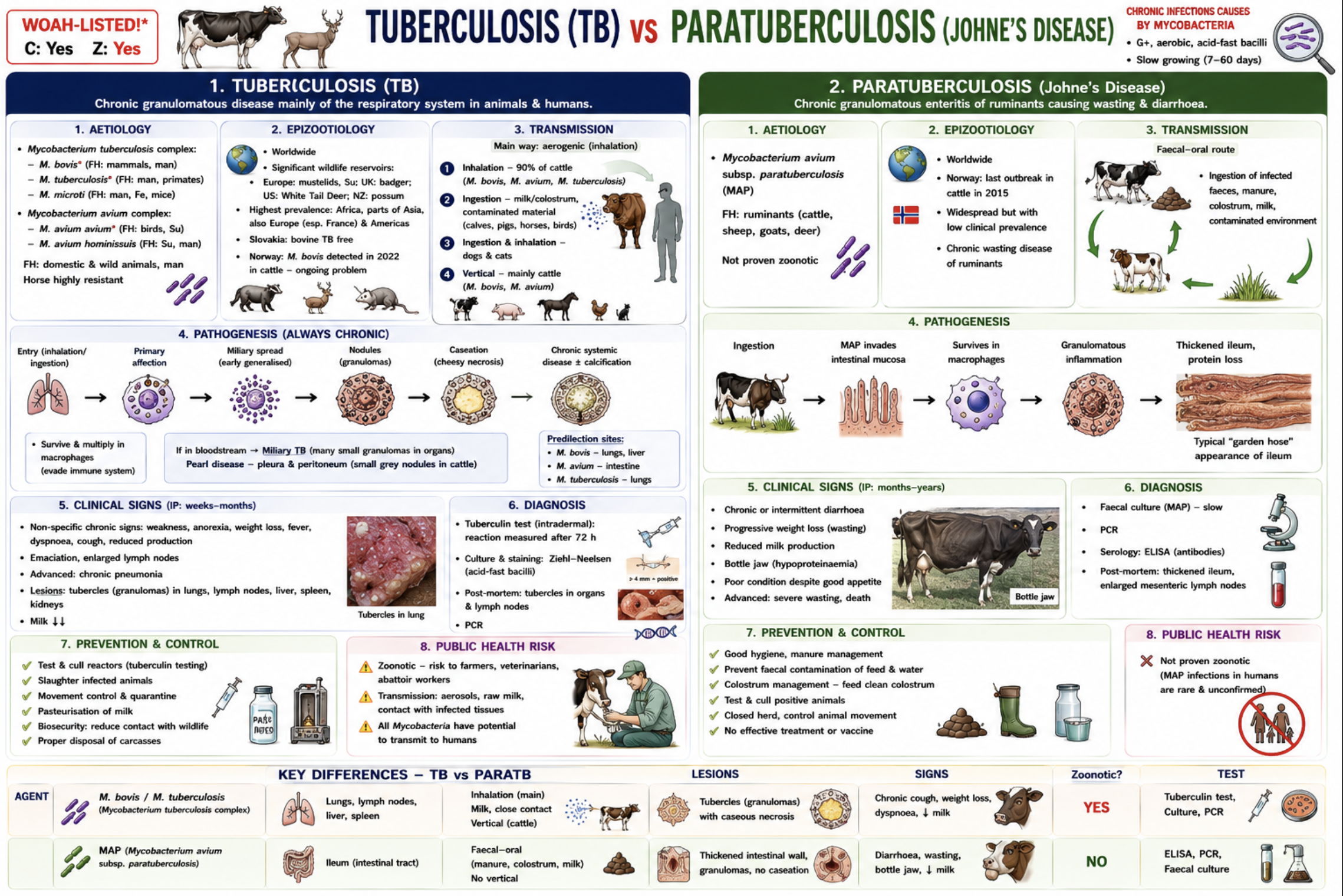
Tuberculosis and paratuberculosis (+ activation of disinfectants)

Etiology

  • TB: M. bovis (mammals, man), M. tuberculosis (man, car, su), M. avium complex (birds, pigs)

  • ParaTB (Johne’s disease): M. avium subsp. paratuberculosis

  • Acid-fast, aerobic, very resistant in environment, chronic granulomatous diseases.

Tuberculosis (TB)

Epizootology

  • Worldwide, zoonotic, WOAH-listed.

  • Controlled in many countries, but wildlife reservoirs persist.

  • Reservoirs: badgers, deer, wild boar, possums.

  • Slovakia TB-free; Norway had M. bovis reappearance in 2022.

Transmission

  • Mainly aerogenic (inhalation).

  • Also milk/feed, vertical transmission, contact with tissues.

Pathogenesis

  • Inhalation/ingestion → granulomas/tubercles → caseous necrosis ± calcification → chronic systemic disease.

Clinical signs

  • Cattle (M.bovis): Chronic cough, Weight loss, Dyspnoea, Fever, Enlarged LN, Often asymptomatic

  • Birds (M. avium): Often asymptomatic, Weight loss, ↓ egg production, Bone lesions

  • Pigs: Usually alimentary form, Intestinal lesions/LN lesions, Often asymptomatic

  • Humans: Pulmonary TB with Productive cough, Hemoptysis, Chest pain, Fever, fatigue

Diagnosis

  • Tuberculin skin test (main test), IFN-γ test, PM inspection, Histology (granulomas), Ziehl-Neelsen staining, Culture, PCR. (Serology is NOT used for tuberculosis)

Public health risk

  • Zoonotic - Infection from raw milk, meat, aerosols. Risk groups: vets, farmers, slaughterhouse workers. M. bovis is resistant to pyrazinamide.

Paratuberculosis (Johne’s disease)

Epizootology

  • Worldwide, chronic wasting disease of ruminants. High economic losses. Not officially zoonotic.

Transmission

  • Ingestion of contaminated milk, colostrum, faeces. Vertical transmission possible. Faecal shedding before clinical signs.

Pathogenesis

  • MAP in ileum/Peyer’s patches → granulomatous enteritis → malabsorption/protein loss.

Clinical signs

  • Adult ruminants: Chronic watery diarrhoea, Progressive weight loss despite appetite, ↓ milk production, Bottle jaw, Cachexia

  • Young animals: Usually infected young, Clinical disease appears in adulthood (long incubation).

Diagnosis

  • History + CS, ELISA (important screening test), PCR, Faecal culture, Histology, Tuberculin test with avian tuberculin

a) Prevention, control, outbreak measures, legislation

Legislation: Regulation (EU) 2016/429, Commission Delegated Regulation (EU) 2020/689, Commission Implementing Regulation (EU) 2020/200

Prevention & control

  • Veterinary supervision of animal movement, Quarantine of healthy animals, Tuberculin testing (TB), Surveillance/screening, PM meat inspection, Pasteurisation of milk, Biosecurity & hygiene, Identification & traceability, Movement restrictions

Outbreak/suspicion

  • Mandatory reporting, Isolation of suspect animals, Testing, Test & slaughter / depopulation, Rendering plant disposal, Epidemiological investigation, Final disinfection, Observation period (~110 d)

b) Sanitation, focal disinfection, activation of disinfectants

Environmental resistance

TB: Soil: 4 y, Pasture: 2 y, Water: 2 y, Slurry: 100–200 d

ParaTB: Soil: 11 mo, Faeces: 240 d

Focal disinfection: Chloramine T 6–10%, Peracetic acid 1%, Alkaline formalin 3%,

Environmental disinfection; Glutaraldehyde 2%, Formaldehyde, lime powder

Disinfection of objects: Sodium hypochlorite 2%

Excrements/slurry: 0.3% peracetic acid, 3% lime, Slurry storage >6 months

Activation of disinfectants:

Activation of disinfectants means increasing disinfectant efficacy by heating, adding alkalis/detergents, and removing organic material before disinfection!

  • Heating: Chloramine T: 50–60°C, NaOH: 70–80°C

  • Addition of detergents: sodium carbonate added to water (Detergents help disinfectants penetrate fat-rich mycobacterial cell walls)

  • Alkaline formaldehyde = NaOH + formaldehyde

Factors decreasing efficacy: Organic matter, Low temperature

<p><u>Etiology</u></p><ul><li><p><strong>TB:</strong> <em><mark data-color="red" style="background-color: red; color: inherit;">M. bovis (mammals, man), M. tuberculosis (man, car, su), M. avium complex</mark></em><mark data-color="red" style="background-color: red; color: inherit;"> (birds, pigs)</mark></p></li><li><p><strong>ParaTB (Johne’s disease):</strong> <em><mark data-color="red" style="background-color: red; color: inherit;">M. avium</mark></em><mark data-color="red" style="background-color: red; color: inherit;"> subsp. </mark><em><mark data-color="red" style="background-color: red; color: inherit;">paratuberculosis</mark></em></p></li></ul><ul><li><p>Acid-fast, aerobic, very resistant in environment, chronic granulomatous diseases.</p></li></ul><p></p><p><strong>Tuberculosis (TB)</strong></p><p><u>Epizootology</u></p><ul><li><p>Worldwide,<span style="color: red;"> <strong>zoonotic, WOAH-listed.</strong></span></p></li><li><p>Controlled in many countries, but <strong>wildlife reservoirs</strong> persist.</p></li><li><p>Reservoirs:<em> badgers, deer, wild boar, possums.</em></p></li><li><p><em>Slovakia TB-free; Norway had </em>M. bovis reappearance in 2022.</p></li></ul><p><u>Transmission</u></p><ul><li><p><strong>Mainly aerogenic (inhalation).</strong></p></li><li><p>Also milk/feed, vertical transmission, contact with tissues.</p></li></ul><p><u>Pathogenesis</u></p><ul><li><p>Inhalation/ingestion → granulomas/tubercles → caseous necrosis ± calcification → chronic systemic disease.</p></li></ul><p><u>Clinical signs</u></p><ul><li><p>Cattle (M.bovis): Chronic cough, Weight loss, Dyspnoea, Fever, Enlarged LN, <strong>Often asymptomatic</strong></p></li><li><p>Birds (<em>M. avium</em>): Often asymptomatic, Weight loss, ↓ egg production, Bone lesions</p></li><li><p>Pigs: Usually alimentary form, Intestinal lesions/LN lesions, Often asymptomatic</p></li><li><p>Humans: Pulmonary TB with Productive cough, Hemoptysis, Chest pain, Fever, fatigue</p></li></ul><p><u>Diagnosis</u></p><ul><li><p>Tuberculin skin test (main test), IFN-γ test, PM inspection, Histology (granulomas), Ziehl-Neelsen staining, Culture, PCR. (Serology is NOT used for tuberculosis)</p></li></ul><p><u>Public health risk</u></p><ul><li><p>Zoonotic <strong>- Infection from raw milk, meat, aerosols</strong>. Risk groups: vets, farmers, slaughterhouse workers. <em>M. bovis</em> is resistant to pyrazinamide.</p></li></ul><p></p><p><strong>Paratuberculosis (Johne’s disease)</strong></p><p><u>Epizootology</u></p><ul><li><p>Worldwide, chronic wasting disease of ruminants. High economic losses<strong>. Not officially zoonotic.</strong></p></li></ul><p><u>Transmission</u></p><ul><li><p>Ingestion of contaminated milk, colostrum, faeces. Vertical transmission possible. Faecal shedding before clinical signs.</p></li></ul><p><u>Pathogenesis</u></p><ul><li><p>MAP in ileum/Peyer’s patches → granulomatous enteritis → malabsorption/protein loss.</p></li></ul><p><u>Clinical signs</u></p><ul><li><p>Adult ruminants: Chronic watery diarrhoea, Progressive weight loss despite appetite, ↓ milk production, Bottle jaw, Cachexia</p></li><li><p>Young animals: Usually infected young, Clinical disease appears in adulthood (long incubation).</p></li></ul><p>Diagnosis</p><ul><li><p>History + CS, ELISA (important screening test), PCR, Faecal culture, Histology, Tuberculin test with avian tuberculin</p></li></ul><p></p><p><strong>a) Prevention, control, outbreak measures, legislation</strong></p><p><u>Legislation:</u> Regulation (EU) 2016/429, Commission Delegated Regulation (EU) 2020/689, Commission Implementing Regulation (EU) 2020/200</p><p><u>Prevention &amp; control</u></p><ul><li><p>Veterinary supervision of animal movement, Quarantine of healthy animals, Tuberculin testing (TB), Surveillance/screening, PM meat inspection, Pasteurisation of milk, Biosecurity &amp; hygiene, Identification &amp; traceability, Movement restrictions</p></li></ul><p><u>Outbreak/suspicion</u></p><ul><li><p>Mandatory reporting, Isolation of suspect animals, Testing, Test &amp; slaughter / depopulation, Rendering plant disposal, Epidemiological investigation, Final disinfection, Observation period (~110 d)</p></li></ul><p></p><p><strong>b) Sanitation, focal disinfection, <mark data-color="purple" style="background-color: purple; color: inherit;">activation of disinfectants</mark></strong></p><p><u>Environmental resistance</u></p><p>TB: Soil: 4 y, Pasture: 2 y, Water: 2 y, Slurry: 100–200 d</p><p>ParaTB: Soil: 11 mo, Faeces: 240 d</p><p><u>Focal disinfection:</u> <strong>Chloramine T</strong> 6–10%, <strong>Peracetic acid</strong> 1%, <strong>Alkaline formalin</strong> 3%,</p><p>Environmental disinfection; Glutaraldehyde 2%, Formaldehyde, lime powder</p><p>Disinfection of objects: <strong>Sodium hypochlorite</strong> 2%</p><p><u>Excrements/slurry: </u><strong>0.3% peracetic acid, 3% lime,</strong> Slurry storage &gt;6 months</p><p><u><mark data-color="purple" style="background-color: purple; color: inherit;">Activation of disinfectants:</mark></u></p><p>Activation of disinfectants means<strong> increasing disinfectant efficacy</strong> by heating, adding alkalis/detergents, and removing organic material before disinfection!</p><ul><li><p><strong>Heating:</strong> Chloramine T: 50–60°C, NaOH: 70–80°C</p></li><li><p><strong>Addition of detergents: </strong>sodium carbonate added to water (Detergents help disinfectants penetrate fat-rich mycobacterial cell walls)</p></li><li><p><strong>Alkaline formaldehyde</strong> = NaOH + formaldehyde</p></li></ul><p>Factors decreasing efficacy: Organic matter, Low temperature</p>
2
New cards

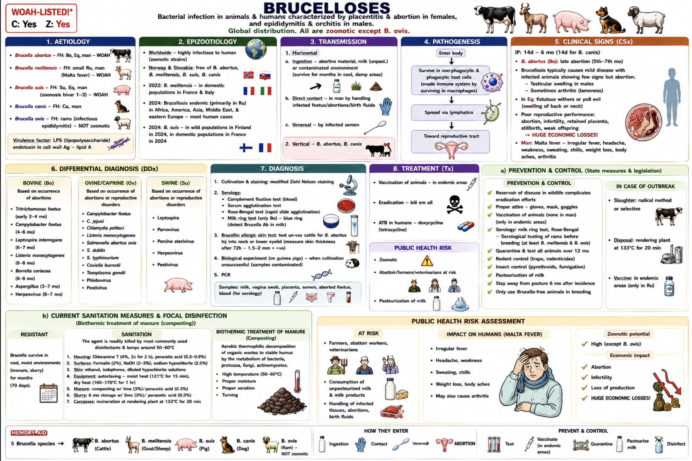
Brucellosis (+ biothermic treatment of manure)

WOAH-listed, Contagious + zoonotic (except B. ovis)

Causes: Abortion, placentitis in females, Epididymitis/orchitis in males, and intracellular bacteria surviving in macrophages. Virulence factor: LPS endotoxin.

Etiology + Main hosts

  • Brucella abortus → cattle

  • Brucella melitensis → sheep/goats (Malta fever in humans)

  • Brucella suis → pigs

  • Brucella canis → dogs

  • Brucella ovis → rams (NOT zoonotic)

Epizootology

  • Worldwide. Endemic mainly in Africa, Asia, Middle East, eastern Europe. Wildlife reservoirs complicate eradication.

  • Norway & Slovakia: Free from: B. abortus, B. melitensis, B. suis, B. canis

  • Recent situation: B. melitensis: France & Italy, B. suis: wild boar in Finland; domestic France

Transmission

  • Horizontal: Ingestion of aborted material/milk, Direct contact with placenta, foetus, birth fluids

  • Venereal transmission

  • Vertical - Especially B. abortus* and B. canis

Important: Brucella survives for months in cool, moist environments.

Pathogenesis

  • Entry → survival in macrophages → spread to reproductive organs → placentitis/abortion.

Clinical signs

  • Cattle (B. abortus): Late abortion (5–7 month), Retained placenta, Infertility, Weak calves

  • Rams (B. ovis): Epididymitis, Orchitis, Infertility

  • Males generally: Testicular swelling, Arthritis/lameness possible

  • Horses: Fistulous withers, Poll evil

  • Humans: Malta fever/Undulating fever, Sweating, Weakness, Weight loss, Arthralgia

Differential diagnosis of abortions

Cattle: Tritrichomonas foetus (2–4 mo), Campylobacter foetus (4–6 mo), Leptospira (6–7 mo), Listeria (6–8 mo), Herpesvirus, Aspergillus

Sheep/goats: Campylobacter, Chlamydia psittaci, Listeria, Salmonella, Coxiella burnetii, Toxoplasma gondii, Pestivirus

Pigs: Leptospira, Parvovirus, Pestivirus, Herpesvirus

Diagnosis

Samples: Placenta, Aborted foetus, Vaginal swab, Milk, Semen, Blood

Tests: Serology (Rose-Bengal test, Complement fixation test, Serum agglutination, Milk ring test (cattle)

Other: Culture + modified Ziehl-Neelsen stain, PCR, Brucellin skin test, Guinea pig bioassay

Public health risk assessment

Important zoonosis. Risk groups are Veterinarians, Farmers, Slaughterhouse workers. Infection via: Raw milk, Contact with aborted material, Aerosols

Prevention in humans: Pasteurisation, PPE (gloves, masks, goggles)

a) Prevention, control, outbreak measures, legislation

Prevention & control

  • Surveillance + serology, Milk ring test/Rose-Bengal, Test breeding animals, Quarantine + testing (>12 mo), Movement control, Pasteurisation, Rodent/insect control, Use Brucella-free breeding animals, Vaccination (Only in endemic areas, mainly ruminants), No human vaccine!

Outbreak/suspicion

  • Mandatory reporting, Isolation/quarantine, Test & slaughter, Selective or radical depopulation, Disposal at rendering plant, Epidemiological investigation, Final disinfection, disposal at rendering plant: 133°C for 20 min

b) Sanitation, focal disinfection, biothermic manure treatment

Resistance: Brucella survives months in Manure, Slurry, Cool/moist environment. But easily destroyed by common disinfectants

and at 50–60°C heat

Focal disinfection

  • Housing: Chloramine T 4%, Peracetic acid 0.3–0.9%

  • Surfaces: Formalin 2%, NaOH 2–3%, Sodium hypochlorite 2.5%

  • Skin: Ethanol, Iodophores

  • Equipment: Autoclave (121°C/15 min), Dry heat (160–170°C/1 h)

  • Manure: composting with lime

  • Slurry: 3% lime, 0.3% peracetic acid (Slurry storage: 6 months storage with lime )

Biothermic treatment of manure (composting)

= Thermophilic aerobic composting producing heat that kills pathogens.

  • Contaminated manture is placed on a surface layer of non-infected manure, straw and dry leaves, and covered with non infected manure.

Conditions:

  • Temperature: 55–70°C

  • minimum for 4 weeks

  • Aeration: O2 for the composting process

  • Moisture: 40–60%

  • C:N ratio: 25:1

  • pH: 5.5–8

Important:

  • Improves soil quality

  • Kills vegetative bacteria, larvae, eggs

  • Spores may survive → combine with disinfectants (3% lime for 4 weeks)

<p><strong><mark data-color="red" style="background-color: red; color: inherit;">WOAH-listed</mark></strong>, <span style="color: red;"><strong><em>Contagious </em></strong></span><strong><em>+</em></strong><span style="color: red;"><strong><em> zoonotic</em></strong></span><em> </em><strong><em>(except </em>B. ovis)</strong></p><p><u>Causes:</u> <strong>Abortion, placentitis</strong> in females,<strong> Epididymitis/orchitis</strong> in males, and <strong>intracellular bacteria surviving in macrophages</strong>. Virulence factor: LPS endotoxin.</p><p><u>Etiology + Main hosts</u></p><ul><li><p><em><mark data-color="red" style="background-color: red; color: inherit;">Brucella abortus</mark></em><mark data-color="red" style="background-color: red; color: inherit;"> </mark>→ cattle</p></li><li><p><em><mark data-color="red" style="background-color: red; color: inherit;">Brucella melitensis</mark></em><mark data-color="red" style="background-color: red; color: inherit;"> </mark>→ sheep/goats (Malta fever in humans)</p></li><li><p><em><mark data-color="red" style="background-color: red; color: inherit;">Brucella suis</mark></em><mark data-color="red" style="background-color: red; color: inherit;"> </mark>→ pigs</p></li><li><p><em><mark data-color="red" style="background-color: red; color: inherit;">Brucella canis</mark></em> → dogs</p></li><li><p><em><mark data-color="red" style="background-color: red; color: inherit;">Brucella ovis</mark></em> → rams (NOT zoonotic)</p></li></ul><p><u>Epizootology</u></p><ul><li><p><strong>Worldwide</strong>. <em>Endemic mainly in </em><strong><em>Africa, Asia, Middle East, eastern Europe</em>.</strong> Wildlife reservoirs complicate eradication.</p></li><li><p><strong>Norway &amp; Slovakia</strong>: Free from: <em>B. abortus, B. melitensis, B. suis, B. canis</em></p></li><li><p>Recent situation: <em>B. melitensis</em>:<strong> France &amp; Italy</strong>, <em>B. suis</em>: wild boar in<strong> Finland</strong>; domestic <strong>France</strong></p></li></ul><p><u>Transmission</u></p><ul><li><p>Horizontal:<strong> Ingestion</strong> of aborted material/milk, <strong>Direct contac</strong>t with placenta, foetus, birth fluids</p></li><li><p><strong>Venereal</strong> transmission</p></li><li><p><strong>Vertical</strong> - <em>Especially </em>B. abortus* and <em>B. canis</em></p></li></ul><p>Important: <mark data-color="blue" style="background-color: blue; color: inherit;">Brucella survives for months in cool, moist environments.</mark></p><p><u>Pathogenesis</u></p><ul><li><p>Entry → survival in macrophages → spread to reproductive organs → placentitis/abortion.</p></li></ul><p><u>Clinical signs</u></p><ul><li><p>Cattle (B. abortus): <strong>Late abortion </strong>(5–7 month), <strong>Retained placenta</strong>, Infertility, Weak calves</p></li><li><p>Rams (B. ovis): <strong>Epididymitis, Orchitis, Infertility</strong></p></li><li><p>Males generally: Testicular swelling, Arthritis/lameness possible</p></li><li><p>Horses: <strong>Fistulous withers, Poll evil</strong></p></li><li><p>Humans: <strong>Malta fever</strong>/Undulating fever, Sweating, Weakness, Weight loss, Arthralgia</p></li></ul><p><u>Differential diagnosis of abortions</u></p><p><strong>Cattle: </strong><em>Tritrichomonas foetus</em> (2–4 mo), <em>Campylobacter foetus</em> (4–6 mo), <em>Leptospira</em> (6–7 mo), <em>Listeria</em> (6–8 mo), Herpesvirus, Aspergillus</p><p><strong>Sheep/goats: </strong><em>Campylobacter, Chlamydia psittaci, Listeria, Salmonella, Coxiella burnetii, Toxoplasma gondii, </em>Pestivirus</p><p><strong>Pigs:</strong> Leptospira, Parvovirus, Pestivirus, Herpesvirus</p><p><u>Diagnosis</u></p><p>Samples: Placenta, Aborted foetus, Vaginal swab, Milk, Semen, Blood</p><p>Tests: Serology (<strong><mark data-color="blue" style="background-color: blue; color: inherit;">Rose-Bengal test</mark></strong>, Complement fixation test, Serum agglutination, <strong><mark data-color="blue" style="background-color: blue; color: inherit;">Milk ring test (cattle)</mark></strong></p><p>Other: Culture + modified Ziehl-Neelsen stain, PCR, Brucellin skin test, Guinea pig bioassay</p><p><u>Public health risk assessment</u></p><p>Important zoonosis. Risk groups are Veterinarians, Farmers, Slaughterhouse workers. Infection via: Raw milk, Contact with aborted material, Aerosols</p><p>Prevention in humans: Pasteurisation, PPE (gloves, masks, goggles)</p><p></p><p><strong>a) Prevention, control, outbreak measures, legislation</strong></p><p><u>Prevention &amp; control</u></p><ul><li><p><strong>Surveillance + serology</strong>, Milk ring test/Rose-Bengal, Test breeding animals, <strong>Quarantine + testing (&gt;12 mo)</strong>, Movement control, Pasteurisation, Rodent/insect control, Use Brucella-free breeding animals, Vaccination (Only in endemic areas, mainly ruminants), No human vaccine!</p></li></ul><p><u>Outbreak/suspicion</u></p><ul><li><p>Mandatory reporting, Isolation/quarantine,<strong> Test &amp; slaughter,</strong> Selective or radical depopulation, Disposal at rendering plant, Epidemiological investigation, Final disinfection, <strong>disposal at rendering plant: 133°C for 20 min</strong></p></li></ul><p></p><p><strong>b) Sanitation, focal disinfection, <mark data-color="purple" style="background-color: purple; color: inherit;">biothermic manure treatment</mark></strong></p><p>Resistance: Brucella survives months in Manure, Slurry, Cool/moist environment. But easily destroyed by common disinfectants</p><p>and at 50–60°C heat</p><p><u>Focal disinfection</u></p><ul><li><p>Housing:<strong> Chloramine T</strong> 4%,<strong> Peracetic acid</strong> 0.3–0.9%</p></li><li><p>Surfaces: <strong>Formalin</strong> 2%, NaOH 2–3%, Sodium hypochlorite 2.5%</p></li><li><p>Skin:<strong> Ethanol,</strong> Iodophores</p></li><li><p>Equipment: <strong>Autoclave </strong>(121°C/15 min),<strong> Dry heat </strong>(160–170°C/1 h)</p></li><li><p>Manure: <strong>composting with lime</strong></p></li><li><p>Slurry: 3% lime, 0.3%<strong> peracetic acid</strong> (Slurry storage: 6 months storage with lime )</p></li></ul><p></p><p><strong><mark data-color="purple" style="background-color: purple; color: inherit;">Biothermic treatment of manure (composting)</mark></strong></p><p>= Thermophilic aerobic composting producing heat that kills pathogens.</p><ul><li><p>Contaminated manture is placed on a surface layer of non-infected manure, straw and dry leaves, and covered with non infected manure. </p></li></ul><p>Conditions:</p><ul><li><p>Temperature:<strong> 55–70°C</strong></p></li><li><p>minimum for <strong>4 weeks</strong></p></li><li><p>Aeration: O2 for the composting process</p></li><li><p>Moisture:<strong><mark data-color="red" style="background-color: red; color: inherit;"> 40–60%</mark></strong></p></li><li><p>C:N ratio: 25:1</p></li><li><p>pH: 5.5–8</p></li></ul><p>Important:</p><ul><li><p>Improves soil quality</p></li><li><p>Kills vegetative bacteria, larvae, eggs</p></li><li><p>Spores may survive → <strong>combine with disinfectants (3% lime for 4 weeks)</strong></p></li></ul><p></p>
3
New cards

Anthrax, tetanus, botulism, and other anaerobic infections (+ conditions for the use of formaldehyde for disinfection with regard to protection of human and animal health)

Anthrax

WOAH-listed | Zoonotic

Etiology:

  • Bacillus anthracis

  • Spore-forming bacterium, produces: lethal toxin + oedema toxin

Epizootology

  • Worldwide except Antarctica, Endemic: Africa, Asia, southern/eastern Europe

  • Spores survive in soil for decades. Rain/flooding may expose spores

  • Norway: no reported cases

Transmission

1. Inhalation (most fatal), 2. Ingestion, 3. Cutaneous inoculation

Pathogenesis

  • Spores → macrophages → lymphatics → septicaemia → sudden death → sporulation after death.

Clinical signs

  • Peracute: Sudden death, No rigor mortis, Cherry-red unclotted blood from orifices, Rapid bloating

  • Acute/subacute: Fever, Dyspnoea, Convulsions, Depression

Human forms

  • Cutaneous: black ulcer

  • Pulmonary: severe pneumonia

  • Intestinal: bloody diarrhoea

Pathology

  • Splenomegaly (“blackberry jam spleen”), Haemorrhages, Congested organs, DO NOT OPEN CARCASS

Diagnosis

  • Report immediately! Blood from a live animal, Ear tissue from a dead animal

  • PCR, ELISA, Culture (“Medusa head” colonies), Ascoli precipitation test, Giemsa/McFadden stain

Treatment/prevention

  • Penicillin, oxytetracycline

  • Annual vaccination in endemic areas

  • Never necropsy!! Incineration preferred

Tetanus (“lockjaw”)

Zoonotic

Etiology:

  • Clostridium tetani Neurotoxins: tetanospasmin + tetanolysin

Transmission

  • Contaminated wounds, Soil/manure contamination

Pathogenesis

Toxin blocks inhibitory neurotransmitters (GABA/glycine) → spastic paralysis

Clinical signs

  • Horse: Sawhorse stance, Third eyelid prolapse, Tail elevated

  • Dog: Muscle rigidity, Retracted lips, Often localised form

  • Humans: Lockjaw, Risus sardonicus, Opisthotonus

Cause of death: Respiratory paralysis

Diagnosis

  • History + wounds, Toxin serology, Culture (“drumstick” appearance)

Treatment/prevention

  • Antitoxin, Metronidazole/penicillin, Wound cleaning + oxygenation, Vaccination (tetanus toxoid)

Botulism (“limber neck”)

WOAH-listed | Zoonotic

Etiology

  • Clostridium botulinum, Toxins A–G, Animals mainly C & D, Humans mainly A, B, E

Epizootology

  • Worldwide, Toxin in decaying food/carcasses, Birds infected through maggots

  • Norway: rakfisk, cured meats (spekemat), contaminated needles

Transmission

  • Ingestion of toxin/spores, Improper canned food, Honey in infants, Iatrogenic injections

Pathogenesis

  • Blocks acetylcholine release → flaccid paralysis.

Clinical signs

  • Birds: Limber neck, Paralysis, Cannot fly/walk

  • Mammals: Dysphagia, Weakness, Drooling, Progressive paralysis

  • Horse: Colic, Ileus, Shaker foal syndrome

Diagnosis

  • Feed history, Toxin ELISA/neutralisation, Serum/GIT samples

Treatment/prevention

  • Antitoxin, Gastric lavage, Remove contaminated feed, Proper food preservation

Other important anaerobic infections

Clostridium perfringens Enterotoxaemia (Diarrhoea, Bloody faeces, Malabsorption)

Clostridium chauvoei → Blackleg (Young cattle, Gas-producing myonecrosis, Lameness + dark muscle)

Clostridium septicum → Malignant edema (Wound contamination, Necrosis + gangrene, Fatal septicaemia)

Clostridium haemolyticum Bacillary haemoglobinuria (Intravascular haemolysis, Haemoglobinuria)

Fusobacterium necrophorum → Necrobacillosis (Calf diphtheria, Hepatic necrosis, Foot rot)

Public health risk

  • Anthrax: inhalation spores = most dangerous

  • Botulism: food poisoning

  • Tetanus: wound contamination

  • PPE + hygiene important

a) Prevention, control, outbreak measures

Anthrax: Do NOT open carcass, Incineration, Vaccination in endemic areas, Rodent/insect control, Disinfection before restocking, Prophylactic ATB for exposed workers

NB! If the carcass accidentally opens → ATB to exposed people, Vaccinate the herd, Soil disinfection

Tetanus: Early wound treatment, Vaccination, Antitoxin therapy

Botulism: Proper canning/storage, Remove dead birds/carcasses, Rodent control, Antiserum

b) Sanitation & focal disinfection, conditions for use of formaldehyde

Anthrax

  • Resistance: Spores survive: heat, UV, desiccation, survives for decades in soil

  • Housing: 10% Chloramine T, 10% chlorinated lime, 10% NaOH, 10% formalin, 4% glutaraldehyde, 1% peracetic acid

  • Soil: Lime, Phosphoric acid, Peracetic acid

  • Manure: Incineration, Composting + formaldehyde/glutaraldehyde

  • Slurry: 3% phosphoric acid, 1% peracetic acid

  • Spores destroyed by Autoclaving

Tetanus: 10% chlorinated lime, 3% formaldehyde, Autoclaving

Botulism: Autoclaving, Gamma radiation, Heating:100°C/10 min or 80°C/30 min

Formaldehyde — conditions of use

Properties

  • Effective against: bacteria, fungi, viruses, mycobacteria, spores (it is not stable)

Usage

  • 30% active formalin

  • Spray: 2% (max10%)

  • Focal: 3% + 3% NaOH (alkaline formaldehyde)

  • Vapour: humidity 70–90%, temperature >15°C

Risks

  • Carcinogenic (Category 1B)

  • Suspected mutagen (class 2 mutagen)

  • Irritating/toxic by inhalation/contact

Important

  • Use only by trained professionals with PPE!!!

  • approved for animal housing, animal feet, vehicles, and eggs in hatcheries in 2015

<p><strong>Anthrax</strong></p><p><span style="color: red;"><strong>WOAH-listed</strong></span> | <span style="color: red;">Zoonotic</span></p><p><u>Etiology:</u></p><ul><li><p><em><mark data-color="red" style="background-color: red; color: inherit;">Bacillus anthracis</mark></em></p></li><li><p><strong>Spore-forming</strong> bacterium, produces: <strong>lethal toxin + oedema toxin</strong></p></li></ul><p><u>Epizootology</u></p><ul><li><p><strong>Worldwide </strong>except Antarctica, <strong><em>Endemic: Africa, Asia, southern/eastern Europe</em></strong></p></li><li><p>Spores survive in soil for decades. Rain/flooding may expose spores</p></li><li><p><strong>Norway:</strong> no reported cases</p></li></ul><p><u>Transmission</u></p><p><strong>1. Inhalation (most fatal), 2. Ingestion, 3. Cutaneous inoculation</strong></p><p><u>Pathogenesis</u></p><ul><li><p>Spores → macrophages → lymphatics → septicaemia → sudden death → sporulation after death.</p></li></ul><p><u>Clinical signs</u></p><ul><li><p><strong>Peracute:</strong> Sudden death,<strong> No rigor mortis</strong>, <span style="color: red;">Cherry-red unclotted blood from orifices</span>, Rapid bloating</p></li><li><p><strong>Acute/subacute: </strong>Fever, Dyspnoea, Convulsions, Depression</p></li></ul><p><u>Human forms</u></p><ul><li><p><strong>Cutaneous</strong>: black ulcer</p></li><li><p><strong>Pulmonary:</strong> severe pneumonia</p></li><li><p><strong>Intestinal:</strong> bloody diarrhoea</p></li></ul><p><u>Pathology</u></p><ul><li><p>Splenomegaly (“blackberry jam spleen”), Haemorrhages, Congested organs, DO NOT OPEN CARCASS</p></li></ul><p><u>Diagnosis</u></p><ul><li><p>Report immediately! <strong>Blood</strong> from a live animal, <strong>Ear tissue</strong> from a dead animal</p></li><li><p>PCR, ELISA, Culture (“Medusa head” colonies),<strong><mark data-color="red" style="background-color: red; color: inherit;"> Ascoli precipitation test</mark></strong>, Giemsa/McFadden stain</p></li></ul><p><u>Treatment/prevention</u></p><ul><li><p> Penicillin, oxytetracycline</p></li><li><p>Annual vaccination in endemic areas</p></li><li><p>Never necropsy!! <strong>Incineration</strong> preferred</p></li></ul><p></p><p><strong>Tetanus (“lockjaw”)</strong></p><p><span style="color: red;">Zoonotic</span></p><p><u>Etiology:</u></p><ul><li><p><em><mark data-color="red" style="background-color: red; color: inherit;">Clostridium tetani </mark>→ </em>Neurotoxins: <strong>tetanospasmin +  tetanolysin</strong></p></li></ul><p><u>Transmission</u></p><ul><li><p>Contaminated wounds, Soil/manure contamination</p></li></ul><p><u>Pathogenesis</u></p><p>Toxin blocks inhibitory neurotransmitters (GABA/glycine) →<strong> spastic paralysis</strong></p><p><u>Clinical signs</u></p><ul><li><p>Horse: <strong>Sawhorse stance, Third eyelid prolapse, Tail elevated</strong></p></li><li><p>Dog: Muscle rigidity, Retracted lips, <strong>Often localised form</strong></p></li><li><p>Humans: Lockjaw, Risus sardonicus, Opisthotonus</p></li></ul><p>Cause of death: <strong>Respiratory paralysis</strong></p><p><u>Diagnosis</u></p><ul><li><p>History + wounds, Toxin serology, Culture (“<strong>drumstick</strong>” appearance)</p></li></ul><p><u>Treatment/prevention</u></p><ul><li><p>Antitoxin, Metronidazole/penicillin, Wound cleaning + oxygenation, Vaccination (tetanus toxoid)</p></li></ul><p></p><p><strong>Botulism (“limber neck”)</strong></p><p><span style="color: red;"><strong>WOAH-listed | Zoonotic</strong></span></p><p><u>Etiology</u></p><ul><li><p><em><mark data-color="red" style="background-color: red; color: inherit;">Clostridium botulinum, </mark></em>Toxins A–G, Animals mainly C &amp; D, Humans mainly A, B, E</p></li></ul><p><u>Epizootology</u></p><ul><li><p><strong>Worldwide</strong>, Toxin in decaying food/carcasses, Birds infected through maggots</p></li><li><p><strong>Norway:</strong> rakfisk, cured meats (spekemat), contaminated needles</p></li></ul><p><u>Transmission</u></p><ul><li><p>Ingestion of toxin/spores, Improper canned food, Honey in infants, Iatrogenic injections</p></li></ul><p><u>Pathogenesis</u></p><ul><li><p>Blocks acetylcholine release → <strong>flaccid paralysis.</strong></p></li></ul><p><u>Clinical signs</u></p><ul><li><p>Birds: <strong>Limber neck</strong>, Paralysis, Cannot fly/walk</p></li><li><p>Mammals: Dysphagia, Weakness, Drooling, <strong>Progressive paralysis</strong></p></li><li><p>Horse: Colic, Ileus, <strong>Shaker foal syndrome</strong></p></li></ul><p><u>Diagnosis</u></p><ul><li><p> Feed history, Toxin ELISA/neutralisation, Serum/GIT samples</p></li></ul><p><u>Treatment/prevention</u></p><ul><li><p>Antitoxin, Gastric lavage, Remove contaminated feed, Proper food preservation</p></li></ul><p></p><p><strong><u>Other important anaerobic infections</u></strong></p><p><em><mark data-color="red" style="background-color: red; color: inherit;">Clostridium perfringens </mark>→ </em>Enterotoxaemia (Diarrhoea, Bloody faeces, Malabsorption)</p><p></p><p><em><mark data-color="red" style="background-color: red; color: inherit;">Clostridium chauvoei</mark> → Blackleg (</em>Young cattle, Gas-producing myonecrosis, Lameness + dark muscle)</p><p></p><p><em><mark data-color="red" style="background-color: red; color: inherit;">Clostridium septicum</mark> → Malignant edema (</em>Wound contamination, Necrosis + gangrene, Fatal septicaemia)</p><p></p><p><mark data-color="red" style="background-color: red; color: inherit;"> </mark><em><mark data-color="red" style="background-color: red; color: inherit;">Clostridium haemolyticum </mark>→ </em>Bacillary haemoglobinuria (Intravascular haemolysis, Haemoglobinuria)</p><p></p><p><em><mark data-color="red" style="background-color: red; color: inherit;">Fusobacterium necrophorum</mark> → Necrobacillosis (</em>Calf diphtheria, Hepatic necrosis, Foot rot)</p><p></p><p><u>Public health risk</u></p><ul><li><p>Anthrax: inhalation spores = most dangerous</p></li><li><p>Botulism: food poisoning</p></li><li><p>Tetanus: wound contamination</p></li><li><p> PPE + hygiene important</p></li></ul><p></p><p><strong> a) Prevention, control, outbreak measures</strong></p><p><strong><u>Anthrax</u></strong>: Do NOT open carcass, Incineration, Vaccination in endemic areas, Rodent/insect control, Disinfection before restocking, Prophylactic ATB for exposed workers</p><p>NB! If the carcass accidentally opens → ATB to exposed people, Vaccinate the herd, Soil disinfection</p><p><strong><u>Tetanus:</u></strong> Early wound treatment, Vaccination, Antitoxin therapy</p><p><strong><u>Botulism: </u></strong>Proper canning/storage, Remove dead birds/carcasses, Rodent control, Antiserum</p><p></p><p><strong> b) Sanitation &amp; focal disinfection, <mark data-color="purple" style="background-color: purple; color: inherit;">conditions for use of formaldehyde</mark></strong></p><p>Anthrax</p><ul><li><p>Resistance: Spores survive: heat, UV, desiccation, survives for decades in soil</p></li></ul><ul><li><p>Housing: <strong>10% Chloramine T, 10% chlorinated lime, 10% NaOH, 10% formalin</strong>, 4% glutaraldehyde, 1% peracetic acid</p></li><li><p>Soil: Lime, Phosphoric acid, Peracetic acid</p></li><li><p>Manure: <strong>Incineration</strong>, Composting + formaldehyde/glutaraldehyde</p></li><li><p>Slurry: 3% phosphoric acid, 1% peracetic acid</p></li><li><p>Spores destroyed by <strong>Autoclaving</strong></p></li></ul><p>Tetanus: 10% chlorinated lime, 3% formaldehyde, Autoclaving</p><p>Botulism: Autoclaving, Gamma radiation, Heating:100°C/10 min or 80°C/30 min</p><p></p><p><strong><mark data-color="purple" style="background-color: purple; color: inherit;">Formaldehyde — conditions of use</mark></strong></p><p> Properties</p><ul><li><p>Effective against: bacteria, fungi, viruses, mycobacteria, spores (it is not stable)</p></li></ul><p> Usage</p><ul><li><p>30% active formalin</p></li></ul><ul><li><p>Spray: 2% (max10%)</p></li><li><p>Focal: 3% + 3% NaOH (alkaline formaldehyde)</p></li><li><p>Vapour: humidity 70–90%, temperature &gt;15°C</p></li></ul><p> Risks</p><ul><li><p><strong>Carcinogenic</strong> (Category 1B)</p></li><li><p><strong>Suspected mutagen</strong> (class 2 mutagen)</p></li><li><p>Irritating/toxic by inhalation/contact</p></li></ul><p> Important</p><ul><li><p><span style="color: red;">Use only by trained professionals with PPE!!!</span></p></li><li><p><span style="color: red;">approved for animal housing, animal feet, vehicles, and eggs in hatcheries in 2015</span></p></li></ul><p></p>
4
New cards

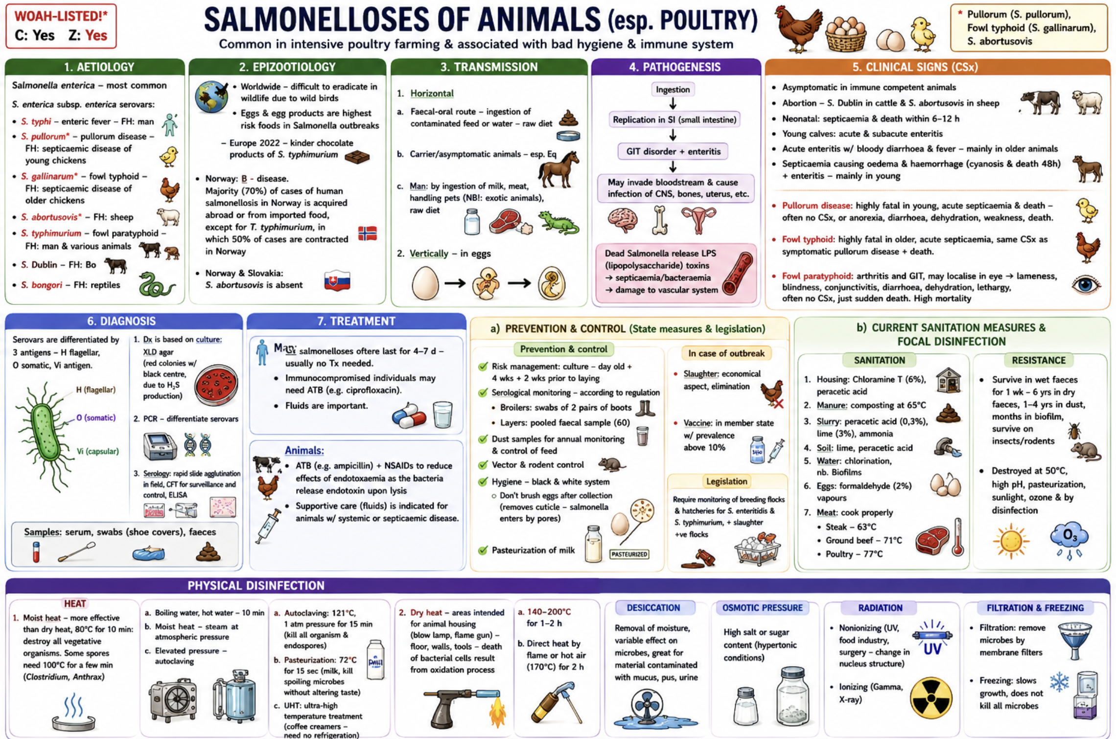
Salmonelloses of animals and poultry (+ physical disinfection)

WOAH-listed* | Contagious | Zoonotic

Etiology

  • Main agent: Salmonella enterica

Important serovars:

  • S. typhi → enteric fever in humans

  • S. pullorum* → pullorum disease (septicemic disease in young chicks)

  • S. gallinarum *→ fowl typhoid (older chickens)

  • S. typhimurium → fowl paratyphoid, zoonotic, not host specific

  • S. dublin → cattle

  • S. abortusovis* → sheep → abortions

  • S. bongori → reptiles

Epizootology

  • Worldwide. Common in intensive poultry farming. Associated with poor hygiene and stress/immunosuppression. Wildlife birds maintain infection.

  • Public health importance: Eggs and egg products = highest-risk foods. Major foodborne zoonosis.

  • Europe 2022: Kinder chocolate products of S. typhimurium

  • Europe January 2023-2025: outbreak with 509 confirmed cases inlcuding Norway, Finland and Sweden, traced to sprouts, especially alfalfa and clover from a supplier in Italy, and the investigation is still ongoing

  • Norway: B-disease. Most human infections are acquired abroad or from imported food, except S. typhimurium 50% of cases contracted in Norway.

  • Slovakia & Norway: S. abortusovis absent!

Transmission

  • Horizontal: Faecal–oral route, Contaminated feed/water, Raw diets, Carrier animals, Rodents/insects

  • Vertical: Through eggs

  • Human infection: Raw eggs, Meat, Milk, handling exotic pets/reptiles

Pathogenesis

  • Ingestion → intestinal replication → enteritis → possible septicaemia → possible infection of CNS, bones, uterus etc.

  • Important: LPS endotoxin released during bacterial lysis causes: endotoxaemia, vascular damage and shock

Clinical signs

  • General: Many healthy adult animals are asymptomatic.

  • Young animals: Septicaemia, Sudden death, Fever, Severe diarrhoea

  • Calves: Acute enteritis, Bloody diarrhoea, Fever

  • Cattle (S. dublin): Abortion, Septicaemia

  • Sheep (S. abortusovis): Abortion

  • Pullorum disease (S. pullorum): Young chicks → White diarrhoea, Weakness, Dehydration, Septicaemia, High mortality

  • Fowl typhoid (S. gallinarum): Older chickens → Septicaemia, Sudden death, High mortality

  • Fowl paratyphoid (S. typhimurium): Arthritis, Lameness, Conjunctivitis, Blindness, Enteritis, Sudden death

Diagnosis

  • Samples: Faeces, Shoe-cover swabs, Serum

  • Methods: Culture (main) XLD agar: red colonies with black centre (H₂S), PCR

  • Serovar differentiation: Serology → Rapid slide agglutination, CFT, ELISA

Treatment

  • Humans: usually self-limiting, fluids, severe cases: ciprofloxacin

  • Animals: Ampicillin/other ATB, NSAIDs, Fluid therapy

Public health risk assessment

  • Important foodborne zoonosis. Eggs/poultry major source. Raw foods and reptiles important reservoirs.

  • Risk groups: Poultry workers, Farmers, Veterinarians, Consumers of raw eggs/meat

a) Prevention, control, outbreak measures, legislation

Prevention & control:

  • There is legislation that requires monitoring of breeding flocks and hatcheries for S.enteritidis and S.typhymurium + slaughter of positive flocks

  • Risk management: culture day-old chicks and 4 + 2weeks before laying

  • Serology: test swabs on 2 pairs of boots in broilers and pooled fecal samples of layers (60 samples)

  • Take dust samples for annual monitoring and control feed

  • Biosecurity: Rodent control, Insect/vector control, Hygiene “Black & white system.”

  • Important: Do NOT brush eggs → removes protective cuticle, bacteria enter pores

  • Food safety: Pasteurisation of milk, Proper cooking

Outbreak measures

  • Slaughter/elimination of positive flocks, Movement restriction, Cleaning/disinfection, Vaccination in countries with prevalence >10%

b) Sanitation, focal disinfection, physical disinfection

  • Resistance: Wet faeces (1 week), Dry faeces (up to 6 years), Dust (1–4 years), Biofilms (months)

  • Destroyed by: Heat, Sunlight, High pH, Ozone, Disinfectants

Sanitation

  • Housing: Chloramine T 6%, Peracetic acid

  • Manure: Composting at 65°C

  • Slurry: 0.3% peracetic acid, 3% lime, Ammonia

  • Water: Chlorination

  • Eggs: 2% formaldehyde vapours

  • Meat cooking temperatures: Steak: 63°C, Ground beef: 71°C, Poultry: 77°C

Physical disinfection

  1. Moist heat (most effective → coagulation of proteins)

  • Boiling/hot water: 80C for 10 min (vegetative organisms), or 100C for 5min (C.perfringens spores), 10min (B.anthracis)

  • Autoclaving (steam): 121°C, 1atm/bar, 15 min → kills spores/endospores

  • Pasteurisation: 72°C for 15 sec

  • UHT: Ultra-high temperature treatment (140C for 2sec)

  1. Dry heat (on animal housings like floor, walls, kill MO by oxidation)

  • 140–200°C for 1–2 h

  • Flame gun/blow lamp for housing/tools

  • Direct heat by using flame or hot air oven with 170C for 2h (mention this one!)

  • NB: some organisms like prions may not be killed by dry heat

  1. Desiccation: Removes moisture, variable effect. great for materials contaminated witg urine, pus, mucus

  1. Osmotic pressure: High salt/sugar

  2. Radiation

  • nonionizing (UV, food industry, surgery), ionizing (Gamma/X-ray)

6. Filtration & freezing

<p><span style="color: red;"><strong>WOAH-listed* </strong>| Contagious | Zoonotic</span></p><p><u>Etiology</u></p><ul><li><p>Main agent: <em><mark data-color="red" style="background-color: red; color: inherit;">Salmonella enterica</mark></em></p></li></ul><p><u>Important serovars:</u></p><ul><li><p><em>S. typhi</em> → enteric fever in <span style="color: red;">humans</span></p></li><li><p><em><mark data-color="red" style="background-color: red; color: inherit;">S. pullorum*</mark></em><mark data-color="red" style="background-color: red; color: inherit;"> </mark>→ pullorum disease (septicemic disease in young chicks)</p></li><li><p><em><mark data-color="red" style="background-color: red; color: inherit;">S. gallinarum</mark></em><mark data-color="red" style="background-color: red; color: inherit;"> *</mark>→ fowl typhoid (older chickens)</p></li><li><p><em>S. typhimurium</em> → fowl paratyphoid, <span style="color: red;">zoonotic</span>, not host specific</p></li><li><p><em>S. dublin</em> → cattle</p></li><li><p><em><mark data-color="red" style="background-color: red; color: inherit;">S. abortusovis*</mark></em><mark data-color="red" style="background-color: red; color: inherit;"> </mark>→ sheep → abortions</p></li><li><p><em>S. bongori</em> → reptiles</p></li></ul><p><u> Epizootology</u></p><ul><li><p><strong>Worldwide</strong>. Common in intensive poultry farming. Associated with<strong> poor hygiene and stress</strong>/immunosuppression. Wildlife birds maintain infection.</p></li><li><p><strong>Public health importance</strong>: Eggs and egg products = highest-risk foods. <strong>Major foodborne zoonosis.</strong></p></li><li><p><strong>Europe 2022: </strong>Kinder chocolate products of S. typhimurium</p></li><li><p><strong>Europe January 2023-2025:</strong> outbreak with 509 confirmed cases inlcuding Norway, Finland and Sweden, traced to sprouts, especially alfalfa and clover from a supplier in Italy, and the investigation is still ongoing</p></li><li><p><strong>Norway:</strong> B-disease. Most <strong>human infections</strong> are acquired abroad or from imported food, except <em>S. typhimurium</em> 50% of cases contracted in Norway.</p></li><li><p><strong>Slovakia &amp; Norway</strong>: <em><mark data-color="red" style="background-color: red; color: inherit;">S. abortusovis</mark></em> absent!</p></li></ul><p><u>Transmission</u></p><ul><li><p><strong>Horizontal:</strong> Faecal–oral route, Contaminated feed/water, Raw diets, Carrier animals, Rodents/insects</p></li><li><p><strong>Vertical: </strong>Through eggs</p></li><li><p>Human infection: Raw eggs, Meat, Milk, <strong>handling exotic pets/reptiles</strong></p></li></ul><p><u>Pathogenesis</u></p><ul><li><p>Ingestion → intestinal replication → enteritis → possible septicaemia → possible infection of <strong>CNS, bones, uterus</strong> etc.</p></li><li><p>Important: <strong>LPS endotoxin </strong>released during bacterial lysis causes:<strong> endotoxaemia, vascular damage and shock</strong></p></li></ul><p><u>Clinical signs</u></p><ul><li><p>General: Many healthy adult animals are <strong>asymptomatic.</strong></p></li><li><p>Young animals: <strong>Septicaemia, Sudden death, Fever, Severe diarrhoea</strong></p></li><li><p>Calves: Acute enteritis, Bloody diarrhoea, Fever</p></li><li><p>Cattle (<em>S. dublin): </em><strong>Abortion</strong>, Septicaemia</p></li><li><p>Sheep (<em>S. abortusovis): </em>Abortion</p></li><li><p>Pullorum disease (<em>S. pullorum): </em>Young chicks → White diarrhoea, Weakness, Dehydration, Septicaemia, High mortality</p></li><li><p>Fowl typhoid (<em>S. gallinarum): </em>Older chickens → Septicaemia, Sudden death, High mortality</p></li><li><p>Fowl paratyphoid (<em>S. typhimurium): </em><strong>Arthritis, Lameness, Conjunctivitis, Blindness</strong>, Enteritis, Sudden death</p></li></ul><p><u>Diagnosis</u></p><ul><li><p>Samples:  Faeces, Shoe-cover swabs, Serum</p></li><li><p>Methods: <strong>Culture</strong> (main) <span style="color: red;">XLD agar</span>: red colonies with black centre (H₂S),  <strong>PCR</strong></p></li><li><p>Serovar differentiation: <strong>Serology</strong> → Rapid slide agglutination, CFT, ELISA</p></li></ul><p><u>Treatment</u></p><ul><li><p>Humans: <strong>usually self-limiting, </strong>fluids, severe cases: ciprofloxacin</p></li><li><p>Animals: Ampicillin/other ATB, NSAIDs, Fluid therapy</p></li></ul><p><u>Public health risk assessment</u></p><ul><li><p>Important foodborne zoonosis. Eggs/poultry major source. Raw foods and reptiles important reservoirs.</p></li><li><p>Risk groups: Poultry workers, Farmers, Veterinarians, Consumers of raw eggs/meat</p></li></ul><p></p><p><strong> a) Prevention, control, outbreak measures, legislation</strong></p><p><u>Prevention &amp; control:</u></p><ul><li><p><mark data-color="red" style="background-color: red; color: inherit;">There is legislation that requires monitoring of breeding flocks and hatcheries for S.enteritidis and S.typhymurium + slaughter of positive flocks</mark></p></li></ul><ul><li><p>Risk management: <strong>culture day-old chicks</strong> and 4 + 2weeks before laying</p></li><li><p>Serology: test <strong>swabs on 2 pairs of boots in broilers </strong>and <strong>pooled fecal samples of layers</strong> (60 samples)</p></li><li><p>Take <strong>dust sample</strong>s for annual monitoring and <strong>control feed</strong></p></li><li><p>Biosecurity: Rodent control, Insect/vector control, Hygiene “Black &amp; white system.”</p></li><li><p>Important: Do NOT brush eggs → removes protective cuticle, bacteria enter pores</p></li><li><p>Food safety: <strong>Pasteurisation of milk, Proper cooking</strong></p></li></ul><p><u>Outbreak measures</u></p><ul><li><p><strong>Slaughter/elimination of positive flocks</strong>, Movement restriction, Cleaning/disinfection, <strong>Vaccination</strong> in countries with prevalence &gt;10%</p></li></ul><p></p><p><strong><u> b) Sanitation, focal disinfection,<mark data-color="purple" style="background-color: purple; color: inherit;"> physical disinfection</mark></u></strong></p><ul><li><p>Resistance: Wet faeces (1 week), Dry faeces (up to 6 years), Dust (1–4 years), Biofilms (months)</p></li><li><p>Destroyed by: Heat, Sunlight, High pH, Ozone, Disinfectants</p></li></ul><p><u>Sanitation</u></p><ul><li><p>Housing: <strong>Chloramine T 6%, Peracetic acid</strong></p></li><li><p>Manure: <strong>Composting at 65°C</strong></p></li><li><p>Slurry: 0.3% peracetic acid, 3% lime, Ammonia</p></li><li><p>Water: Chlorination</p></li><li><p>Eggs: 2%<strong> formaldehyde vapours</strong></p></li><li><p>Meat cooking temperatures: Steak: 63°C, Ground beef: 71°C, Poultry: 77°C</p></li></ul><p></p><p><u><mark data-color="purple" style="background-color: purple; color: inherit;">Physical disinfection</mark></u></p><ol><li><p><strong>Moist heat </strong>(most effective → coagulation of proteins)</p></li></ol><ul><li><p>Boiling/hot water: 80C for 10 min (vegetative organisms), or 100C for 5min (C.perfringens spores), 10min (B.anthracis)</p></li><li><p>Autoclaving (steam): 121°C, 1atm/bar, 15 min → kills spores/endospores</p></li><li><p>Pasteurisation: 72°C for 15 sec</p></li><li><p>UHT: Ultra-high temperature treatment (140C for 2sec)</p></li></ul><ol start="2"><li><p><strong>Dry heat (on animal housings like floor, walls, kill MO by oxidation)</strong></p></li></ol><ul><li><p>140–200°C for 1–2 h</p></li><li><p>Flame gun/blow lamp for housing/tools</p></li><li><p>Direct heat by using flame or <span style="color: red;">hot air oven with 170C for 2h (mention this one!)</span></p></li><li><p>NB: some organisms like prions may not be killed by dry heat</p></li></ul><ol start="3"><li><p><strong>Desiccation: </strong>Removes moisture, variable effect. great for materials contaminated witg urine, pus, mucus</p></li></ol><ol start="4"><li><p><strong>Osmotic pressure</strong>: High salt/sugar</p></li><li><p><strong>Radiation</strong></p></li></ol><ul><li><p>nonionizing (UV, food industry, surgery), ionizing (Gamma/X-ray)</p></li></ul><p><strong> 6. Filtration &amp; freezing</strong></p>
5
New cards

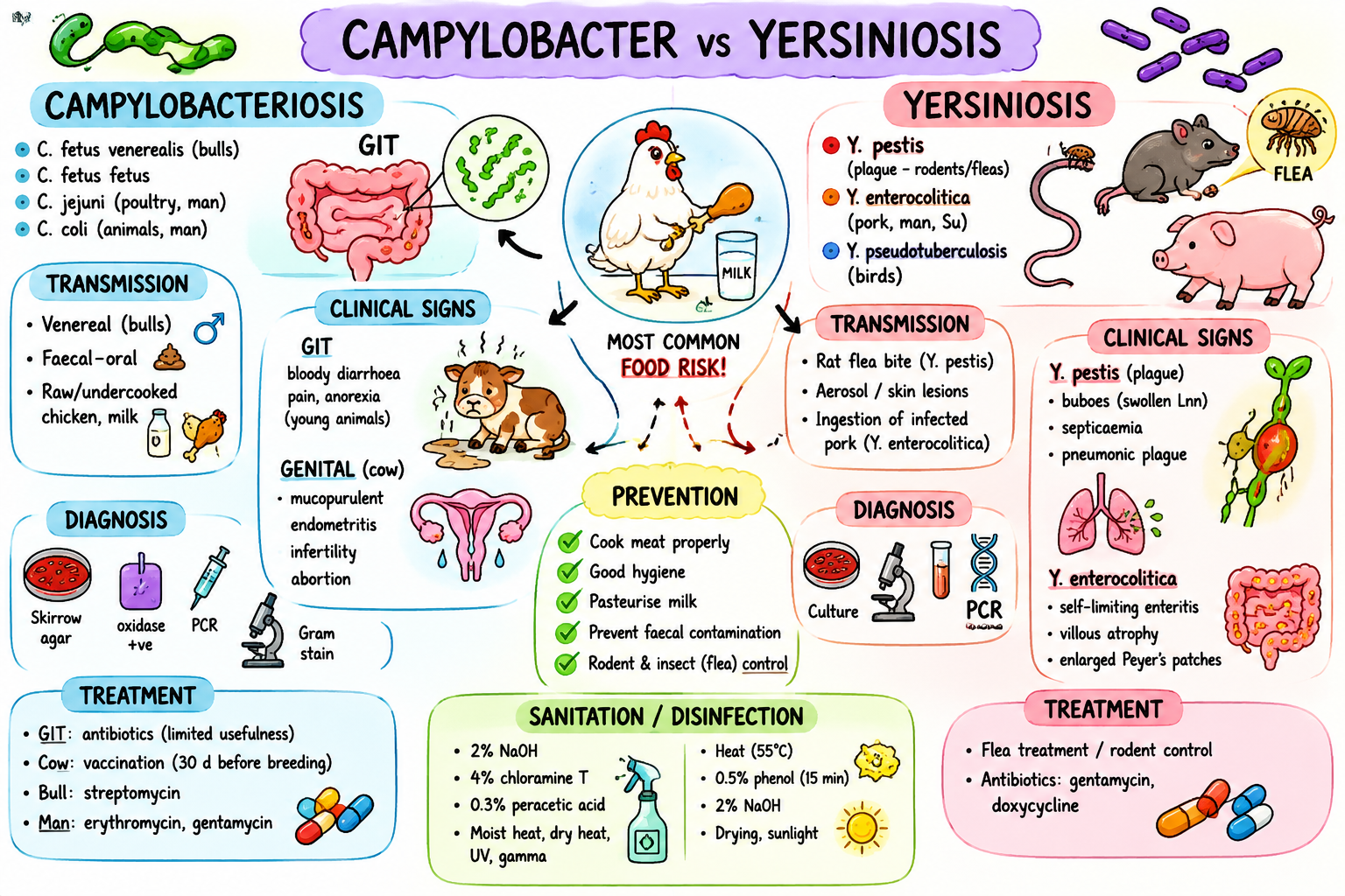
Campylobacterioses, yersinioses (+ disinfection in food production)

Campylobacteriosis

WOAH-listed | Contagious | Zoonotic

Etiology

* Campylobacter fetus venerealis → Ru → bovine genital campylobacteriosis

* Campylobacter fetus fetus→ Ru

* Campylobacter jejuni → poultry, mammals, man

* Campylobacter coli → animals, man

Important: C. jejuni and C. coli are normal intestinal flora in many animals and poultry. Poultry = major reservoir.

Epizootology

  • Worldwide. Most commonly reported food-borne zoonosis in Europe.

  • The main human source = undercooked poultry.

  • Europe: C. fetus venerealis reported in cattle in France and Ireland.

  • Norway: C. fetus venerealis = B-listed disease.

Transmission

  • Genital forms (C. fetus): Venereal transmission (Bulls are asymptomatic carriers → Spread during natural breeding/AI)

  • Enteric forms (C. jejuni, C. coli): Faecal–oral route, Contaminated food/water, Raw poultry, Unpasteurised milk

Pathogenesis

  • GIT form → Colonisation of jejunum/ileum/colon → enteritis. Toxin: Cytolethal distending toxin inflammation + villous atrophy.

  • Genital form → Catarrhal inflammation of the reproductive tract.

Clinical signs

  • GIT form: Diarrhoea (may be bloody), Enteritis, Abdominal pain, Anorexia, Often asymptomatic adults

  • Genital form: Bulls → Usually asymptomatic carriers. Cows → Endometritis, Metritis, Infertility, Irregular oestrus, Early embryonic death, Abortion

Diagnosis

  • GIT: Faecal culture (Skirrow selective agar and blood agar), Oxidase positive (reagent turn purple), PCR, Gram stain

  • Genital: Vaginal/preputial wash culture (skirrow agar), PCR, ELISA (use on vaginal mucus), VMAT (vaginal mucosal agglutination test, a field test)

Treatment

  • GIT: ATB, often limited usefulness

  • Genital: Vaccination, Streptomycin in bulls

  • Humans: Erythromycin, Gentamicin

Public health risk

  • Major food-borne zoonosis. Poultry = highest risk. Infection mainly from undercooked chicken/raw milk.

Yersiniosis

Contagious | Zoonotic

Etiology

* Yersinia pestis → black death, pneumonic plague (pest) in man. mammals are PH.

* Yersinia enterocolitica → enteritis in man and pigs.

* Yersinia pseudotuberculosis → birds, rodents, swine, car, cervids

Epizootology

  • Y. pestis: Historical Black Death pandemic (bubonic plaque pandemic in europe and asia in 1300)

  • Endemic in East Africa, Asia, South America

  • Cats are highly susceptible. Rodents = reservoir.

  • 2009 china: pneumonic plague outbreak

  • 2021 madagascar: pneumonic and bubonic plaque

Transmission

  • Y. pestis: Rat fleas, Aerosols, Skin wounds/contact with infected animal

  • Y. enterocolitica: Ingestion of contaminated pork

Pathogenesis

  • Y. pestis: Skin inoculation → buboes (lymph node abscesses) → septicaemia/pneumonia.

  • Severe forms: Bubonic plague, Septicaemic plague, Pneumonic plague

  • Y. enterocolitica: Colonises ileum/cecum → enteritis + lymph node enlargement.

Clinical signs

  • Y. pestis

→ Bubonic: Painful enlarged lymph nodes

→ Pneumonic: Respiratory failure

→ Septicaemic: DIC and tissue necrosis

  • Y. enterocolitica: Usually self-limiting enteritis

Diagnosis

  • History + CS, Blood agar culture (Blood/nasal swabs, LN aspirates), PCR, ELISA, Agglutination tests, Gram stain

Treatment

  • Rodent control, Flea treatment, Gentamicin, Doxycycline

Public health risk assessment

  • Campylobacter: Major zoonotic foodborne disease. Poultry = main source.

  • Yersinia: Third most important zoonosis after Campylobacteriosis and Salmonellosis

a) Prevention, control, outbreak measures

Campylobacter:

  • Prevention: Cook meat properly, Pasteurise milk, Hygiene/biosecurity, Remove faeces, Rodent & insect control, Bull examination, Vaccination before breeding

  • Outbreak: Treatment or culling/selection

Yersinia:

  • Prevention: Cook pork properly, Milk hygiene, Rodent control, Flea treatment, Water hygiene

  • Outbreak: Treatment or selection/culling, No vaccine

b) Sanitation, focal disinfection, food production disinfection

  • Resistance: Low environmental resistance, Survive best in moist conditions

Campylobacter sanitation

  • Housing: 4% chloramine T, 0.3% peracetic acid, 2% formalin, 2% NaOH, 20% slaked lime, Cresol-sulphuric mixture

  • Water: Chlorination

  • Physical methods: Moist heat, Dry heat, UV, Gamma radiation

Yersinia sanitation

  • Easily destroyed by: Heat, Drying, Sunlight, Disinfectants

  • Disinfectants: 2% NaOH, 2% chloramine T, 0.5% formaldehyde, Other: Drying for 2 days, Moist/dry heat

Disinfection in food production

HACCP (Hazard Analysis Critical Control Points)

  • identifies contamination risks in food production.

Cleaning/disinfection steps:

1. Remove food

2. Mechanical cleaning

3. Pre-rinse: high pressure water

4. Detergent cleaning: remove biofilm (washing soda, Na2CO3).

  • alkaline cleaning agent: Removes fats/proteins

  • acid cleaning agent: Removes encrusted residues like milkstone/waterstone

  • neutral cleaning agent: Smooth surfaces wo dirt/manual cleaning

5. Rinse

6. Drying (to not dilute the disinfectant)

7. Disinfection

Disinfection methods:

  • Physical: Hot water, Steam, UV, Radiation, Ultrasound

  • Chemical: Iodonal A, Sodium hypochlorite, Nitric acid, QUACs (Despon). MUST NOT LEAVE TOXIC RESIDUES!

<p><strong>Campylobacteriosis</strong></p><p><span style="color: red;"><strong>WOAH-listed</strong> | Contagious | Zoonotic</span></p><p><u>Etiology</u></p><p><mark data-color="red" style="background-color: red; color: inherit;">* </mark><em><mark data-color="red" style="background-color: red; color: inherit;">Campylobacter fetus venerealis</mark></em><mark data-color="red" style="background-color: red; color: inherit;"> </mark>→ Ru → bovine genital campylobacteriosis</p><p><mark data-color="red" style="background-color: red; color: inherit;">* </mark><em><mark data-color="red" style="background-color: red; color: inherit;">Campylobacter fetus fetus</mark>→ Ru</em></p><p><mark data-color="red" style="background-color: red; color: inherit;">* </mark><em><mark data-color="red" style="background-color: red; color: inherit;">Campylobacter jejuni</mark> → poultry, mammals, man</em></p><p><mark data-color="red" style="background-color: red; color: inherit;">* </mark><em><mark data-color="red" style="background-color: red; color: inherit;">Campylobacter coli </mark>→ animals, man</em></p><p>Important: <em>C. jejuni</em> and <em>C. coli</em> are normal intestinal flora in many animals and poultry. Poultry = major reservoir.</p><p><u>Epizootology</u></p><ul><li><p><strong>Worldwide</strong>. <span style="color: red;">Most commonly reported food-borne zoonosis in Europe.</span></p></li><li><p>The main human source =<strong> undercooked poultry.</strong></p></li><li><p><strong>Europe:</strong> <em>C. fetus venerealis</em> reported in cattle in<strong> France and Ireland.</strong></p></li><li><p><strong>Norway:</strong> <em>C. fetus venerealis</em> = B-listed disease.</p></li></ul><p><u>Transmission</u></p><ul><li><p><strong>Genital forms</strong> (C. fetus): Venereal transmission (Bulls are asymptomatic carriers → Spread during natural breeding/AI)</p></li><li><p><strong>Enteric forms</strong> (C. jejuni, C. coli): Faecal–oral route, Contaminated food/water, Raw poultry, Unpasteurised milk</p></li></ul><p><u>Pathogenesis</u></p><ul><li><p>GIT form → Colonisation of jejunum/ileum/colon → enteritis. Toxin: <strong>Cytolethal distending toxin</strong> →<strong> inflammation + villous atrophy.</strong></p></li><li><p>Genital form → <strong>Catarrhal inflammation</strong> of the reproductive tract.</p></li></ul><p><u>Clinical signs</u></p><ul><li><p>GIT form: Diarrhoea (may be bloody), Enteritis, Abdominal pain, Anorexia, Often asymptomatic adults</p></li><li><p>Genital form: Bulls → Usually asymptomatic carriers. Cows → Endometritis, Metritis, Infertility, Irregular oestrus, Early embryonic death, Abortion</p></li></ul><p><u>Diagnosis</u></p><ul><li><p>GIT: <strong>Faecal culture</strong> (<span style="color: red;">Skirrow selective agar and blood agar</span>), <span style="color: red;">Oxidase positive</span><span style="color: purple;"> (reagent turn purple),</span> PCR, Gram stain</p></li><li><p>Genital: <strong>Vaginal/preputial wash culture</strong> (skirrow agar), PCR, <strong>ELISA </strong>(use on vaginal mucus), <strong>VMAT </strong>(vaginal mucosal agglutination test, a field test)</p></li></ul><p><u>Treatment</u></p><ul><li><p>GIT: ATB, often limited usefulness</p></li><li><p>Genital: Vaccination, Streptomycin in bulls</p></li><li><p>Humans: Erythromycin, Gentamicin</p></li></ul><p><u>Public health risk</u></p><ul><li><p>Major food-borne zoonosis. Poultry = highest risk. Infection mainly from undercooked chicken/raw milk.</p></li></ul><p></p><p><strong>Yersiniosis</strong></p><p><span style="color: red;">Contagious | Zoonotic</span></p><p><u>Etiology</u></p><p><mark data-color="red" style="background-color: red; color: inherit;">* </mark><em><mark data-color="red" style="background-color: red; color: inherit;">Yersinia pestis</mark></em><mark data-color="red" style="background-color: red; color: inherit;"> </mark>→ black death, pneumonic plague (pest) in man. mammals are PH.</p><p><mark data-color="red" style="background-color: red; color: inherit;">* </mark><em><mark data-color="red" style="background-color: red; color: inherit;">Yersinia enterocolitica</mark></em> → enteritis in man and pigs.</p><p><mark data-color="red" style="background-color: red; color: inherit;">* </mark><em><mark data-color="red" style="background-color: red; color: inherit;">Yersinia pseudotuberculosis </mark>→ birds, rodents, swine, car, cervids</em></p><p><u>Epizootology</u></p><ul><li><p><strong><em>Y. pestis: </em></strong>Historical <span style="color: red;"><strong>Black Death</strong></span> pandemic (bubonic plaque pandemic in europe and asia in 1300)</p></li><li><p><em>Endemic in East Africa, Asia, South America</em></p></li><li><p>Cats are highly susceptible. Rodents = reservoir.</p></li><li><p><strong>2009 china: </strong>pneumonic plague outbreak</p></li><li><p><strong>2021 madagascar:</strong> pneumonic and bubonic plaque</p></li></ul><p><u>Transmission</u></p><ul><li><p><em>Y. pestis: </em>Rat fleas, Aerosols, Skin wounds/contact with infected animal</p></li><li><p><em>Y. enterocolitica:</em> Ingestion of contaminated pork</p></li></ul><p><u>Pathogenesis</u></p><ul><li><p><em>Y. pestis: </em>Skin inoculation → buboes (lymph node abscesses) → septicaemia/pneumonia.</p></li><li><p>Severe forms:<strong> Bubonic plague, Septicaemic plague, Pneumonic plague</strong></p></li></ul><ul><li><p><em>Y. enterocolitica: </em>Colonises ileum/cecum → enteritis + lymph node enlargement.</p></li></ul><p><u>Clinical signs</u></p><ul><li><p><em>Y. pestis</em></p></li></ul><p>→ Bubonic: Painful enlarged lymph nodes</p><p>→ Pneumonic: Respiratory failure</p><p>→ Septicaemic: DIC and tissue necrosis</p><ul><li><p><em>Y. enterocolitica: </em>Usually self-limiting enteritis</p></li></ul><p><u>Diagnosis</u></p><ul><li><p>History + CS, <span style="color: red;">Blood agar culture</span> (Blood/nasal swabs, LN aspirates), PCR, ELISA, Agglutination tests, Gram stain</p></li></ul><p><u>Treatment</u></p><ul><li><p>Rodent control, Flea treatment, Gentamicin, Doxycycline</p></li></ul><p><u>Public health risk assessment</u></p><ul><li><p>Campylobacter: Major zoonotic foodborne disease. Poultry = main source.</p></li><li><p>Yersinia: Third most important zoonosis after Campylobacteriosis and Salmonellosis</p></li></ul><p></p><p><strong><u>a) Prevention, control, outbreak measures</u></strong></p><p>Campylobacter:</p><ul><li><p>Prevention: Cook meat properly, Pasteurise milk, Hygiene/biosecurity, Remove faeces, Rodent &amp; insect control, Bull examination, Vaccination before breeding</p></li><li><p>Outbreak: Treatment or culling/selection</p></li></ul><p>Yersinia:</p><ul><li><p>Prevention: Cook pork properly, Milk hygiene, Rodent control, Flea treatment, Water hygiene</p></li><li><p>Outbreak: Treatment or selection/culling, No vaccine</p></li></ul><p></p><p><strong><u>b) Sanitation, focal disinfection, <mark data-color="purple" style="background-color: purple; color: inherit;">food production disinfection</mark></u></strong></p><ul><li><p>Resistance: Low environmental resistance, Survive best in moist conditions</p></li></ul><p>Campylobacter sanitation</p><ul><li><p>Housing: 4% chloramine T, 0.3% peracetic acid, 2% formalin, 2% NaOH, 20% slaked lime, Cresol-sulphuric mixture</p></li><li><p>Water: Chlorination</p></li><li><p>Physical methods: Moist heat, Dry heat, UV, Gamma radiation</p></li></ul><p>Yersinia sanitation</p><ul><li><p>Easily destroyed by: Heat, Drying, Sunlight, Disinfectants</p></li><li><p>Disinfectants: 2% NaOH, 2% chloramine T, 0.5% formaldehyde, Other: Drying for 2 days, Moist/dry heat</p></li></ul><p></p><p><strong><mark data-color="purple" style="background-color: purple; color: inherit;">Disinfection in food production</mark></strong></p><p><strong>HACCP </strong>(Hazard Analysis Critical Control Points)</p><ul><li><p>identifies contamination risks in food production.</p></li></ul><p>Cleaning/disinfection steps:</p><p><strong>1. Remove food</strong></p><p><strong>2. Mechanical cleaning</strong></p><p><strong>3. Pre-rinse: high pressure water</strong></p><p><strong>4. Detergent cleaning: </strong>remove biofilm (washing soda, Na2CO3).</p><ul><li><p>alkaline cleaning agent: Removes fats/proteins</p></li><li><p>acid cleaning agent: Removes encrusted residues like milkstone/waterstone</p></li><li><p>neutral cleaning agent: Smooth surfaces wo dirt/manual cleaning</p></li></ul><p><strong>5. Rinse</strong></p><p><strong>6. Drying </strong>(to not dilute the disinfectant)</p><p><strong>7. Disinfection</strong></p><p>Disinfection methods:</p><ul><li><p>Physical: Hot water, Steam, UV, Radiation, Ultrasound</p></li><li><p>Chemical: Iodonal A, Sodium hypochlorite, Nitric acid, QUACs (Despon). MUST NOT LEAVE TOXIC RESIDUES!</p></li></ul><p></p>
6
New cards

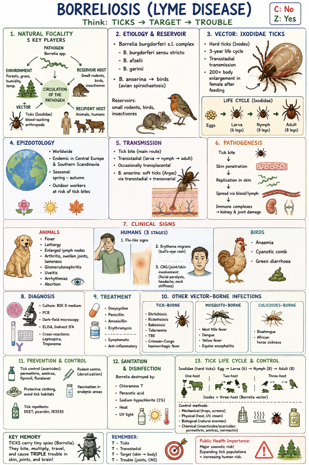
Borreliosis and other tickborne diseases (+life cycle of ticks and their prevention and control)

Borreliosis (Lyme disease)Vector-borne | Zoonotic | Natural focal disease

Etiology

* Borrelia burgdorferi sensu stricto Lyme disease

* Borrelia afzelii, Borrelia garinii

* Borrelia anserina avian spirochaetosis

Important: Borrelia burgdorferi s.l. complex causes Lyme disease in mammals and humans.

Reservoirs = small rodents, birds, and insectivores.

Natural focality

Natural focus includes:

  • Pathogen (pathoergont), Reservoir host, Vector (blood-sucking arthropod), Recipient host (animal/man) and Suitable environment

  • Other natural focal diseases: leptospirosis, rabies, TBE, ehrlichiosis, babesiosis.

Epizootology

  • Worldwide, endemic in Central Europe and Southern Scandinavia

  • Seasonal: spring–autumn. Professional risk for outdoor workers

  • Norway: endemic areas in the southern regions

Transmission

Ixodidae hard ticks (Ixodes*)

  • Transstadial transmission, occasionally transplacental

  • B. anserina is transmitted by soft ticks (Argas) via transstadial + transovarial transmission

Pathogenesis

Tick bite → skin penetration → bacterial replication → spread via blood/lymph → immune complex formation → kidney/joint damage.

Clinical signs

Animals:

  • Fever, Lethargy, Enlarged lymph nodes, Arthritis, swollen joints, lameness, Glomerulonephritis, Uveitis, Arrhythmias, Abortion

Humans:

1. Flu-like signs, 2. Erythema migrans, 3. CNS/joint involvement (facial paralysis, headache, neck stiffness)

Birds:

  • Anaemia, Cyanotic comb, Green diarrhoea

Diagnosis

  • Culture: Barbour–Stoenner–Kelly II medium at 33C

  • PCR

  • Dark-field microscopy

  • ELISA, indirect IFA

Cross-reactions with Leptospira* and Treponema

Treatment

  • Doxycycline, Penicillin, Amoxicillin, Erythromycin

  • Symptomatic + anti-inflammatory therapy

Other vector-borne infections

Tick-borne:

  • Ehrlichiosis, Rickettsiosis, Babesiosis, Tularaemia, Tick-borne encephalitis (TBE), Crimean–Congo haemorrhagic fever

Mosquito-borne:

  • West Nile fever, Dengue, Yellow fever, Equine encephalitis

Culicoides-borne:

  • Bluetongue, African horse sickness

Public health risk assessment

Major zoonotic vector-borne disease. Expanding tick populations and climate change increase risk to animals and humans.

a) Prevention, control, outbreak measures

  • Tick control with acaricides (Permethrin): Amitraz collar for dogs, Fipronil spot on, Fluralaner (bravecto)

  • Rodent control (deratization)

  • Protective clothing, avoid tick habitats

  • Vaccination in endemic areas

  • Tick repellents: DEET, Picaridin, IR3535

Outbreak measures

  • Antibiotic treatment, Tick elimination, Monitoring of exposed animals/humans

b) Sanitation, focal disinfection, tick life cycle & control

Borrelia destroyed by:

  • Chloramine T, Peracetic acid, Sodium hypochlorite, 70% ethanol

  • UV light

  • Heat

Tick life cycle (Ixodidae – hard ticks)

  • incomplete metamorphosis

Egg → larva (6 legs) → nymph (8 legs) → adult (8 legs)

Types:

  • One-host ticks, Two-host ticks, Three-host ticks (Ixodes = Borrelia vector)

  • Ixodes larva and nymphal stage feed on reservoirs (small mammals and birds) and get infected by the pathogen, then they feed on man/animal and spread the pathogen.

Soft ticks (Argasidae)

  • Multi-host life cycle, Can survive years without feeding

  • they have several nymphal stages and may feed on several hosts like rodents and birds, and transmit borrelia at anyh developmental stage

Tick/insect control methods

  • Mechanical: traps, screens

  • Physical: heat, UV, steam

  • Biological: natural enemies

  • Chemical: insecticides/acaricides (permethrin, amitraz, ivermectin)

<p><strong>Borreliosis (Lyme disease)</strong> — <span style="color: red;">Vector-borne | <strong>Zoonotic</strong> | Natural focal disease</span></p><p><strong>Etiology</strong></p><p><mark data-color="red" style="background-color: red; color: inherit;">* </mark><em><mark data-color="red" style="background-color: red; color: inherit;">Borrelia burgdorferi sensu stricto </mark>→ </em><strong><em>Lyme disease</em></strong></p><p><mark data-color="red" style="background-color: red; color: inherit;">* </mark><em><mark data-color="red" style="background-color: red; color: inherit;">Borrelia afzelii, Borrelia garinii</mark></em></p><p><mark data-color="red" style="background-color: red; color: inherit;">* </mark><em><mark data-color="red" style="background-color: red; color: inherit;">Borrelia anserina</mark></em><mark data-color="red" style="background-color: red; color: inherit;"> </mark>→ <strong>avian spirochaetosis</strong></p><p><u>Important:</u> <em>Borrelia burgdorferi s.l.</em> complex causes <span style="color: red;"><strong>Lyme disease</strong></span> in mammals and humans.</p><p>Reservoirs = small rodents, birds, and insectivores.</p><p><strong>Natural focality</strong></p><p>Natural focus includes:</p><ul><li><p>Pathogen (pathoergont), Reservoir host, Vector (blood-sucking arthropod), Recipient host (animal/man) and Suitable environment</p></li><li><p>Other natural focal diseases: leptospirosis, rabies, TBE, ehrlichiosis, babesiosis.</p></li></ul><p><strong>Epizootology</strong></p><ul><li><p><span style="color: red;"><strong>Worldwide</strong></span>, <strong>endemic in Central Europe and Southern Scandinavia</strong></p></li><li><p>Seasonal: <strong>spring–autumn.</strong> Professional risk for outdoor workers</p></li><li><p><strong>Norway:</strong> endemic areas in the southern regions</p></li></ul><p><strong>Transmission</strong></p><p><em>Ixodidae hard ticks </em><strong><em><mark data-color="blue" style="background-color: blue; color: inherit;">(</mark></em><mark data-color="blue" style="background-color: blue; color: inherit;">Ixodes</mark></strong>*)</p><ul><li><p>Transstadial transmission, occasionally transplacental</p></li><li><p><em>B. anserina</em> is transmitted by soft ticks (Argas) via transstadial + transovarial transmission</p></li></ul><p><strong>Pathogenesis</strong></p><p>Tick bite → skin penetration → bacterial replication → spread via blood/lymph → immune complex formation → <strong>kidney/joint damage.</strong></p><p><strong>Clinical signs</strong></p><p><strong>Animals:</strong></p><ul><li><p>Fever, Lethargy, Enlarged lymph nodes, <strong>Arthritis, swollen joints, lameness</strong>, <strong>Glomerulonephritis</strong>, Uveitis, Arrhythmias, Abortion</p></li></ul><p><strong>Humans:</strong></p><p>1. <strong>Flu-like signs, 2. Erythema migrans, 3. CNS/joint involvement</strong> (facial paralysis, headache, neck stiffness)</p><p><strong>Birds:</strong></p><ul><li><p>Anaemia, Cyanotic comb, Green diarrhoea</p></li></ul><p><strong>Diagnosis</strong></p><ul><li><p>Culture: <strong>Barbour–Stoenner–Kelly II</strong> medium at 33C</p></li><li><p>PCR</p></li><li><p>Dark-field microscopy</p></li><li><p>ELISA, indirect IFA</p></li></ul><p><em>Cross-reactions with </em>Leptospira* and <em>Treponema</em></p><p><strong>Treatment</strong></p><ul><li><p>Doxycycline, Penicillin, Amoxicillin, Erythromycin</p></li><li><p>Symptomatic + anti-inflammatory therapy</p></li></ul><p><strong>Other vector-borne infections</strong></p><p>Tick-borne:</p><ul><li><p>Ehrlichiosis, Rickettsiosis, Babesiosis, Tularaemia, Tick-borne encephalitis (TBE), Crimean–Congo haemorrhagic fever</p></li></ul><p>Mosquito-borne:</p><ul><li><p>West Nile fever, Dengue, Yellow fever, Equine encephalitis</p></li></ul><p>Culicoides-borne:</p><ul><li><p>Bluetongue, African horse sickness</p></li></ul><p><strong>Public health risk assessment</strong></p><p><span style="color: red;">Major zoonotic vector-borne disease. </span>Expanding tick populations and climate change increase risk to animals and humans.</p><p></p><p><strong><u>a) Prevention, control, outbreak measures</u></strong></p><ul><li><p>Tick control with acaricides (Permethrin): Amitraz collar for dogs, Fipronil spot on, Fluralaner (bravecto)</p></li><li><p>Rodent control (deratization)</p></li><li><p>Protective clothing, avoid tick habitats</p></li><li><p>Vaccination in endemic areas</p></li><li><p>Tick repellents: DEET, Picaridin, IR3535</p></li></ul><p><strong>Outbreak measures</strong></p><ul><li><p>Antibiotic treatment, Tick elimination, Monitoring of exposed animals/humans</p></li></ul><p></p><p><strong><u>b) Sanitation, focal disinfection,<mark data-color="purple" style="background-color: purple; color: inherit;"> tick life cycle &amp; control</mark></u></strong></p><p><strong>Borrelia destroyed by:</strong></p><ul><li><p><span style="color: red;">Chloramine T, Peracetic acid, Sodium hypochlorite, 70% ethanol</span></p></li><li><p>UV light</p></li><li><p>Heat</p></li></ul><p><strong><mark data-color="purple" style="background-color: purple; color: inherit;">Tick life cycle</mark> (Ixodidae – hard ticks)</strong></p><ul><li><p>incomplete metamorphosis </p></li></ul><p>Egg → larva (6 legs) → nymph (8 legs) → adult (8 legs)</p><p>Types:</p><ul><li><p>One-host ticks, Two-host ticks, <strong><em>Three-host ticks</em></strong><em> (</em>Ixodes = Borrelia vector)</p></li><li><p>Ixodes larva and nymphal stage feed on reservoirs (small mammals and birds) and get infected by the pathogen, then they feed on man/animal and spread the pathogen.</p></li></ul><p><strong>Soft ticks (Argasidae)</strong></p><ul><li><p>Multi-host life cycle, Can survive years without feeding</p></li><li><p>they have several nymphal stages and may feed on several hosts like rodents and birds, and transmit borrelia at anyh developmental stage</p></li></ul><p><strong>Tick/insect control methods</strong></p><ul><li><p>Mechanical: traps, screens</p></li><li><p>Physical: heat, UV, steam</p></li><li><p>Biological: natural enemies</p></li><li><p>Chemical: insecticides/acaricides (permethrin, amitraz, ivermectin)</p></li></ul><p></p>
7
New cards

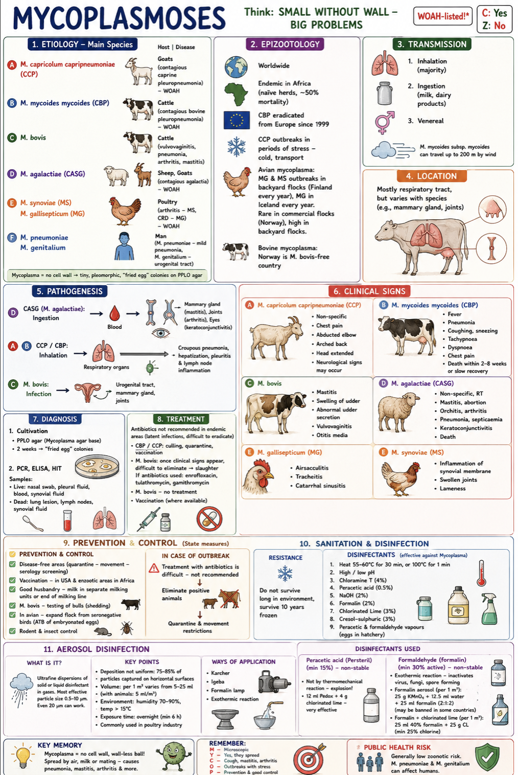
Mycoplasmosis (+ aerosol disinfection)

WOAH-listed | Contagious | Mostly respiratory diseases | Zoonotic risk low/no in animals

Etiology

  • Mycoplasma capricolum subsp. capripneumoniae → Contagious caprine pleuropneumonia (CCP)

  • Mycoplasma mycoides mycoides → Contagious bovine pleuropneumonia (CBP)

  • Mycoplasma bovis → mastitis, pneumonia, arthritis, vulvovaginitis

  • Mycoplasma agalactiae → contagious agalactia in sheep/goats

  • M. gallisepticum → chronic respiratory disease in poultry

  • M. synoviae → arthritis/synovitis in poultry

  • M.hyopneumonia, M.hyosynovia, M.hyogenitalia (PIGS)

  • M.genitalium, M.pneumonia (mild “walking pneumonia” in humans)

Important: Mycoplasma lack a cell wall → fragile in environment but resistant to some antibiotics.

Epizootology

  • Worldwide: Africa: endemic CBP with high mortality, Europe: CBP eradicated since 1999

  • Norway: M. bovis free country

  • Backyard poultry in Nordic countries is commonly affected by avian mycoplasmosis (yearly in Iceland), and in Finland in 2024)

  • CCP outbreaks associated with stress, transport, cold

Transmission

  • Horizontal transmission: Inhalation (main route), Milk/dairy products, Venereal transmission

  • Wind spread possible up to 200 m (M. mycoides*)

Location

  • Mainly respiratory tract, but also Mammary gland, Joints, Eyes, Urogenital tract

Pathogenesis

  • Inhalation → pneumonia, pleuritis, hepatization of lungs

  • Ingestion → spread via blood to mammary gland, joints, eyes

  • Chronic latent infections are common

Clinical signs

Cattle (CBP / M. bovis):

  • Fever, Pneumonia, Dyspnoea, chest pain, Mastitis (“sand-like” milk), Vulvovaginitis, Otitis media

Goats/sheep:

  • Mastitis, Arthritis, Keratoconjunctivitis, Abortion, Septicaemia

Poultry:

  • Airsacculitis, Tracheitis, Sinusitis, Swollen joints

Diagnosis

  • Culture on PPLO agar (“fried egg” colonies) and PCR

  • ELISA, HIT

  • Samples: Nasal swabs, Pleural fluid, Synovial fluid, Lung lesions, Lymph nodes

Treatment

  • Often difficult/not recommended due to latent carriers

  • Culling + quarantine preferred

  • Vaccination in endemic areas

  • Antibiotics: Enrofloxacin, Tulathromycin, Gamithromycin

Public health risk assessment

Animal mycoplasmoses are mainly veterinary/economic problems. Human mycoplasmas exist (*M. pneumoniae*, M. genitalium), but animal strains are generally not zoonotic.

a) Prevention, control, outbreak measures

Prevention & control

  • Quarantine + movement restrictions

  • Serological screening and Vaccination in endemic areas

  • Good husbandry and milking hygiene

  • Test breeding bulls

  • Expand poultry flocks from seronegative birds

  • Rodent/insect control

Outbreak measures

  • Eliminate positive animals, Quarantine affected herds/flocks

  • Antibiotic treatment is usually not sufficient

b) Sanitation, focal disinfection, aerosol disinfection

Resistance

  • Poor survival in environment, but can survive for years when frozen

Destroyed by:

  • Heat: 55–60°C for 30 min or 100°C for 1 min

  • Extreme pH

  • Chloramine T, Peracetic acid, NaOH, Formalin, Chlorinated lime, cresol-sulphuric, peracetic and formaldehyde vapours for eggs in hatcheries.

Aerosol disinfection

  • Common in the poultry industry.

  • Ultrafine dispersions of solid of solid or liquid disinfectant in gases

  • not uniform deposition: 75-85% of aerosol particles is captured on horizontal surfaces

Conditions:

  • Particle size: 0.5–10 µm (max 20)

  • Humidity: 70–90%

  • Temperature: >15°C

  • Exposure: overnight (≥6 h)

  • Volume: 5-25ml per 1m3. 5ml/m3 in presence of animals.

Methods:

  • Karcher, Igeba, Formalin lamp, Exothermic reaction

Disinfectants used:

  • Peracetic acid vapours (non-stable, min.15%. not by thermomechanical reaction → explosion. 12mil Pedox + 4g chlorinated lime is very effective)

  • Formaldehyde aerosols (non-stable, min 30%. exothermic reaction → inactivate virus, fungi, spore forming. use Formalin + potassium permanganate mixtures or mixed with chlorinated lime)

<p><span style="color: red;"><strong>WOAH-listed</strong> | Contagious | Mostly respiratory diseases | Zoonotic risk <u>low/no </u>in animals</span></p><p><strong>Etiology</strong></p><ul><li><p><em><mark data-color="red" style="background-color: red; color: inherit;">Mycoplasma capricolum subsp. capripneumoniae</mark></em> → Contagious caprine pleuropneumonia (CCP)</p></li><li><p><em><mark data-color="red" style="background-color: red; color: inherit;">Mycoplasma mycoides mycoides</mark></em> → Contagious bovine pleuropneumonia (CBP)</p></li><li><p><em><mark data-color="red" style="background-color: red; color: inherit;">Mycoplasma bovis</mark></em><mark data-color="red" style="background-color: red; color: inherit;"> </mark>→ mastitis, pneumonia, arthritis, vulvovaginitis</p></li><li><p><em><mark data-color="red" style="background-color: red; color: inherit;">Mycoplasma agalactiae</mark></em> → contagious agalactia in sheep/goats</p></li><li><p><em><mark data-color="red" style="background-color: red; color: inherit;">M. gallisepticum</mark></em> → chronic respiratory disease in poultry</p></li><li><p><em><mark data-color="red" style="background-color: red; color: inherit;">M. synoviae</mark></em><mark data-color="red" style="background-color: red; color: inherit;"> </mark>→ arthritis/synovitis in poultry</p></li><li><p><mark data-color="red" style="background-color: red; color: inherit;">M.hyopneumonia, M.hyosynovia, M.hyogenitalia</mark> (PIGS)</p></li><li><p><mark data-color="red" style="background-color: red; color: inherit;">M.genitalium, M.pneumonia</mark> (mild “walking pneumonia” in humans)</p></li></ul><p>Important: Mycoplasma<strong> lack a cell wall </strong>→ fragile in environment but<strong> resistant to some antibiotics.</strong></p><p><strong>Epizootology</strong></p><ul><li><p><span style="color: red;"><strong>Worldwide:</strong></span><u> Africa:</u> <strong>endemic CBP with high mortality</strong>, <u>Europe:</u> <strong>CBP eradicated since 1999</strong></p></li><li><p><em><u>Norway: </u></em><strong>M. bovis free</strong> country</p></li><li><p><strong>Backyard poultry in Nordic countries</strong> is commonly affected by <strong>avian mycoplasmosis </strong>(yearly in Iceland), and in Finland in 2024)</p></li><li><p>CCP outbreaks associated with stress, transport, cold</p></li></ul><p><strong>Transmission</strong></p><ul><li><p> Horizontal transmission: Inhalation (main route), Milk/dairy products, Venereal transmission</p></li><li><p><em>Wind spread possible up to 200 m (</em>M. mycoides*)</p></li></ul><p><strong>Location</strong></p><ul><li><p>Mainly respiratory tract, but also Mammary gland, Joints, Eyes, Urogenital tract</p></li></ul><p><strong>Pathogenesis</strong></p><ul><li><p>Inhalation → pneumonia, pleuritis, hepatization of lungs</p></li><li><p>Ingestion → spread via blood to mammary gland, joints, eyes</p></li><li><p>Chronic latent infections are common</p></li></ul><p><strong>Clinical signs</strong></p><p><strong>Cattle (CBP / M. bovis):</strong></p><ul><li><p>Fever, <span style="color: red;">Pneumonia, Dyspnoea, chest pain, Mastitis </span>(“sand-like” milk), Vulvovaginitis, Otitis media</p></li></ul><p><strong>Goats/sheep:</strong></p><ul><li><p><span style="color: red;">Mastitis, Arthritis, Keratoconjunctivitis</span>, Abortion, Septicaemia</p></li></ul><p><strong>Poultry:</strong></p><ul><li><p>Airsacculitis, Tracheitis, Sinusitis, Swollen joints</p></li></ul><p><strong>Diagnosis</strong></p><ul><li><p>Culture on<span style="color: red;"> PPLO agar</span> (“fried egg” colonies) and PCR</p></li><li><p>ELISA, HIT</p></li><li><p>Samples: Nasal swabs, Pleural fluid, Synovial fluid, Lung lesions, Lymph nodes</p></li></ul><p><strong>Treatment</strong></p><ul><li><p>Often difficult/<span style="color: red;">not recommended</span> due to latent carriers</p></li><li><p><strong>Culling + quarantine preferred</strong></p></li><li><p><span style="color: red;">Vaccination</span> in endemic areas</p></li><li><p>Antibiotics: Enrofloxacin, Tulathromycin, Gamithromycin</p></li></ul><p><strong>Public health risk assessment</strong></p><p>Animal mycoplasmoses are mainly veterinary/economic problems. Human mycoplasmas exist (*M. pneumoniae*, <em>M. genitalium</em>), but <span style="color: red;"><strong>animal strains are generally not zoonotic.</strong></span></p><p></p><p><strong><u> a) Prevention, control, outbreak measures</u></strong></p><p><strong>Prevention &amp; control</strong></p><ul><li><p>Quarantine + movement restrictions</p></li><li><p>Serological screening and Vaccination in endemic areas</p></li><li><p>Good husbandry and milking hygiene</p></li><li><p><strong>Test breeding bulls</strong></p></li><li><p><strong>Expand poultry flocks from seronegative birds</strong></p></li><li><p>Rodent/insect control</p></li></ul><p><strong>Outbreak measures</strong></p><ul><li><p><span style="color: red;"><strong> Eliminate positive animals, </strong></span>Quarantine affected herds/flocks</p></li><li><p>Antibiotic treatment is usually not sufficient</p></li></ul><p></p><p> b<strong><u>) Sanitation, focal disinfection, <mark data-color="purple" style="background-color: purple; color: inherit;">aerosol disinfection</mark></u></strong></p><p><strong>Resistance</strong></p><ul><li><p>Poor survival in environment, but can survive for years when frozen</p></li></ul><p><strong>Destroyed by:</strong></p><ul><li><p>Heat: 55–60°C for 30 min or 100°C for 1 min</p></li><li><p>Extreme pH</p></li><li><p><span style="color: red;"><strong>Chloramine T, Peracetic acid, NaOH, Formalin, Chlorinated lime, cresol-sulphuric, peracetic and formaldehyde vapours for eggs in hatcheries.</strong></span></p></li></ul><p><strong><mark data-color="purple" style="background-color: purple; color: inherit;">Aerosol disinfection</mark></strong></p><ul><li><p>Common in the poultry industry.</p></li><li><p>Ultrafine dispersions of solid of solid or liquid disinfectant in gases</p></li><li><p>not uniform deposition: 75-85% of aerosol particles is captured on horizontal surfaces</p></li></ul><p>Conditions:</p><ul><li><p>Particle size: 0.5–10 µm (max 20)</p></li><li><p>Humidity: 70–90%</p></li><li><p>Temperature: &gt;15°C</p></li><li><p>Exposure: overnight (≥6 h)</p></li><li><p>Volume: 5-25ml per 1m3. 5ml/m3 in presence of animals.</p></li></ul><p>Methods:</p><ul><li><p>Karcher, Igeba, Formalin lamp, Exothermic reaction</p></li></ul><p>Disinfectants used:</p><ul><li><p><span style="color: red;"><strong>Peracetic acid </strong></span>vapours (non-stable, min.15%. not by thermomechanical reaction → explosion. 12mil Pedox + 4g chlorinated lime is very effective)</p></li><li><p><span style="color: red;"><strong>Formaldehyde </strong></span>aerosols (non-stable, min 30%. exothermic reaction → inactivate virus, fungi, spore forming. use Formalin + potassium permanganate mixtures or mixed with chlorinated lime)</p></li></ul><p></p>
8
New cards

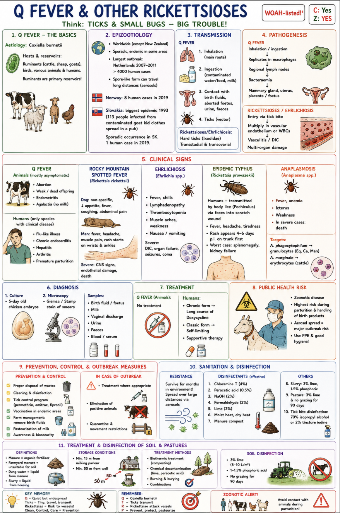
Q fever & other rickettsioses (+Treatment and disinfection of soil and pastures)

WOAH-listed | Vector-borne | Zoonotic

Etiology

* Coxiella burnetii Q fever

* Reservoirs: ruminants, birds, various mammals

* Humans are infected mainly through farm animal exposure

Other rickettsioses:

* Rickettsia rickettsii → Rocky Mountain spotted fever

* Ehrlichia canis → canine ehrlichiosis

* Ehrlichia ruminantium → heartwater disease

* Rickettsia prowazekii → epidemic typhus

* Anaplasma phagocytophilum → granulocytic anaplasmosis

Epizootology

  • Q fever worldwide except New Zealand

  • Large outbreak: Netherlands 2007–2011 (>4000 human cases)

  • Norway: 8 human cases in 2019

  • Slovakia: largest outbreak in 1993 after men handled aborted goat kid. 2011 - goat cheese. 1 human case in 2019.

  • Spread possibly over long distances by aerosolized spore-like forms

  • Tick-borne diseases are increasing due to climate change/global warming

  • Seasonal tick activity mainly spring–autumn

Transmission

Q fever:

  • Inhalation (main route)

  • Ingestion of contaminated milk/food

  • Exposure to birth fluids, placenta, aborted foetuses

  • Tick transmission possible

Rickettsioses/ehrlichiosis:

  • Hard ticks (Dermacentor, Amblyomma, Rhipicephalus*)

  • Transstadial/transovarial transmission

Pathogenesis

Q fever:

  • Inhalation → replication in macrophages → lymph nodes → bacteraemia → uterus, placenta, mammary gland.

Rickettsioses:

  • Attack vascular endothelium or WBCs → vasculitis/DIC → multiorgan damage.

Clinical signs

Animals (Q fever):

  • Often asymptomatic, Abortions, Weak offspring, Endometritis, Agalactia

Humans (Q fever):

  • Flu-like disease, Chronic endocarditis, Hepatitis, Arthritis, Premature birth

Rocky Mountain spotted fever:

  • Fever, Rash, CNS signs, Endothelial damage

Ehrlichiosis:

  • Fever, Lymphadenopathy, Thrombocytopenia, DIC, Seizures/coma in severe cases

Diagnosis

  • PCR, ELISA / IFA, Blood smears, Cell culture / embryonated eggs, Giemsa or Stamp staining

Samples: Blood, Birth fluids, Placenta/foetus, Milk, Vaginal discharge

Treatment

  • Doxycycline (main antibiotic), Supportive therapy, Q fever in animals: usually no treatment

Public health risk assessment

  • Important zoonotic diseases. The highest risk during animal parturition and handling of contaminated birth materials.

  • Aerosol spread creates major outbreak potential.

a) Prevention, control, outbreak measures

Prevention & control

  • Proper disposal of birth products/manure

  • Cleaning and disinfection

  • Tick control: Permethrin, Amitraz, Fipronil

  • Pasteurization of milk

  • Vaccination in endemic areas

  • Awareness and biosecurity

  • Remove contaminated birth fluids immediately

Outbreak measures

  • Treatment where appropriate

  • Elimination/isolation of positive animals

  • Movement restrictions

b) Sanitation, focal disinfection, treatment and disinfection of soil/pasture

Resistance

  • Survive for months in environment. Aerosol spread over large distances

Disinfection

  • Chloramine T (4%), Peracetic acid (0.5%), NaOH (2%), Formaldehyde (2%), Lime (3%), Moist/dry heat

  • Manure compost

  • Slurry:  3% lime, 1.5% phosphoric

  • Pasture: 3% lime & no grazing for 90 days

  • Tick bite disinfection: 70% isopropyl alcohol and 2% tincture iodine

Manure/slurry treatment

* Composting (biothermic treatment)

* Lime or phosphoric acid treatment

* Burning/burying if necessary

  • Storage condition: minimum 15m from milking parlour & 50m from well

Pasture/soil disinfection

  • 3% lime

  • 1–1.5% phosphoric acid

  • 0.3-0.5% peracetic acid

  • all 10L per 1m2

* No grazing for 90 days

<p><span style="color: red;"><strong>WOAH-listed</strong> | Vector-borne | Zoonotic</span></p><p><strong>Etiology</strong></p><p><strong><mark data-color="red" style="background-color: red; color: inherit;">* </mark><em><mark data-color="red" style="background-color: red; color: inherit;">Coxiella burnetii</mark></em><mark data-color="red" style="background-color: red; color: inherit;"> </mark></strong>→ <strong>Q fever</strong></p><p>* Reservoirs: <em>ruminants, birds, various mammals</em></p><p>* Humans are infected mainly through farm animal exposure</p><p><u>Other rickettsioses:</u></p><p><mark data-color="red" style="background-color: red; color: inherit;">* </mark><em><mark data-color="red" style="background-color: red; color: inherit;">Rickettsia rickettsii</mark></em><mark data-color="red" style="background-color: red; color: inherit;"> </mark>→ Rocky Mountain spotted fever</p><p><mark data-color="red" style="background-color: red; color: inherit;">* </mark><em><mark data-color="red" style="background-color: red; color: inherit;">Ehrlichia canis</mark></em><mark data-color="red" style="background-color: red; color: inherit;"> </mark>→ canine ehrlichiosis</p><p><mark data-color="red" style="background-color: red; color: inherit;">* </mark><em><mark data-color="red" style="background-color: red; color: inherit;">Ehrlichia ruminantium</mark></em><mark data-color="red" style="background-color: red; color: inherit;"> </mark>→ heartwater disease</p><p><mark data-color="red" style="background-color: red; color: inherit;">* </mark><em><mark data-color="red" style="background-color: red; color: inherit;">Rickettsia prowazekii</mark></em><mark data-color="red" style="background-color: red; color: inherit;"> </mark>→ epidemic typhus</p><p><mark data-color="red" style="background-color: red; color: inherit;">* </mark><em><mark data-color="red" style="background-color: red; color: inherit;">Anaplasma phagocytophilum</mark></em><mark data-color="red" style="background-color: red; color: inherit;"> </mark>→ granulocytic anaplasmosis</p><p><strong>Epizootology</strong></p><ul><li><p>Q fever <span style="color: red;"><strong>worldwide</strong></span> <em>except New Zealand</em></p></li><li><p>Large outbreak: <strong>Netherlands 2007–2011</strong> (&gt;4000 human cases)</p></li><li><p>Norway: 8 human cases in 2019</p></li><li><p>Slovakia: largest outbreak in 1993 after men handled aborted goat kid. 2011 - goat cheese. 1 human case in 2019.</p></li><li><p><strong>Spread possibly over long distances</strong> by aerosolized spore-like forms</p></li><li><p>Tick-borne diseases are increasing due to <strong>climate change/global warming</strong></p></li><li><p>Seasonal tick activity mainly <strong>spring–autumn</strong></p></li></ul><p><strong>Transmission</strong></p><p><strong>Q fever:</strong></p><ul><li><p>Inhalation (main route)</p></li><li><p>Ingestion of contaminated milk/food</p></li><li><p>Exposure to birth fluids, placenta, aborted foetuses</p></li><li><p>Tick transmission possible</p></li></ul><p><strong>Rickettsioses/ehrlichiosis:</strong></p><ul><li><p><em>Hard ticks (</em>Dermacentor, Amblyomma, Rhipicephalus*)</p></li><li><p>Transstadial/transovarial transmission</p></li></ul><p><strong>Pathogenesis</strong></p><p><strong>Q fever:</strong></p><ul><li><p>Inhalation → replication in macrophages → lymph nodes → bacteraemia → uterus, placenta, mammary gland.</p></li></ul><p><strong>Rickettsioses:</strong></p><ul><li><p>Attack vascular endothelium or WBCs → vasculitis/DIC → multiorgan damage.</p></li></ul><p><strong>Clinical signs</strong></p><p><strong>Animals (Q fever):</strong></p><ul><li><p>Often asymptomatic, Abortions, Weak offspring, Endometritis, Agalactia</p></li></ul><p><strong>Humans (Q fever):</strong></p><ul><li><p>Flu-like disease, Chronic endocarditis, Hepatitis, Arthritis, Premature birth</p></li></ul><p><strong>Rocky Mountain spotted fever:</strong></p><ul><li><p>Fever, Rash, CNS signs, Endothelial damage</p></li></ul><p><strong>Ehrlichiosis:</strong></p><ul><li><p>Fever, Lymphadenopathy, Thrombocytopenia, DIC, Seizures/coma in severe cases</p></li></ul><p><strong>Diagnosis</strong></p><ul><li><p>PCR, ELISA / IFA, Blood smears, Cell culture / embryonated eggs, Giemsa or Stamp staining</p></li></ul><p>Samples: Blood, Birth fluids, Placenta/foetus, Milk, Vaginal discharge</p><p><strong>Treatment</strong></p><ul><li><p>Doxycycline (main antibiotic), Supportive therapy, Q fever in animals: usually no treatment</p></li></ul><p><strong>Public health risk assessment</strong></p><ul><li><p>Important zoonotic diseases. The highest risk during animal parturition and handling of contaminated birth materials.</p></li><li><p>Aerosol spread creates major outbreak potential.</p></li></ul><p></p><p><strong><u>a) Prevention, control, outbreak measures</u></strong></p><p><strong>Prevention &amp; control</strong></p><ul><li><p>Proper disposal of birth products/manure</p></li><li><p>Cleaning and disinfection</p></li><li><p>Tick control: Permethrin, Amitraz, Fipronil</p></li><li><p>Pasteurization of milk</p></li><li><p>Vaccination in endemic areas</p></li><li><p>Awareness and biosecurity</p></li><li><p>Remove contaminated birth fluids immediately</p></li></ul><p><strong>Outbreak measures</strong></p><ul><li><p>Treatment where appropriate</p></li><li><p>Elimination/isolation of positive animals</p></li><li><p>Movement restrictions</p></li></ul><p></p><p><strong><u>b) Sanitation, focal disinfection, <mark data-color="purple" style="background-color: purple; color: inherit;">treatment and disinfection of soil/pasture</mark></u></strong></p><p><strong>Resistance</strong></p><ul><li><p>Survive for months in environment. Aerosol spread over large distances</p></li></ul><p><strong>Disinfection</strong></p><ul><li><p>Chloramine T (4%), Peracetic acid (0.5%), NaOH (2%), Formaldehyde (2%), Lime (3%), Moist/dry heat</p></li><li><p><span>Manure compost</span></p></li><li><p><span>Slurry:&nbsp; 3% lime, 1.5% phosphoric</span></p></li><li><p><span>Pasture: 3% lime &amp; no grazing for 90 days</span></p></li><li><p><strong>Tick bite disinfection: </strong>70% isopropyl alcohol and 2% tincture iodine</p></li></ul><p><strong><mark data-color="purple" style="background-color: purple; color: inherit;">Manure/slurry treatment</mark></strong></p><p>* Composting (biothermic treatment)</p><p>* Lime or phosphoric acid treatment</p><p>* Burning/burying if necessary</p><ul><li><p><span style="line-height: 15.333332px;"><strong><em>Storage condition</em></strong>: minimum 15m from milking parlour &amp; 50m from well</span></p></li></ul><p><strong>Pasture/soil disinfection</strong></p><ul><li><p>3% lime</p></li></ul><ul><li><p>1–1.5% phosphoric acid</p></li></ul><ul><li><p>0.3-0.5% peracetic acid</p></li><li><p>all 10L per 1m2</p></li></ul><p>* No grazing for 90 days</p>
9
New cards

Chlamydiosis (+hygiene measures to reduce airborne diseases)

WOAH-listed: Yes*  C: Yes  Z: Yes*

Aetiology

* Chlamydia psittaci → birds (avian chlamydiosis), zoonotic

* C. abortus → sheep/goats, enzootic abortion, zoonotic

* C. felis → cats, conjunctivitis

* C. suis → pigs, respiratory/enteric/reproductive disease

* C. trachomatis → humans (STD)

* C. pneumoniae → mild respiratory infection in humans

Unique reproductive cycle

* Elementary body (EB) = infectious, small, metabolically inert

* Reticulate body (RB) = intracellular replicative form

EB enters host cell → RB replicates in inclusion bodies → cell lysis → spread

Epizootiological situation

  • Worldwide distribution

  • Avian chlamydiosis common in psittacines & poultry

  • Reported in Norway in 2019

  • Enzootic abortion of sheep is endemic in UK, France, Spain, Germany

  • An important cause of abortion in sheep in Europe

Transmission

Horizontal:

  • Inhalation of dust/faeces/discharges

  • Ingestion

  • Direct contact with afterbirth/vaginal discharge

  • Venereal transmission

  • Milk ingestion

Vertical:

  • Placenta & milk (especially C. abortus)

Pathogenesis

  • Inhalation/ingestion → EB enters host cell → RB replication → cell lysis → infection of mucosal epithelial cells & macrophages → possible septicaemia

Clinical signs

Birds:

  • Anorexia, lethargy, weight loss, Yellow droppings, Conjunctivitis, sinusitis, sneezing, Hepatosplenomegaly, airsacculitis, pericarditis

Mammals:

  • Late-term abortion, Stillborn/weak offspring, Inflamed placenta

  • Cats: conjunctivitis, rhinitis, bronchopneumonia

Humans:

  • Flu-like illness to severe pneumonia/encephalitis

  • Pregnant women at risk from C. abortus

Diagnosis

  • History + clinical signs

  • PCR = method of choice

  • McCoy/BHK cell culture

  • Giemsa, Ziehl-Neelsen, Macchiavello staining

  • ELISA, CFT serology

Treatment

  • Long-course doxycycline/tetracycline

a) Prevention & control

Avian:

  • No vaccine, Monitoring, All-in/all-out, Remove dead birds

Mammalian:

  • Vaccination (C. abortus, C. felis), Quarantine & isolation, Disinfection after abortions, Veterinary surveillance, Doxycycline treatment

General:

  • Pasteurise milk, Control rodents, insects & wild birds, Avoid contact with aborted material

Outbreak measures

  • Depopulation/slaughter, Incineration of carcasses/material, Vaccination in mammals

b) Sanitation & disinfection

  • Sensitive to most disinfectants: Sodium hypochlorite, Ethanol, Peracetic acid, Glutaraldehyde, Hydrogen peroxide, Lime, Heat (autoclave 121°C/15 min), chlorination of water, quick lime on pasture, manure composting

Hygiene measures against airborne spread

  • Max gases in animal housings: CO2 (0,25%), H2S (10ppm), methane, ammonia (25ppm), N2O (nitrous oxide)

  • Air of animal house differ from atmospheric: No UV (disinfection), high humidity (no dehydration of MO), higher dust (carriers of MO), aerosol droplets (carry MO)

  • Reduce dust & aerosols

  • Good ventilation

  • Proper humidity: Most airborne bacteria survive better at RH either higher or glower than 50-80% RH range

  • Regular manure/bedding removal

  • Adequate animal spacing

  • Biosecurity distances between farms

Public health risk assessment: Important zoonosis. Avoid contact with aborted foetuses, placentas, discharges, contaminated dust, and unpasteurised milk. Pregnant women should not handle suspected cases.

<p><span style="color: red;"><strong>WOAH-listed:</strong> Yes*  <strong>C:</strong> Yes  <strong>Z:</strong> Yes*</span></p><p>Aetiology</p><p>* <strong>Chlamydia psittaci</strong> → birds (avian chlamydiosis), zoonotic</p><p>* <strong>C. abortus</strong> → sheep/goats, enzootic abortion, zoonotic</p><p>* <strong>C. felis</strong> → cats, conjunctivitis</p><p>* <strong>C. suis</strong> → pigs, respiratory/enteric/reproductive disease</p><p>* <strong>C. trachomatis</strong> → humans (STD)</p><p>* <strong>C. pneumoniae</strong> → mild respiratory infection in humans</p><p>Unique reproductive cycle</p><p>* <strong>Elementary body (EB)</strong> = infectious, small, metabolically inert</p><p>* <strong>Reticulate body (RB)</strong> = intracellular replicative form</p><p>EB enters host cell → RB replicates in inclusion bodies → cell lysis → spread</p><p><u>Epizootiological situation</u></p><ul><li><p>Worldwide distribution</p></li><li><p>Avian chlamydiosis common in psittacines &amp; poultry</p></li><li><p>Reported in <strong>Norway in 2019</strong></p></li><li><p>Enzootic abortion of sheep is endemic in UK, France, Spain, Germany</p></li><li><p>An important cause of<strong> abortion in sheep in Europe</strong></p></li></ul><p>Transmission</p><p><strong>Horizontal:</strong></p><ul><li><p>Inhalation of dust/faeces/discharges</p></li><li><p> Ingestion</p></li><li><p>Direct contact with afterbirth/vaginal discharge</p></li><li><p>Venereal transmission</p></li><li><p>Milk ingestion</p></li></ul><p><strong>Vertical:</strong></p><ul><li><p> Placenta &amp; milk (especially C. abortus)</p></li></ul><p> Pathogenesis</p><ul><li><p>Inhalation/ingestion → EB enters host cell → RB replication → cell lysis → infection of mucosal epithelial cells &amp; macrophages → possible septicaemia</p></li></ul><p> Clinical signs</p><p><strong>Birds:</strong></p><ul><li><p>Anorexia, lethargy, weight loss, Yellow droppings, Conjunctivitis, sinusitis, sneezing,  Hepatosplenomegaly, airsacculitis, pericarditis</p></li></ul><p><strong>Mammals:</strong></p><ul><li><p>Late-term abortion, Stillborn/weak offspring, Inflamed placenta</p></li><li><p>Cats: conjunctivitis, rhinitis, bronchopneumonia</p></li></ul><p><strong>Humans:</strong></p><ul><li><p>Flu-like illness to severe pneumonia/encephalitis</p></li><li><p>Pregnant women at risk from C. abortus</p></li></ul><p>Diagnosis</p><ul><li><p>History + clinical signs</p></li><li><p>PCR = method of choice</p></li><li><p>McCoy/BHK cell culture</p></li><li><p>Giemsa, Ziehl-Neelsen, Macchiavello staining</p></li><li><p>ELISA, CFT serology</p></li></ul><p>Treatment</p><ul><li><p> Long-course doxycycline/tetracycline</p></li></ul><p><strong><u>a) Prevention &amp; control</u></strong></p><p><strong>Avian:</strong></p><ul><li><p>No vaccine, Monitoring, All-in/all-out, Remove dead birds</p></li></ul><p><strong>Mammalian:</strong></p><ul><li><p>Vaccination (C. abortus, C. felis), Quarantine &amp; isolation, Disinfection after abortions, Veterinary surveillance, Doxycycline treatment</p></li></ul><p>General:</p><ul><li><p>Pasteurise milk, Control rodents, insects &amp; wild birds, Avoid contact with aborted material</p></li></ul><p>Outbreak measures</p><ul><li><p>Depopulation/slaughter, Incineration of carcasses/material, Vaccination in mammals</p></li></ul><p></p><p><strong><u>b) Sanitation &amp; disinfection</u></strong></p><ul><li><p>Sensitive to most disinfectants: Sodium hypochlorite, Ethanol, Peracetic acid, Glutaraldehyde, Hydrogen peroxide, Lime, Heat (autoclave 121°C/15 min), chlorination of water, quick lime on pasture, manure composting</p></li></ul><p><strong><mark data-color="purple" style="background-color: purple; color: inherit;">Hygiene measures against airborne spread</mark></strong></p><ul><li><p><span>Max gases in animal housings: CO2 (0,25%), H2S (10ppm), methane, ammonia (25ppm), N2O (nitrous oxide)</span></p></li></ul><ul><li><p class="MsoNormal" style="text-align: justify;"><span>Air of animal house differ from atmospheric: No UV (disinfection), high humidity (no dehydration of MO), higher dust (carriers of MO), aerosol droplets (carry MO)</span></p></li></ul><ul><li><p class="MsoNormal" style="text-align: justify;">Reduce dust &amp; aerosols</p></li><li><p class="MsoNormal" style="text-align: justify;">Good ventilation</p></li><li><p>Proper humidity: <span> Most airborne bacteria survive better at RH either higher or glower than 50-80% RH range</span></p></li><li><p>Regular manure/bedding removal</p></li><li><p>Adequate animal spacing</p></li><li><p>Biosecurity distances between farms</p></li></ul><p><u>Public health risk assessment:</u> <strong>Important zoonosis. </strong>Avoid contact with aborted foetuses, placentas, discharges, contaminated dust, and unpasteurised milk. Pregnant women should not handle suspected cases.</p><p></p>
10
New cards

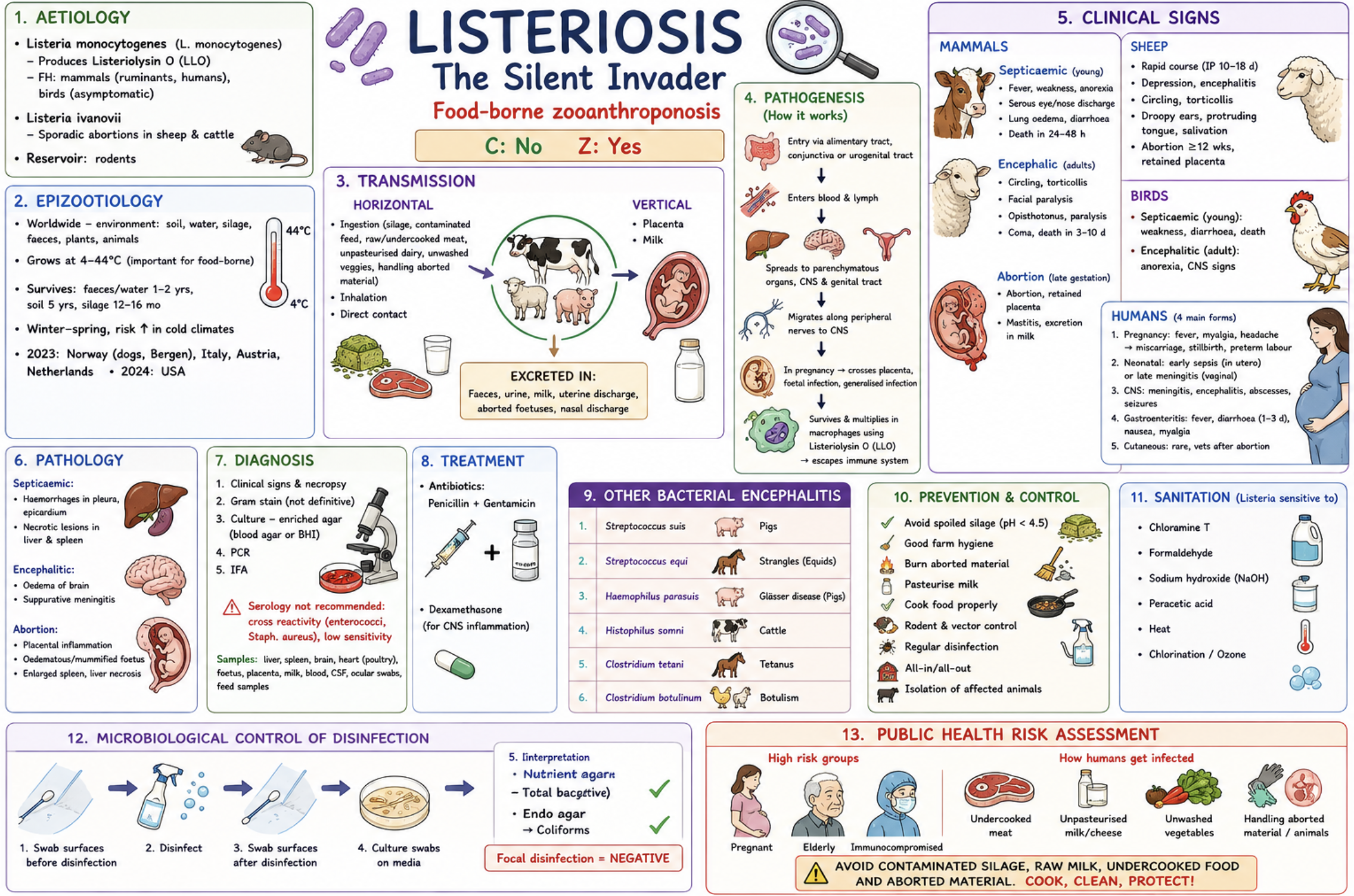
Listeriosis and other bacterial encephalitis (+microbiological control of disinfection)

Listeria:

C: No  Z: Yes

Important food-borne zooanthroponosis, especially in immunocompromised individuals.

Etiology

  • Listeria monocytogenes

Produces Listeriolysin O (LLO)

  • FH: mammals (ruminants, humans), birds asymptomatic

  • Listeria ivanovii Sporadic abortions in sheep & cattle.

  • Reservoir: rodents → urine contaminates silage, vegetables etc. (tror e ho sa på epizoo eksamen)

Epizootiological situation

  • Worldwide: soil, water, silage, faeces, plants

  • Grows at 4–44°C (important food-borne pathogen)

  • Survives: Soil: up to 5 years, Silage: 12–16 month, Faeces/water: 1–2 years, Winter–spring disease in colder climates

  • 2023: Norway (dogs in Bergen), Italy, Austria, Netherlands

  • 2024: USA

Transmission

Horizontal:

  • Contaminated silage/feed, Unpasteurised dairy products, Undercooked meat, Direct contact/inhalation

Vertical:

  • Placenta, milk

Excreted in:

  • Faeces, urine, milk, uterine discharge, aborted foetuses

Pathogenesis

  • Entry via alimentary tract/conjunctiva → blood & lymph → CNS, liver, spleen, genital tract

  • Migrates along peripheral nerves to CNS

  • Survives in macrophages using *Listeriolysin O**

  • Pregnancy/immunosuppression predispose disease

Clinical signs

Mammals

Septicaemic form (young):

  • Fever, diarrhoea, weakness, Nasal/ocular discharge, Death within 24–48 h

Encephalic form (adults):

  • Circling, Facial paralysis, Torticollis, Opisthotonus, Paralysis/coma

Abortion form:

  • Late gestation abortion, Retained placenta, Mastitis

Sheep

  • Rapid encephalitis, Droopy ears, protruding tongue, Excess salivation, Circling

Birds

  • Septicaemia in young, CNS signs in adults

Humans

  • Pregnancy infections, Neonatal sepsis/meningitis, Gastroenteritis, Encephalitis/meningitis, Cutaneous listeriosis in veterinarians

Pathology

  • Liver & spleen necrosis, Brain oedema, Suppurative meningitis, Placental inflammation, Oedematous/mummified foetus

Diagnosis

  • Clinical signs + necropsy, Blood agar/BHI culture, PCR, IFA, Gram stain, Serology not recommended

Treatment

* Penicillin + gentamicin

* Dexamethasone for CNS inflammation

Other bacterial encephalitis

* Streptococcus suis — pigs

* Streptococcus equi — strangles

* Haemophilus parasuis — Glässer disease

* Histophilus somni — cattle

a) Prevention & control

* Avoid spoiled silage (pH 4.5), Good farm hygiene, Burn aborted material, Pasteurise milk, Cook food properly, Rodent/vector control, Regular disinfection, All-in/all-out, Isolation of affected animals

outbreak: Isolation of affected animals – Tx (if CNS is not involved)

Vaccine: in sheep – but not very effective 

b) Sanitation and disinfection

Sensitive to:

* Chloramine T, Formaldehyde, NaOH, Peracetic acid, Heat, Chlorination/ozone

Microbiological control of disinfection efficiency

1.          Chemical control – determines concentration of active substance by chemical swabs

2.          Microbial control – the most objective test – based on microbial swabs (area – 10cm2)

a.         Total no. of bacteria or presence of indicator microbes is established

b.         Preventative (quantitative), focal (qualitative), direct or indirect

Before disinfection - Evaluation of effect of disinfectant

  1. Take sterile swabs from 10cm^2

  2. Total no. of Mo on nutritive agar and indicator on endo agar

  3. CFU = colonies grown on plate x 00 (dilution)

-        e.g.: 86 colonies x 00= 8.6x10^3CFU/cm^2

 After disinfection

  1. Preventive

    1. 30 sterile swabs on 10cm^2 area into physiological solution

    2. Measures total no. of MO & indicator MO

    3. Make decimal dilutions 10^1, 10^2, 10^3, 10^4

    4. On nutritive agar

    5. Max 10% positive indicator MO

  2. Focal

    1. 30 sterile swabs on 100cm^2 area & into selective liquid broth

    2. Make decimal dilutions

    3. On endo agar

    4. Should be negative!!

    5. CFU: Colonies x 00(dilution) x 0

    6. Total no. can be 1.2x10^3CFU/10cm^2

Public health risk assessment

Major zoonotic food-borne disease.

High risk for:

  • Pregnant women, Elderly, Immunocompromised people

  • Avoid contaminated silage, raw milk, undercooked food, unwashed vegetables, and aborted material

<p><strong>Listeria:</strong></p><p><strong>C:</strong> No  <span style="color: red;"><strong>Z:</strong> Yes</span></p><p>Important food-borne <strong>zooanthroponosis</strong>, especially in immunocompromised individuals.</p><p>Etiology</p><ul><li><p><strong><mark data-color="red" style="background-color: red; color: inherit;">Listeria monocytogenes</mark></strong></p></li></ul><p><em>Produces </em><strong><mark data-color="blue" style="background-color: blue; color: inherit;">Listeriolysin O</mark></strong> (LLO)</p><ul><li><p> FH: mammals (ruminants, humans), birds asymptomatic</p></li><li><p><mark data-color="red" style="background-color: red; color: inherit;"> </mark><strong><mark data-color="red" style="background-color: red; color: inherit;">Listeria ivanovii </mark>→</strong> Sporadic abortions in sheep &amp; cattle. </p></li><li><p><em>Reservoir: </em><strong>rodents </strong>→ urine contaminates silage, vegetables etc. (tror e ho sa på epizoo eksamen)</p></li></ul><p>Epizootiological situation</p><ul><li><p><span style="color: red;"><strong>Worldwide:</strong></span> soil, water, silage, faeces, plants</p></li><li><p><em>Grows at </em>4–44°C (important food-borne pathogen)</p></li><li><p>Survives: Soil: up to 5 years, Silage: 12–16 month, Faeces/water: 1–2 years, Winter–spring disease in colder climates</p></li><li><p>2023: <strong>Norway </strong>(dogs in Bergen), Italy, Austria, Netherlands</p></li><li><p>2024: <strong>USA</strong></p></li></ul><p>Transmission</p><p><strong>Horizontal:</strong></p><ul><li><p><span style="color: red;">Contaminated silage/feed</span>, Unpasteurised dairy products, Undercooked meat, Direct contact/inhalation</p></li></ul><p><strong>Vertical:</strong></p><ul><li><p>Placenta, milk</p></li></ul><p><strong>Excreted in:</strong></p><ul><li><p>Faeces, urine, milk, uterine discharge, aborted foetuses</p></li></ul><p>Pathogenesis</p><ul><li><p>Entry via alimentary tract/conjunctiva → blood &amp; lymph → <span style="color: red;">CNS, liver, spleen, genital tract</span></p></li><li><p>Migrates along peripheral nerves to CNS</p></li><li><p><em>Survives in macrophages using </em>*Listeriolysin O**</p></li><li><p>Pregnancy/immunosuppression predispose disease</p></li></ul><p>Clinical signs</p><p>Mammals</p><p><strong><mark data-color="yellow" style="background-color: yellow; color: inherit;">Septicaemic form (young):</mark></strong></p><ul><li><p>Fever, diarrhoea, weakness, Nasal/ocular discharge, Death within 24–48 h</p></li></ul><p><strong><mark data-color="yellow" style="background-color: yellow; color: inherit;">Encephalic form (adults):</mark></strong></p><ul><li><p>Circling, Facial paralysis, Torticollis, Opisthotonus, Paralysis/coma</p></li></ul><p><strong><mark data-color="yellow" style="background-color: yellow; color: inherit;">Abortion form:</mark></strong></p><ul><li><p>Late gestation abortion, Retained placenta, Mastitis</p></li></ul><p>Sheep</p><ul><li><p>Rapid encephalitis, Droopy ears, protruding tongue, Excess salivation, Circling</p></li></ul><p>Birds</p><ul><li><p>Septicaemia in young,  CNS signs in adults</p></li></ul><p>Humans</p><ul><li><p>Pregnancy infections, Neonatal sepsis/meningitis, Gastroenteritis, Encephalitis/meningitis, Cutaneous listeriosis in veterinarians</p></li></ul><p>Pathology</p><ul><li><p>Liver &amp; spleen necrosis, Brain oedema, Suppurative meningitis, Placental inflammation, Oedematous/mummified foetus</p></li></ul><p> Diagnosis</p><ul><li><p>Clinical signs + necropsy, <span style="color: red;"><strong>Blood agar</strong></span>/BHI culture, PCR, IFA, Gram stain,<span style="color: red;"> Serology not recommended</span></p></li></ul><p>Treatment</p><p>* Penicillin + gentamicin</p><p>* Dexamethasone for CNS inflammation</p><p>Other bacterial encephalitis</p><p>* <strong>Streptococcus suis</strong> — pigs</p><p>* <strong>Streptococcus equi</strong> — strangles</p><p>* <strong>Haemophilus parasuis</strong> — Glässer disease</p><p>* <strong>Histophilus somni</strong> — cattle</p><p></p><p><strong><u>a) Prevention &amp; control</u></strong></p><p>* Avoid spoiled silage (pH 4.5), Good farm hygiene, Burn aborted material, Pasteurise milk, Cook food properly, Rodent/vector control, Regular disinfection, All-in/all-out, Isolation of affected animals</p><p>outbreak: <span style="line-height: 15.333332px;">Isolation of affected animals – Tx (if CNS is not involved)</span></p><p><span style="line-height: 15.333332px;">Vaccine: in sheep – but not very effective&nbsp;</span></p><p></p><p><strong><u>b) Sanitation and disinfection</u></strong></p><p>Sensitive to:</p><p>* Chloramine T, Formaldehyde, NaOH, Peracetic acid, Heat, Chlorination/ozone</p><p><strong><mark data-color="purple" style="background-color: purple; color: inherit;">Microbiological control of disinfection efficiency</mark></strong></p><p class="MsoListParagraphCxSpFirst" style="text-align: justify;"><span><strong>1.</strong></span><span style="font-family: &quot;Times New Roman&quot;; line-height: normal; font-size: 7pt;"><strong>&nbsp;&nbsp;&nbsp;&nbsp;&nbsp;&nbsp;&nbsp;&nbsp;&nbsp; </strong></span><span><strong><em>Chemical control</em></strong> – determines concentration of active substance by chemical swabs</span></p><p class="MsoListParagraphCxSpMiddle" style="text-align: justify;"><span><strong>2.</strong></span><span style="font-family: &quot;Times New Roman&quot;; line-height: normal; font-size: 7pt;"><strong>&nbsp;&nbsp;&nbsp;&nbsp;&nbsp;&nbsp;&nbsp;&nbsp;&nbsp; </strong></span><span><strong><em>Microbial control</em></strong> – the most objective test – based on microbial swabs (area – 10cm2)</span></p><p class="MsoListParagraphCxSpMiddle" style="text-align: justify;"><span><strong>a.</strong></span><span style="font-family: &quot;Times New Roman&quot;; line-height: normal; font-size: 7pt;"><strong>&nbsp;&nbsp;&nbsp;&nbsp;&nbsp;&nbsp;&nbsp;&nbsp; </strong></span><span>Total no. of bacteria or presence of indicator microbes is established</span></p><p class="MsoListParagraphCxSpLast" style="text-align: justify;"><span><strong>b.</strong></span><span style="font-family: &quot;Times New Roman&quot;; line-height: normal; font-size: 7pt;"><strong>&nbsp;&nbsp;&nbsp;&nbsp;&nbsp;&nbsp;&nbsp;&nbsp; </strong></span><span>Preventative (quantitative), focal (qualitative), direct or indirect</span></p><p class="MsoNormal" style="text-align: justify;"><span><strong><em>Before disinfection - </em></strong>Evaluation of effect of disinfectant</span></p><ol type="1"><li><p><span>Take sterile swabs from 10cm^2</span></p></li><li><p><span>Total no. of Mo on nutritive agar and indicator on endo agar</span></p></li><li><p><span>CFU = colonies grown on plate x 00 (dilution)</span></p></li></ol><p class="MsoListParagraph" style="text-align: justify;"><span>-&nbsp;&nbsp;&nbsp;&nbsp;&nbsp;&nbsp;&nbsp; e.g.: 86 colonies x 00= 8.6x10^3CFU/cm^2</span></p><p class="MsoNormal" style="text-align: justify;"><strong><em>&nbsp;</em></strong><span><strong><em>After disinfection</em></strong></span></p><ol type="1"><li><p><span><u>Preventive</u></span></p><ol type="a"><li><p><span>30 sterile swabs on <strong>10cm^2</strong> area into physiological solution</span></p></li><li><p><span>Measures total no. of MO &amp; indicator MO</span></p></li><li><p><span>Make decimal dilutions 10^1, 10^2, 10^3, 10^4</span></p></li><li><p><span>On nutritive agar</span></p></li><li><p><span style="color: red;">Max 10% positive indicator MO</span></p></li></ol></li><li><p><span><u>Focal</u></span></p><ol type="a"><li><p><span>30 sterile swabs on <strong>100cm^2</strong> area &amp; into selective liquid broth</span></p></li><li><p><span>Make decimal dilutions</span></p></li><li><p><span>On endo agar</span></p></li><li><p><span style="color: red;">Should be negative!!</span></p></li><li><p><span>CFU: Colonies x 00(dilution) x 0</span></p></li><li><p><span>Total no. can be 1.2x10^3CFU/10cm^2</span></p></li></ol></li></ol><p></p><p><strong><u>Public health risk assessment</u></strong></p><p><span style="color: red;">Major zoonotic food-borne disease</span>.</p><p>High risk for:</p><ul><li><p>Pregnant women, Elderly, Immunocompromised people</p></li><li><p>Avoid contaminated silage, raw milk, undercooked food, unwashed vegetables, and aborted material</p></li></ul><p></p>
11
New cards

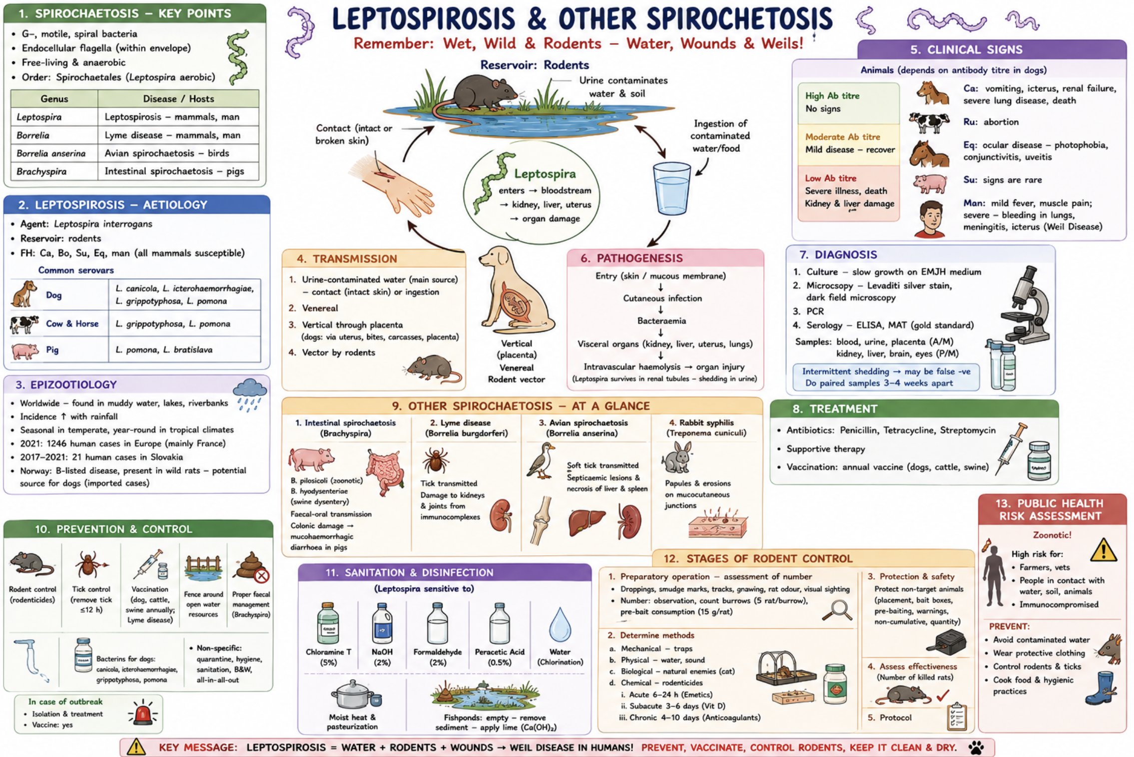
Leptospirosis and other spirochetosis (+stages of rodent control)

Spirochaetosis

* Gram-negative, motile, spiral bacteria

* Endocellular flagella

* Mostly anaerobic

Order: *Spirochaetales**

Includes:

* Leptospira → leptospirosis

* Borrelia → Lyme disease

* Borrelia anserina → avian spirochaetosis

* Brachyspira → intestinal spirochaetosis

Leptospirosis

C: No  Z: Yes

Aetiology

* Leptospira interrogans

Reservoir: *rodents**

Common serovars:

* Dogs: canicola, icterohaemorrhagiae, grippotyphosa, pomona

* Cattle/horses: grippotyphosa, pomona

* Pigs: pomona, bratislava

FH:

* Dogs, cattle, pigs, horses, humans

Epizootiological situation

  • Worldwide

  • Associated with muddy water, lakes, riverbanks

  • Natural focal disease - More common after rainfall, Seasonal in temperate climates

Europe:

  • 2021: 1246 human cases in Europe (mainly France)

  • Slovakia: 21 cases (2017–2021)

  • Norway: B-listed disease, mainly in wild rats/imported cases

Transmission

* Urine-contaminated water (main source)

* Contact through skin or mucous membranes

* Ingestion

* Venereal

* Placental transmission

* Rodent vectors

Dogs:

* Infection often from bites, carcasses, placenta

Pathogenesis

  • Skin/MM contact → bloodstream → bacteraemia → kidney, liver, uterus

→ intravascular haemolysis & liver injury

  • Leptospira persist in renal tubules → shedding in urine

Clinical signs

Dogs

  • Vomiting, Icterus, Renal failure, Severe lung disease, Death

Ruminants

  • Abortion

Horses

  • Uveitis, Conjunctivitis, Photophobia

Pigs

  • Usually mild/subclinical

Humans

  • Fever, myalgia, Meningitis, Pulmonary haemorrhage, Icterus - Weil disease

Diagnosis

  • EMJH culture medium, Dark-field microscopy, Levaditi silver stain, PCR

  • Serology: ELISA, MAT = gold standard

Samples:

  • Blood, urine, placenta, Kidney, liver, brain, eye

NB:

  • Intermittent shedding → paired samples after 3–4 weeks

Treatment

  • Penicillin, Tetracycline, Streptomycin, Supportive therapy

  • Annual vaccines: Dogs, Cattle, Swine

Other Spirochaetoses

  • Brachyspira (Swine dysentery → Mucohaemorrhagic diarrhoea)

  • Borrelia burgdorferi (Lyme disease - Tick-borne → Kidney & joint damage)

  • Borrelia anserina (Avian spirochaetosis - Soft tick transmission → Septicaemia, liver/spleen necrosis)

  • Treponema cuniculi (Rabbit syphilis → Papules & erosions)

a) Prevention & control

  • Rodent control, Tick removal (<12 h), Vaccination, Fence open water sources, Proper sanitation, Quarantine, Black & white system, All-in/all-out

  • Outbreak: Isolation, Treatment, Vaccination

b) Sanitation, focal disinfection and rodent control stages

Sensitive to:

  • Chloramine T, NaOH, Formaldehyde, Peracetic acid, Chlorination of water, Heat/pasteurisation

Rodent control stages

1. Assessment of rodent population (number based on feces etc)

2. Choose control method:

  • Mechanical, Physical, Biological, Chemical

3. Protection of non-target animals (bait box etc)

4. Assess effectiveness

5. Protocol/documentation

Public health risk assessment

  • Important zoonosis. Humans infected mainly from contaminated water or urine.

  • High-risk groups: Farmers, Veterinarians, People exposed to floodwater or rodents.

<p><strong><u>Spirochaetosis</u></strong></p><p>* Gram-negative, motile, spiral bacteria</p><p>* Endocellular flagella</p><p>* Mostly anaerobic</p><p><em>Order: </em>*Spirochaetales**</p><p>Includes:</p><p>* <strong>Leptospira</strong> → leptospirosis</p><p>* <strong>Borrelia</strong> → Lyme disease</p><p>* <strong>Borrelia anserina</strong> → avian spirochaetosis</p><p>* <strong>Brachyspira</strong> → intestinal spirochaetosis</p><p></p><p><strong><u>Leptospirosis</u></strong></p><p><span style="color: red;"><strong>C:</strong> No  <strong>Z:</strong> Yes</span></p><p>Aetiology</p><p><mark data-color="red" style="background-color: red; color: inherit;">* </mark><strong><mark data-color="red" style="background-color: red; color: inherit;">Leptospira interrogans</mark></strong></p><p><em>Reservoir: </em>*<mark data-color="blue" style="background-color: blue; color: inherit;">rodents</mark>**</p><p>Common serovars:</p><p>* <strong>Dogs:</strong><em> canicola, icterohaemorrhagiae, grippotyphosa, pomona</em></p><p>* <strong>Cattle/horses:</strong> <em>grippotyphosa, pomona</em></p><p>* <strong>Pigs:</strong> <em>pomona, bratislava</em></p><p>FH:</p><p>* Dogs, cattle, pigs, horses, humans</p><p></p><p>Epizootiological situation</p><ul><li><p><span style="color: red;"><strong>Worldwide</strong></span></p></li><li><p>Associated with muddy water, lakes, riverbanks</p></li><li><p>Natural focal disease - More common after rainfall, Seasonal in temperate climates</p></li></ul><p>Europe:</p><ul><li><p>2021: 1246 human cases in Europe (mainly France)</p></li><li><p><strong>Slovakia: </strong>21 cases (2017–2021)</p></li><li><p><strong>Norway:</strong> B-listed disease, mainly in wild rats/imported cases</p></li></ul><p></p><p>Transmission</p><p>* <span style="color: red;">Urine-contaminated water</span> (main source)</p><p>* Contact<strong> through skin or mucous membranes</strong></p><p>* Ingestion</p><p>* Venereal</p><p>* Placental transmission</p><p>* Rodent vectors</p><p>Dogs:</p><p>* Infection often from bites, carcasses, placenta</p><p></p><p>Pathogenesis</p><ul><li><p>Skin/MM contact → bloodstream → bacteraemia → kidney, liver, uterus</p></li></ul><p>→ intravascular haemolysis &amp; liver injury</p><ul><li><p>Leptospira persist in renal tubules → shedding in urine</p></li></ul><p></p><p>Clinical signs</p><p>Dogs</p><ul><li><p>Vomiting, Icterus, Renal failure, Severe lung disease, Death</p></li></ul><p>Ruminants</p><ul><li><p>Abortion</p></li></ul><p>Horses</p><ul><li><p>Uveitis, Conjunctivitis, Photophobia</p></li></ul><p>Pigs</p><ul><li><p>Usually mild/subclinical</p></li></ul><p>Humans</p><ul><li><p> Fever, myalgia, Meningitis, Pulmonary haemorrhage, Icterus -  <strong>Weil disease</strong></p></li></ul><p></p><p>Diagnosis</p><ul><li><p>EMJH culture medium, Dark-field microscopy, Levaditi silver stain, PCR</p></li><li><p>Serology: ELISA, <span style="color: red;">MAT = gold standard</span></p></li></ul><p>Samples:</p><ul><li><p>Blood, urine, placenta, Kidney, liver, brain, eye</p></li></ul><p>NB:</p><ul><li><p>Intermittent shedding → paired samples after 3–4 weeks</p></li></ul><p></p><p>Treatment</p><ul><li><p>Penicillin, Tetracycline, Streptomycin, Supportive therapy</p></li><li><p>Annual vaccines: Dogs, Cattle, Swine</p></li></ul><p></p><p>Other Spirochaetoses</p><ul><li><p><mark data-color="red" style="background-color: red; color: inherit;">Brachyspira</mark> (Swine dysentery → Mucohaemorrhagic diarrhoea)</p></li><li><p><mark data-color="red" style="background-color: red; color: inherit;">Borrelia burgdorferi</mark> (Lyme disease - Tick-borne → Kidney &amp; joint damage)</p></li><li><p><mark data-color="red" style="background-color: red; color: inherit;">Borrelia anserina </mark>(Avian spirochaetosis - Soft tick transmission → Septicaemia, liver/spleen necrosis)</p></li><li><p><mark data-color="red" style="background-color: red; color: inherit;">Treponema cuniculi</mark> (Rabbit syphilis → Papules &amp; erosions)</p></li></ul><p></p><p><strong><u>a) Prevention &amp; control</u></strong></p><ul><li><p>Rodent control, Tick removal (&lt;12 h), Vaccination, Fence open water sources, Proper sanitation, Quarantine, Black &amp; white system, All-in/all-out</p></li><li><p>Outbreak: Isolation, Treatment, Vaccination</p></li></ul><p></p><p><strong><u>b) Sanitation, focal disinfection and rodent control stages</u></strong></p><p>Sensitive to:</p><ul><li><p>Chloramine T, NaOH, Formaldehyde, Peracetic acid, Chlorination of water, Heat/pasteurisation</p></li></ul><p></p><p><strong><mark data-color="purple" style="background-color: purple; color: inherit;">Rodent control stages</mark></strong></p><p>1. Assessment of rodent population (number based on feces etc)</p><p>2. Choose control method:</p><ul><li><p>Mechanical, Physical, Biological, Chemical</p></li></ul><p>3. Protection of non-target animals (bait box etc)</p><p>4. Assess effectiveness</p><p>5. Protocol/documentation</p><p></p><p><strong><u>Public health risk assessment</u></strong></p><ul><li><p>Important zoonosis. Humans infected mainly from contaminated water or urine.</p></li><li><p>High-risk groups: Farmers, Veterinarians, People exposed to floodwater or rodents.</p></li></ul><p></p>
12
New cards

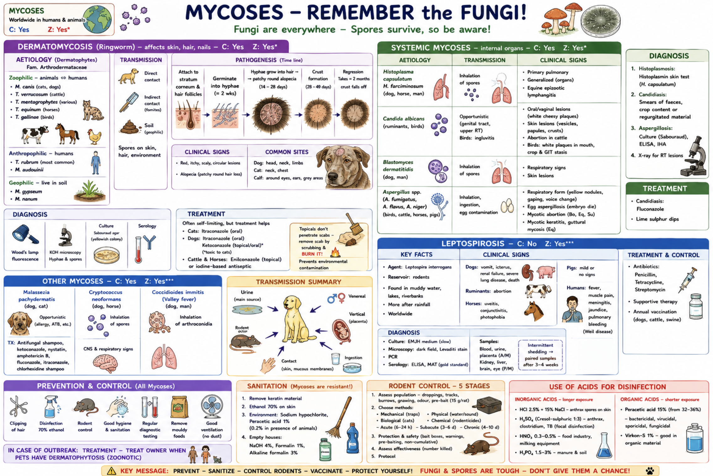
Mycoses (+ use of acids for disinfection)

  • Mycoses = fungal diseases of humans & animals

  • Worldwide distribution

Cause:

  • Dermatomycoses (skin)

  • Systemic mycoses (internal organs)

Dermatomycosis (Ringworm)

C: Yes  Z: Yes*

Key facts

  • Affect skin, hair, nails, Caused by dermatophytes, Colonise stratum corneum

  • Hyphae & spores highly resistant in environment

Types of dermatophytes

  1. Anthropophilic: Human hosts, Do not survive in soil. T. rubrum

  1. Zoophilic: Animal → human transmission. Microsporum canis, Trichophyton verrucosum, T. mentagrophytes

  1. Geophilic: Soil fungi. M. gypseum

Aetiology

Family: Arthrodermataceae

Important species:

  • Trichophyton verrucosum,T. mentagrophytes, T. equinum,T. gallinae

  • Microsporum canis, M. nanum, M. gypseum

Transmission

  • Direct contact, Indirect contact with spores, Soil contamination, Animal/vector transmission

Pathogenesis

  • Spores attach to skin/hair follicles

→ hyphae develop (~2 weeks)

→ invade hair centre

→ circular alopecia/crusts

→ regression after ~2 months

Clinical signs

  • Red, itchy, scaly circular lesions + Alopecia

Typical locations:

* Dog: head, neck, limbs

* Cat: neck, chest

* Calves: around eyes & ears

Diagnosis

  • Wood’s lamp, KOH microscopy, Sabouraud agar culture, Serology

Treatment

Usually self-limiting.

Cats: Itraconazole

Dogs: Itraconazole, Ketoconazole (toxic in cats)

Cattle/Horses: Enilconazole, Iodine antiseptics

NB: Remove crusts before topical therapy, Burn removed scabs

Systemic Mycoses

C: Yes  Z: Yes*

Aetiology

  • Histoplasmosis: Histoplasma capsulatumDogs, horses, humans

  • Candidiasis: Candida albicansRuminants, birds

  • Blastomycosis: Blastomyces dermatitidis Dogs, humans

  • Aspergillosis: Aspergillus fumigatus, A. flavus, A. niger

Transmission

* Inhalation of spores

* Opportunistic infection

* Egg contamination (Aspergillus)

Clinical signs

  • Histoplasmosis: Pulmonary/generalised disease, Equine lymphangitis

  • Candidiasis: White cheesy plaques, Oral/vaginal lesions, Crop stasis in birds

  • Blastomycosis: Respiratory & skin lesions

  • Aspergillosis: Respiratory disease, Yellow nodules, Gaping, Voice changes, Embryo death, Mycotic abortion

Diagnosis

  • Histoplasmin skin test, Smears/culture, Sabouraud agar, ELISA, X-ray

Treatment

  • Fluconazole, Lime sulphur dips

Other mycoses

  • Malassezia pachydermatis: Dogs/cats, Opportunistic dermatitis/otitis

  • Cryptococcus neoformans: Inhaled spores → CNS/respiratory disease

  • Coccidioides immitis → Valley fever, Inhalation of arthroconidia

a) Prevention & control

  • Clip hair, 70% ethanol disinfection, Rodent control, Good hygiene, Remove mouldy feed, Good ventilation, Avoid dusty environment, Regular testing

  • Outbreak: Treat animals and owners (zoonotic)

b) Resistance & sanitation

  • Fungal spores survive: 12 months–2 years

  • Disinfection: Sodium hypochlorite, Peracetic acid, NaOH, Formalin, Ethanol

Use of acids for disinfection

Inorganic acids: Hydrochloric acid, Sulphuric acid, Nitric acid, Phosphoric acid

Organic acids: Peracetic acid, Virkon-S

Public health risk assessment

Many mycoses are zoonotic. Transmission mainly through:

  • Direct animal contact, Spores in environment, Contaminated dust/hair/soil

High risk for:

* Immunocompromised people, Veterinarians, Animal handlers.

<ul><li><p>Mycoses = fungal diseases of humans &amp; animals</p></li></ul><ul><li><p><span style="color: red;">Worldwide distribution</span></p></li></ul><p>Cause:</p><ul><li><p><strong>Dermatomycoses</strong> (skin)</p></li><li><p><strong>Systemic mycoses</strong> (internal organs)</p></li></ul><p></p><p><strong><u>Dermatomycosis </u></strong>(Ringworm)</p><p><span style="color: red;"><strong>C:</strong> Yes  <strong>Z:</strong> Yes*</span></p><p>Key facts</p><ul><li><p>Affect skin, hair, nails, Caused by dermatophytes, Colonise stratum corneum</p></li><li><p>Hyphae &amp; spores highly resistant in environment</p></li></ul><p>Types of dermatophytes</p><ol><li><p><strong>Anthropophilic:</strong> Human hosts, Do not survive in soil. <mark data-color="red" style="background-color: red; color: inherit;">T. rubrum</mark></p></li></ol><ol start="2"><li><p><strong>Zoophilic</strong>: Animal → human transmission. <em><mark data-color="red" style="background-color: red; color: inherit;">Microsporum canis, Trichophyton verrucosum, T. mentagrophytes</mark></em></p></li></ol><ol start="3"><li><p><strong>Geophilic:</strong> Soil fungi. <em><mark data-color="red" style="background-color: red; color: inherit;">M. gypseum</mark></em></p></li></ol><p>Aetiology</p><p>Family: <strong>Arthrodermataceae</strong></p><p>Important species:</p><ul><li><p><em>Trichophyton verrucosum,T. mentagrophytes, T. equinum,T. gallinae</em></p></li></ul><ul><li><p><em>Microsporum canis, M. nanum, M. gypseum</em></p></li></ul><p>Transmission</p><ul><li><p>Direct contact, Indirect contact with spores, Soil contamination, Animal/vector transmission</p></li></ul><p></p><p>Pathogenesis</p><ul><li><p>Spores attach to skin/hair follicles</p></li></ul><p>→ hyphae develop (~2 weeks)</p><p>→ invade hair centre</p><p>→ circular alopecia/crusts</p><p>→ regression after ~2 months</p><p></p><p>Clinical signs</p><ul><li><p>Red, itchy, scaly circular lesions + Alopecia</p></li></ul><p>Typical locations:</p><p>* Dog: head, neck, limbs</p><p>* Cat: neck, chest</p><p>* Calves: around eyes &amp; ears</p><p></p><p>Diagnosis</p><ul><li><p>Wood’s lamp, KOH microscopy, Sabouraud agar culture, Serology</p></li></ul><p></p><p>Treatment</p><p>Usually self-limiting.</p><p>Cats: Itraconazole</p><p>Dogs: Itraconazole, Ketoconazole (toxic in cats)</p><p>Cattle/Horses: Enilconazole, Iodine antiseptics</p><p>NB: Remove crusts before topical therapy, Burn removed scabs</p><p></p><p><strong><u>Systemic Mycoses</u></strong></p><p><span style="color: red;"><strong>C:</strong> Yes  <strong>Z:</strong> Yes*</span></p><p>Aetiology</p><ul><li><p>Histoplasmosis: <em><mark data-color="red" style="background-color: red; color: inherit;">Histoplasma capsulatum</mark> → </em>Dogs, horses, humans</p></li><li><p>Candidiasis: <em><mark data-color="red" style="background-color: red; color: inherit;">Candida albicans</mark> → </em>Ruminants, birds</p></li><li><p>Blastomycosis: <em><mark data-color="red" style="background-color: red; color: inherit;">Blastomyces dermatitidis </mark>→</em> Dogs, humans</p></li><li><p>Aspergillosis: <em><mark data-color="red" style="background-color: red; color: inherit;">Aspergillus fumigatus,</mark></em><mark data-color="red" style="background-color: red; color: inherit;"> </mark><em><mark data-color="red" style="background-color: red; color: inherit;">A. flavus,</mark></em><mark data-color="red" style="background-color: red; color: inherit;"> </mark><em><mark data-color="red" style="background-color: red; color: inherit;">A. niger</mark></em></p></li></ul><p></p><p>Transmission</p><p>* Inhalation of spores</p><p>* Opportunistic infection</p><p>* Egg contamination (Aspergillus)</p><p></p><p>Clinical signs</p><ul><li><p>Histoplasmosis: Pulmonary/generalised disease, Equine lymphangitis</p></li><li><p>Candidiasis: White cheesy plaques, Oral/vaginal lesions, Crop stasis in birds</p></li><li><p>Blastomycosis: Respiratory &amp; skin lesions</p></li><li><p>Aspergillosis: Respiratory disease, Yellow nodules, Gaping, Voice changes, Embryo death, Mycotic abortion</p></li></ul><p>Diagnosis</p><ul><li><p>Histoplasmin skin test, Smears/culture, Sabouraud agar, ELISA, X-ray</p></li></ul><p>Treatment</p><ul><li><p>Fluconazole, Lime sulphur dips</p></li></ul><p></p><p>Other mycoses</p><ul><li><p><mark data-color="red" style="background-color: red; color: inherit;">Malassezia pachydermatis</mark>: Dogs/cats, Opportunistic dermatitis/otitis</p></li><li><p>Cryptococcus neoformans: Inhaled spores → CNS/respiratory disease</p></li><li><p>Coccidioides immitis → Valley fever, Inhalation of arthroconidia</p></li></ul><p></p><p>a) Prevention &amp; control</p><ul><li><p>Clip hair, 70% ethanol disinfection, Rodent control, Good hygiene, Remove mouldy feed, Good ventilation, Avoid dusty environment, Regular testing</p></li><li><p>Outbreak: Treat animals and owners (zoonotic)</p></li></ul><p></p><p>b) Resistance &amp; sanitation</p><ul><li><p>Fungal spores survive: 12 months–2 years</p></li><li><p>Disinfection: Sodium hypochlorite, Peracetic acid, NaOH, Formalin, Ethanol</p></li></ul><p></p><p><strong><mark data-color="purple" style="background-color: purple; color: inherit;">Use of acids for disinfection</mark></strong></p><p>Inorganic acids: Hydrochloric acid, Sulphuric acid, Nitric acid, Phosphoric acid</p><p>Organic acids: Peracetic acid, Virkon-S</p><p></p><p><strong><u>Public health risk assessment</u></strong></p><p>Many mycoses are zoonotic. Transmission mainly through:</p><ul><li><p>Direct animal contact, Spores in environment, Contaminated dust/hair/soil</p></li></ul><p>High risk for:</p><p>* Immunocompromised people, Veterinarians, Animal handlers.</p>