VIVA Revision: Anatomy of Pelvis, Limbs, and Upper Body
 Call Kai
 Learn
Learn Practice Test
 Spaced Repetition
 Match
Match Flashcards
 Knowt Play
1/90

There's no tags or description
Looks like no tags are added yet.
Name | Mastery | Learn | Test | Matching | Spaced | Call with Kai |
|---|
No analytics yet
Send a link to your students to track their progress
91 Terms
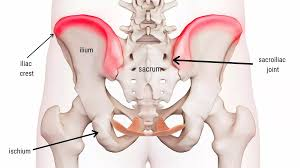
Iliac crest
The iliac crest is the superior component of the ilium. Several muscles attach to the iliac crest such as the gluteal muscles, the abdominal muscles and the erector spinae muscles.
Anterior superior iliac spine
Bony prominence on the ilium, part of the pelvis. Sartorius, inguinal ligament and tensor fascia latae all originate on the anterior superior iliac spine.
Posterior superior iliac spine
Bony prominence on the posterior aspect of the ilium. Gluteus maximus, erector spinae muscles and latissimus dorsi all attach at this prominence.
Sacroiliac joint
Joint plays a crucial role in absorbing shock and transferring weight from the upper body to the lower limbs. It is an extremely strong synovial joint which has thick ligaments.
Ischial tuberosities
The ischial tuberosities is what we sit on when we sit down. Several muscles attach here including the hamstring muscles and adductor magnus.
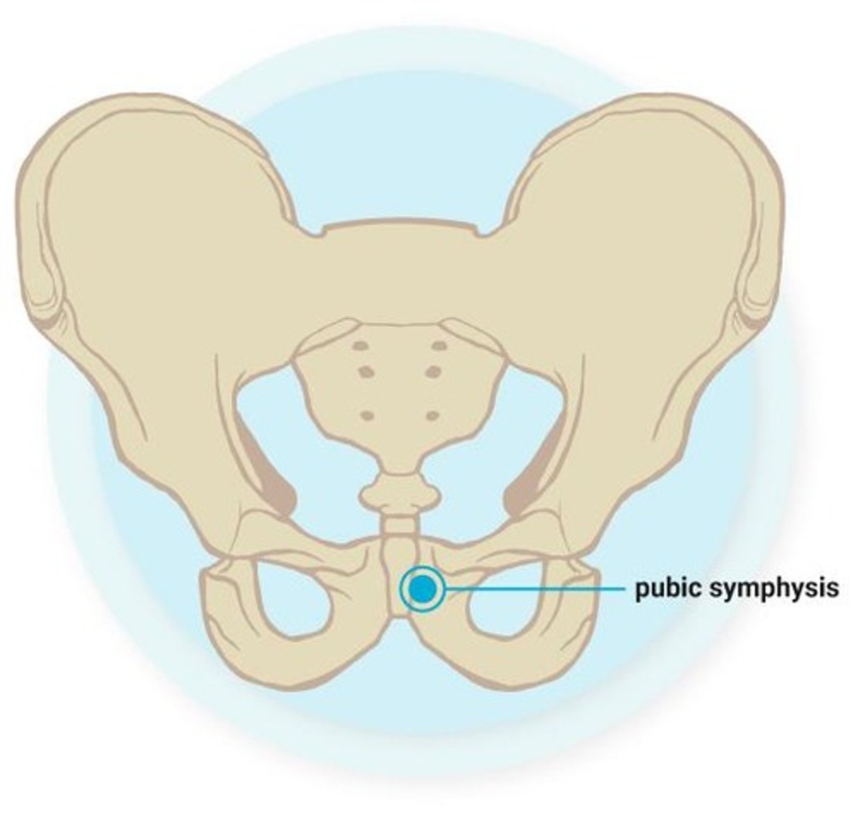
Symphysis pubis
A fibrocartilaginous disc sits between the two sections of the pubis bone allowing them to connect. Stabilises the pelvis and absorbs shock during movement.
Greater trochanter of femur
Bony prominence on the upper lateral aspect of the femur. Key attachment point for many muscles such as gluteus maximus (hip extension) and gluteus minimus and medius (hip abduction).
Lateral tibial condyle/Gerdy's tubercle
One of two condyles of the tibia along with the medial condyle. The lateral meniscus also sits on the lateral condyle, acting as a shock absorber and providing stability for the knee.
Head of fibula
Articulates with the lateral tibial condyle to form the proximal tibiofibular joint. A bony projection called the styloid process extends upwards from the lower part of the head.
Tibial tuberosity
Serves as an attachment point for the patella tendon which connects the kneecap to the shin bone. Facilitates knee extension by transmitting the force of the quadriceps muscle.
Medial malleolus
Distal part of the tibia, works with the talus to form the talocrural joint. It is an attachment point for the deltoid ligament.
tarsal tunnel.
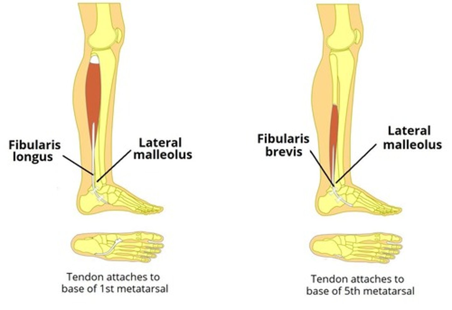
Lateral malleolus
Part of the fibula, works with the talus to form the ankle joint. It is an attachment point for the lateral ankle ligaments.
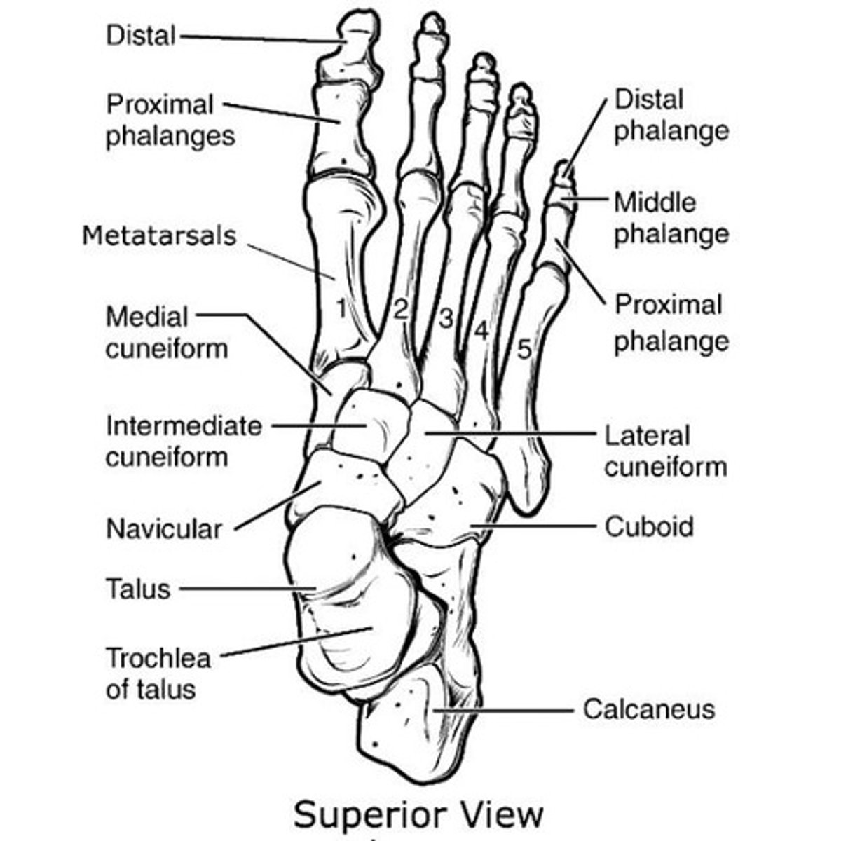
Navicular
It articulates with the talus, cuboid and the three cuneiform bones.
Calcaneus
It forms the heel and is located at the proximal aspect of the foot. It is the largest bone in the foot and articulates with the talus bone. The achilles tendon attaches to the back of the calcaneus.
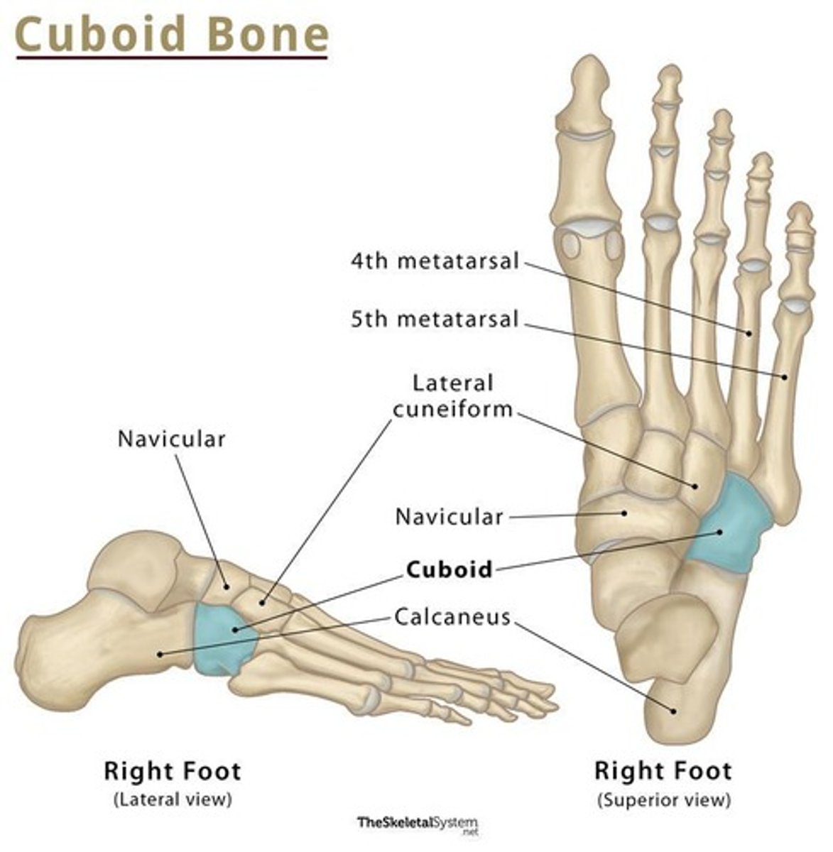
Cuboid
It is located towards the lateral side of the foot. It articulates with the calcaneus, the lateral cuneiform, the 4th and 5th metatarsals. It forms a tarsometatarsal joint with the 4th and 5th metatarsals.
Base of 5th metatarsal
It is located on the 5th toe (laterally). It is a bony prominence between the heel and the little toe. It is the most proximal of the aspects of the metatarsal. It supports the weight of the foot as well as facilitating movement.
Metatarsals 1-5
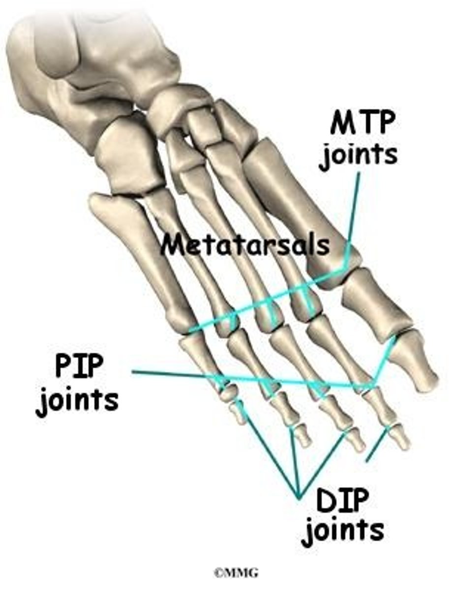
Located in the middle of the foot. Each metatarsal has a base, shaft and head. The base articulates with the tarsal bones and the head articulates with the proximal phalanges. Metatarsals are named from 1-5 with number 1 being the most medial and number 5 being the most lateral. They form tarsometatarsal joints, intermetatarsal joints and metatarsophalangeal joints.
Metatarsophalangeal joint
Located in between the metatarsals and the phalanges. There are five metatarsophalangeal joints. These joints allow flexion, extension, abduction and adduction. They are condyloid joints.
Proximal phalanges (feet)
They are the part of the phalanges that are closest to the heel. Located between the metatarsals and the middle phalanges. They allows flexion and extension of the toes to occur.
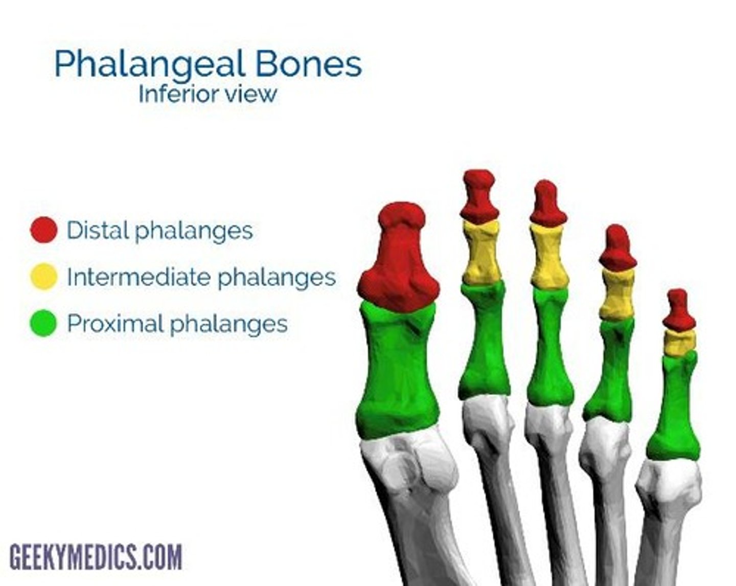
Middle phalanges (feet)
They are located between the proximal and distal phalanges. Only present in toes 2-5, not the big toe. They are smaller than the proximal phalanges. They help the foot to push off when moving.
Distal phalanges (feet)
Located at the most distal point of each toe. The distal phalanges of toes 2-5 all articulate with the middle phalanges. The hallux only has two phalanges.
Proximal and distal interphalangeal joints (feet)
Located between the phalanges. They are synovial hinge joints as they allow movement in one plane. The joint allows flexion and extension of the toes. The hallux only has one interphalangeal joint while the other toes have two.
Femoral triangle
The femoral triangle is located in the anterior upper thigh. Superior: Inguinal ligament. Lateral: Sartorius. Medial: Adductor longus. Contains femoral nerve, artery and vein.
Popliteal fossa
On the posterior side of the knee joint. Superior: Semimembranosus and semitendinosus. Inferior: Lateral and medial heads of the gastrocnemius. Contains popliteal artery and vein and tibial nerve.
Tarsal tunnel
On the medial aspect of the ankle. Contains flexor retinaculum, medial malleolus, talus and calcaneus. Contains posterior tibial artery and vein, tibial nerve, tibialis posterior, flexor hallucis longus and flexor digitorum longus. tom dick and very nervous harry.
Anterior thigh
Muscle group consisting of Rectus femoris, vastus intermedius, vastus medialis, vastus lateralis, sartorius. Innervated by the femoral nerve. Action: Knee extension and hip flexion.
Posterior thigh
Muscle group consisting of Biceps femoris, semitendinosus, semimembranosus. Tibial nerve innervates the semitendinosus, semimembranosus and the long head of biceps femoris. Common fibular nerve innervates the short head of biceps femoris. Action: Knee flexion and hip extension.
Anterior leg
Muscle group consisting of Tibialis anterior, extensor digitorum longus, extensor hallucis longus, fibularis tertius. Innervated by deep fibular nerve. Action: Ankle dorsiflexion and toe extension.
Posterior leg
Muscle group consisting of Gastrocnemius, soleus, plantaris, flexor digitorum longus, flexor hallucis longus, tibialis posterior. Innervated by tibial nerve. Action: Ankle plantarflexion and toe flexion.
Lateral leg
Muscle group consisting of Fibularis longus, fibularis brevis. Innervated by superficial fibular nerve. Action: Foot eversion.
Acromioclavicular joint
First, find the acromion process which is a bony prominence on the top of the shoulder. Then follow the clavicle laterally to where they meet. The AC joint connects the acromion, a bony projection of the scapula, to the clavicle. A cartilage disc is present between the two bones, allowing for slight gliding movement.
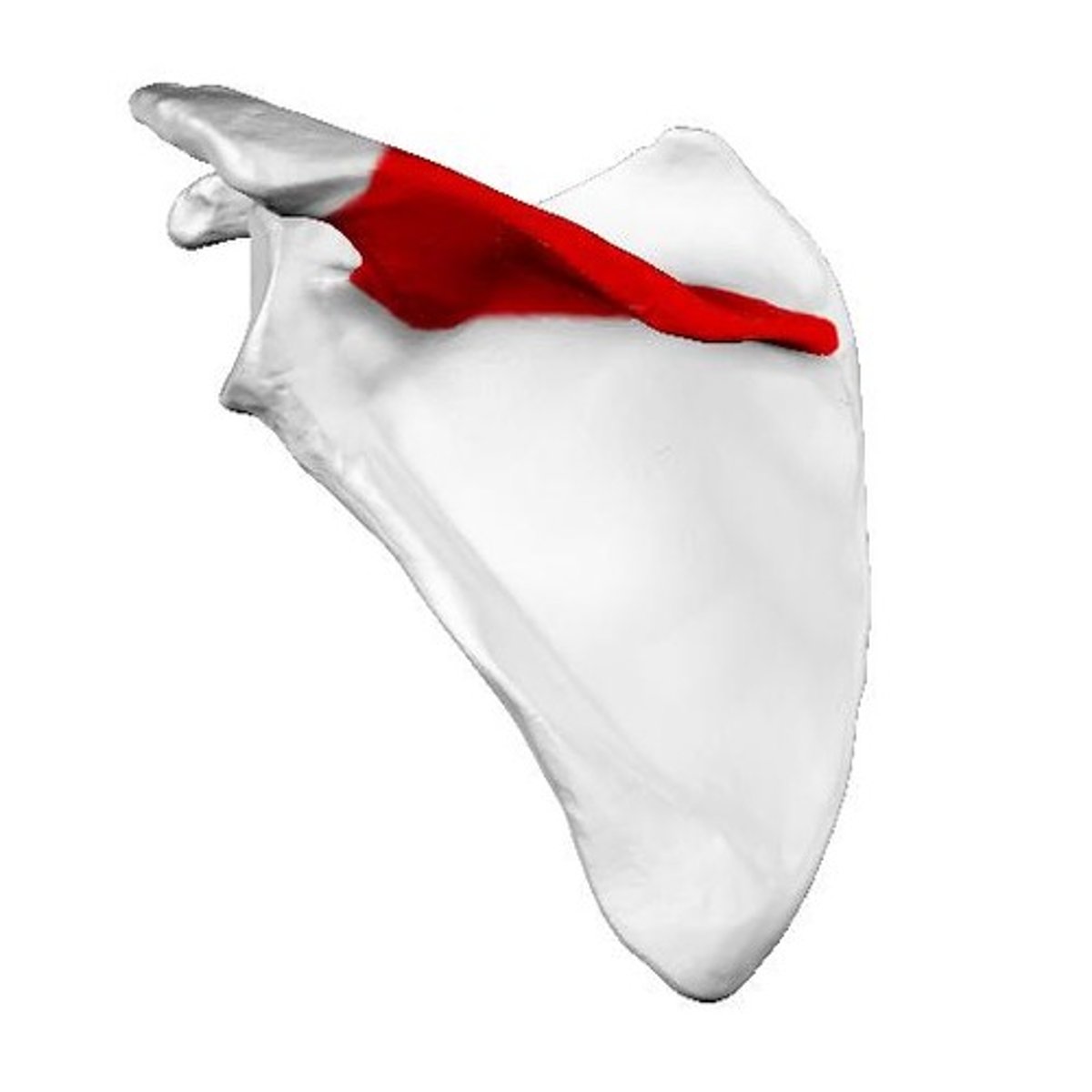
Acromioclavicular ligaments
Ligaments that support the acromioclavicular joint.
Coracoclavicular ligaments
Ligaments that provide additional support to the shoulder joint.
Spine of scapula
Divides the scapula into the supraspinous and infraspinous fossas and serves as an attachment point for muscles.
Acromion process
Bony prominence on the scapula that forms the bony tip of the shoulder and serves as an attachment point for muscles.
Inferior angle of the scapula
The most inferior point of the shoulder blade, formed by the meeting of the medial and lateral borders.
Medial border of the scapula
Runs parallel to the spine, thinner of the two borders, and attaches to the rhomboid muscles.
Lateral border of the scapula
Thickest of the borders, extends towards the axilla, and contains the glenoid fossa which articulates with the humerus.
Greater tubercle of the humerus
The most lateral projection of the proximal humerus and primary attachment site for three muscles of the rotator cuff.
Lesser tubercle of the humerus
Located on the anterior, medial aspect of the proximal humerus and serves as an attachment point for the subscapularis muscle.
Medial epicondyle of the humerus
Located on the medial side of the distal humerus and is the origin of muscles that flex the wrist and fingers.
Lateral epicondyle of the humerus
Located on the lateral side of the distal humerus and is the origin of muscles that extend the fingers and wrist.
Olecranon process
Prominent bump on the posterior aspect of the elbow that serves as a lever for the triceps muscle.
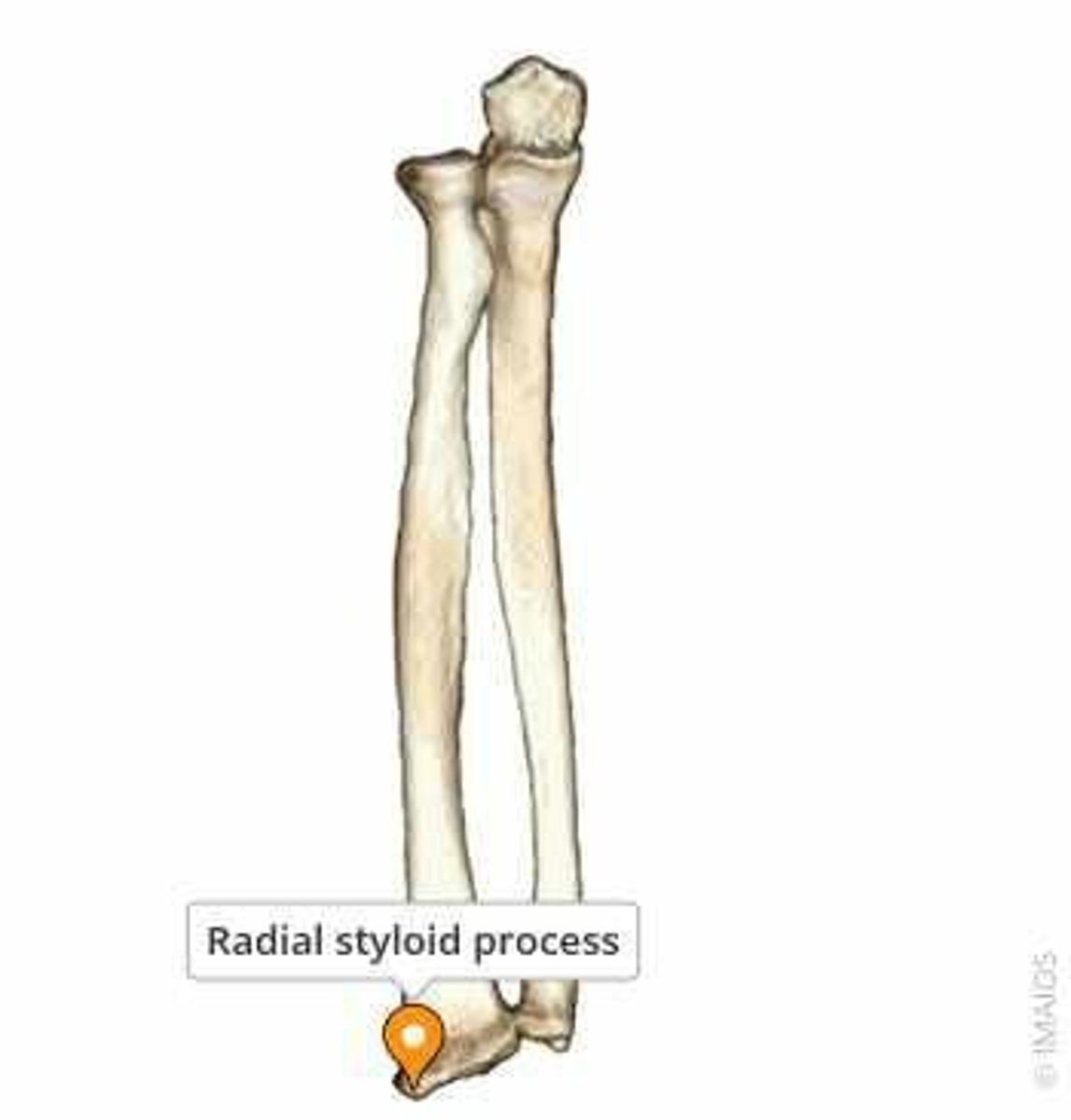
Radial styloid process
Bony prominence on the lateral side of the wrist that serves as an attachment point for the radial collateral ligament. brachioradialis originates on the lateral epicondyle, and inserts onto the radial styloid process.
anatomical snuff box
Scaphoid
Shaped like a boat, it articulates with the lunate and trapezium bones in the wrist and acts as a bridge between the bones of the forearm and the hand.
Trapezius muscle
Muscle that attaches to the spine of the scapula and assists in shoulder movements.
Deltoid muscle
Muscle that attaches to the acromion process and assists in shoulder movements.
Latissimus dorsi
Muscle that attaches to the inferior angle of the scapula.
Teres major
Muscle that attaches to the inferior angle of the scapula.
Rhomboid muscles
Muscles that attach to the medial border of the scapula.
Glenoid fossa
Part of the lateral border of the scapula that articulates with the humerus to form the glenohumeral joint.
Intertubercular sulcus
Located between the greater and lesser tubercles of the humerus.
Ulnar collateral ligament
Helps to stabilize the elbow joint at the medial epicondyle.
Ulnar nerve
Runs behind the medial epicondyle and supplies sensation to the medial one and a half fingers.
Radial collateral ligament
Prevents lateral displacement of the elbow at the lateral epicondyle.
Branches of the radial nerve
Run close to the lateral epicondyle before supplying sensation to the posterior hand.
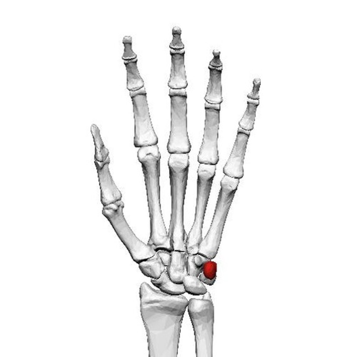
Pisiform
Acts as a bridge between the bones of the forearm and the hand.
Metacarpals
Long bones in the palm that form the knuckles.
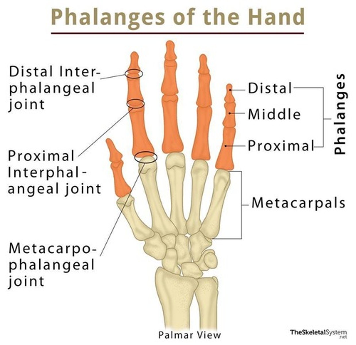
Metacarpophalangeal joints
Located between the metacarpals and the phalanges.
Carpometacarpal joint of the thumb
Located between the carpal bones and the 1st metacarpal.
Phalanges (proximal, middle, distal) *hand
Located on the distal end of the hand.
Interphalangeal joints (proximal and distal) *hand
Can be felt on the creases of the finger.
Axillary zone
Transition zone located in armpit.
borders: anterior: pec major and minor, posterior: latissimus dorsi, medial: serratus anterior and upper ribs, lateral: humerus.
Contents: brachial plexus nerves, axillary artery and vein, and lymph nodes
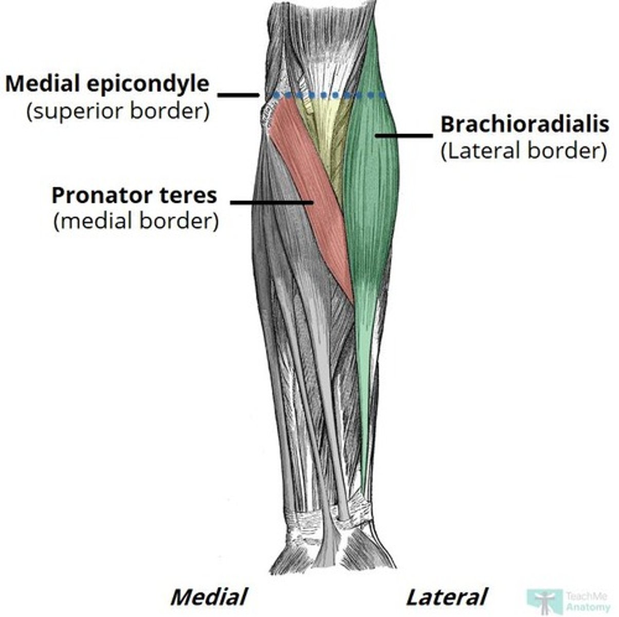
Cubital tunnel
Posterior aspect of the elbow. Passageway between the medial epicondyle of the humerus and the olecranon process of the ulna. Contains the ulnar nerve.
Cubital fossa
transition zone in anterior aspect of the elbow.
Borders: medial: brachioradialis muscle, lateral: pronator teres, epicondyles of humerus.
Contents: median nerve, radial nerve, brachial artery (splits into radial and ulnar), tendon of biceps brachii
Carpal tunnel
Anterior aspect of the wrist. contains median nerve, and tendons of flexor pollicis longus, flexor digitorum superificialis, flexor digitorum profundus.
Flexor compartment of arm
Brachialis, brachioradialis, biceps brachii.
Extensor compartment of arm
Triceps brachii.
Flexor compartment of forearm
Flexor carpi radialis, flexor carpi ulnaris, pronator teres, pronator quadratis, flexor digitorum profundis, flexor digitorum superficialis.
Extensor compartment of forearm
Extensor carpi ulnaris, extensor carpi radialis longus, extensor carpi radialis brevis, extensor digitorum.
First dorsal interosseus muscles
N/A.
Thenar muscles
Flexor pollicis brevis, abductor pollicis brevis, adductor pollicis brevis, opponens pollicis.
Hypothenar muscles
Flexor digiti minimi, abductor digiti minimi, opponens digiti minimi.
Heart
Located slightly to the left of the midline.
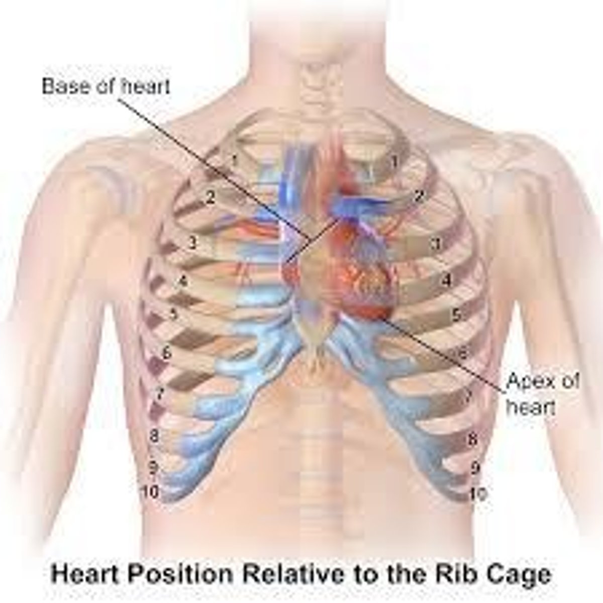
Lungs/pleura
Lungs extend two fingers above the clavicle.
Lung
The lung extends laterally to the 8th rib (axillary line) and posteriorly to the 10th rib.
Parietal pleura
The parietal pleura extends two ribs further down than the lung.
Liver
Located in the upper right portion of the abdomen, superior to the stomach and inferior to the diaphragm, the liver has many functions including bile secretion, detoxification and metabolism.
Spinous process of C7
Located at the bottom of the neck, it is a thick and nearly horizontal prominence that can be palpated as a bony prominence. C7 is not bifid like the other cervical vertebrae but is a single process.
Manubrium
Located at the top of the sternum, it has a depression called the suprasternal notch and articulates with the clavicle at the sternoclavicular joint and the sternum at the sternal angle. It also articulates with the 1st ribs on both sides. 3 fingers down from notch
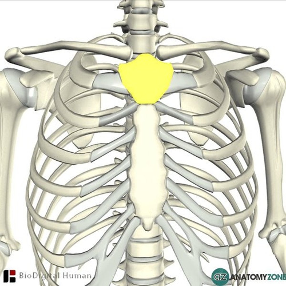
Sternal angle
Located between the manubrium and the body of the sternum at the level of T4-5, it is where the manubrium and sternum articulate and the second ribs articulate with this joint.
Body of sternum
Located inferiorly to the manubrium, it is the central and longest part of the breastbone that articulates with the manubrium and the xiphoid. Costal cartilage of ribs 3-7 attaches to the body. pec major originates here
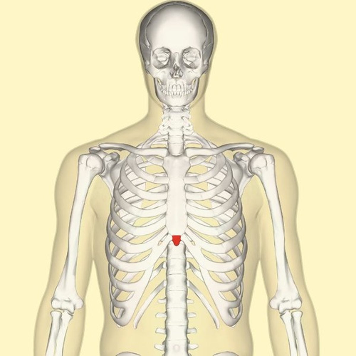
Xiphoid process
Located inferiorly to the sternum, it is a small, cartilaginous, bony extension of the sternum that serves as an attachment point for rectus abdominus and articulates with the body of the sternum.
Sternoclavicular joints
Located on the medial side of the clavicle, it is a synovial joint that allows a range of movements and is separated by a fibrocartilaginous disc. The joint is supported by several ligaments.
Erector spinae
A muscle group consisting of spinalis, longissimus, and iliocostalis, innervated by the dorsal rami of spinal nerves, responsible for spinal extension (bilateral) and lateral flexion (unilateral).
Abdominal muscles
Includes internal and external obliques, rectus abdominus, and transversus abdominus, innervated by T7-12 and L1, with actions such as compressing the trunk, flexing the trunk, and unilaterally rotating the trunk.
Pelvic floor
Located between the coccyx and the pubis, it is essential for holding up the bladder, bowel, and uterus, and stabilizing the spine. It has three openings for women (urethra, vagina, and anus) and two openings for men (urethra and anus). levator ani: iliococcygeous, pubococcygeus, puborectalis. and coccygeous
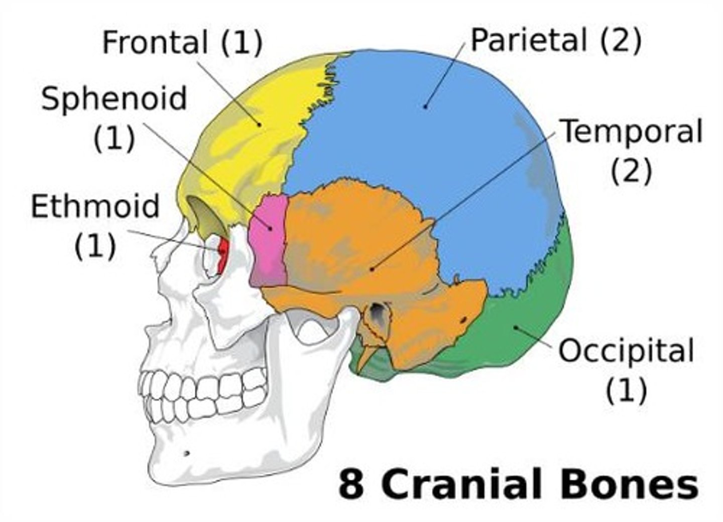
Cranial bones
Includes the frontal bone (forehead), parietal bones (two flat bones on either side of the head), temporal bones (side of the skull), occipital bone (back of the skull), sphenoid bone (butterfly shaped at the base of the cranium), and ethmoid bone (between the eyes and nasal cavity), protecting and encasing the brain.
Facial bones
Includes nasal bones (bridge of the nose), lacrimal bones (medial orbit), zygomatic bones (cheekbones), inferior nasal conchae (nasal cavity), vomer (nasal septum), maxilla (upper jaw), and mandible (lower jaw), forming the framework of the face.
Temporomandibular joint
Located on each side of the head, just in front of the ears, it is a synovial joint that moves in two parts, allowing for a wide range of jaw movements such as talking, chewing, and facial expressions.
Medial thigh compartment
Hip adductor muscles: pectineus, adductor brevis, adductor longus, gracilis, adductor magnus.
O: on the pelvis
All except for pectineus are innervated by obturator nerve.