Biol 3333 Module 2 Chp 2 and 3
1/84
There's no tags or description
Looks like no tags are added yet.
Name | Mastery | Learn | Test | Matching | Spaced | Call with Kai |
|---|
No analytics yet
Send a link to your students to track their progress
85 Terms
Standard measurement for microorganisms
Measured in micrometers (μm) and nanometers (nm)
stain used for capsules
india ink or nigrosin is commonly used to visualize the presence of capsules around bacteria.
what are eukaryotic cell walls made up of
polysaccharides
2 types of glycocalyx
capsules and slime layer
function of glycocalyx
protects against dehydration and nutrient loss as well as prevents phagocytosis
Magnification
An increase in the apparent size of an image to resolve smaller separations between objects.
Resolution
The smallest distance by which two objects can be separated and still be distinguished.
Limit of microscopy
Resolution, not magnification, limits the ability of what we can see
Refractive Index
A measure of the light-bending ability of a medium
Purpose of Immersion Oil
Used with 100X objectives to keep light from refracting/scattering, which maintains high resolution.
Brightfield Microscopy
Most widely used; specimen is darker than the surrounding bright field; used for live and preserved stained specimens
Darkfield Microscopy
Brightly illuminated specimens surrounded by a dark field; used for live and unstained specimens
Phase-contrast Microscopy
Transforms subtle changes in light waves into differences in intensity; best for observing intracellular structures
Differential Interference Contrast (DIC)
Uses two light beams and prisms to provide higher contrast, color, and a 3-D appearance to live specimens.
Fluorescence Microscopy
Uses UV radiation to excite specimens stained with fluorochromes, which then emit visible light; useful for diagnosing infections
Confocal Microscopy
Uses a laser to scan multiple "z-planes" to construct a 3-D image; useful for thick specimens like biofilms.
Electron waves vs. Light waves
Electron waves are 100,000 times shorter than visible light, providing much greater resolution
Transmission Electron Microscope (TEM)
Transmits electrons through a specimen to reveal detailed internal structures; magnification up to 1,000,000X.
Scanning Electron Microscope (SEM)
Bombards the surface of a metal-coated specimen with electrons to provide a detailed 3-D view of external structures
Purpose of Wet Mounts and Hanging Drop
Used to examine live cells to observe size, motility, shape, and arrangement.
Fixed Mounts (Smears)
Created by drying and heating a film of specimen; kills the microbes and attaches them to the slide.
Heat Fixation
Passing a slide through a Bunsen burner flame to attach the specimen and preserve it.
Chemical Fixation
Using fixatives like 10% formalin or methanol to preserve tissue structure.
Basic Dyes
Cationic, positively charged dyes attracted to negatively charged microbial surfaces.
Acidic Dyes
Anionic, negatively charged dyes repelled by microbes, resulting in a stained background
Positive Staining
The surface of the microbe is colored by the dye while the background remains white/clear.
Negative Staining
The microbe remains clear while the background is stained dark; useful for viewing capsules.
Simple Stain
Uses a single dye to reveal cell shape, size, and arrangement.
Differential Stain
Uses a primary stain and a counterstain to distinguish between cell types or parts.
Gram-Positive Bacteria
Retain crystal violet due to a thicker cell wall and appear purple
Gram-Negative Bacteria
Lose crystal violet during alcohol wash; take up safranin counterstain and appear red/pink.
Gram Stain Steps
1. Crystal violet (Primary), 2. Iodine (Mordant), 3. Alcohol (Decolorizer), 4. Safranin (Counterstain) .
Acid-Fast Stain use and what species does it identify
binds to waxy mycolic acids in cell walls; used to identify Mycobacterium and Nocardia
Endospore Stain
Uses malachite green (primary) and safranin (counterstain); spores appear green within red/pink vegetative cells
Flagellar Stain
Uses a mordant (tannic acid) to increase the diameter of flagella so they can be seen via light microscopy.
Three main types of Cell shapes:
Pleomorphic vs. Monomorphic
Monomorphic bacteria species maintain a single shape; pleomorphic bacteria species can have many shapes.
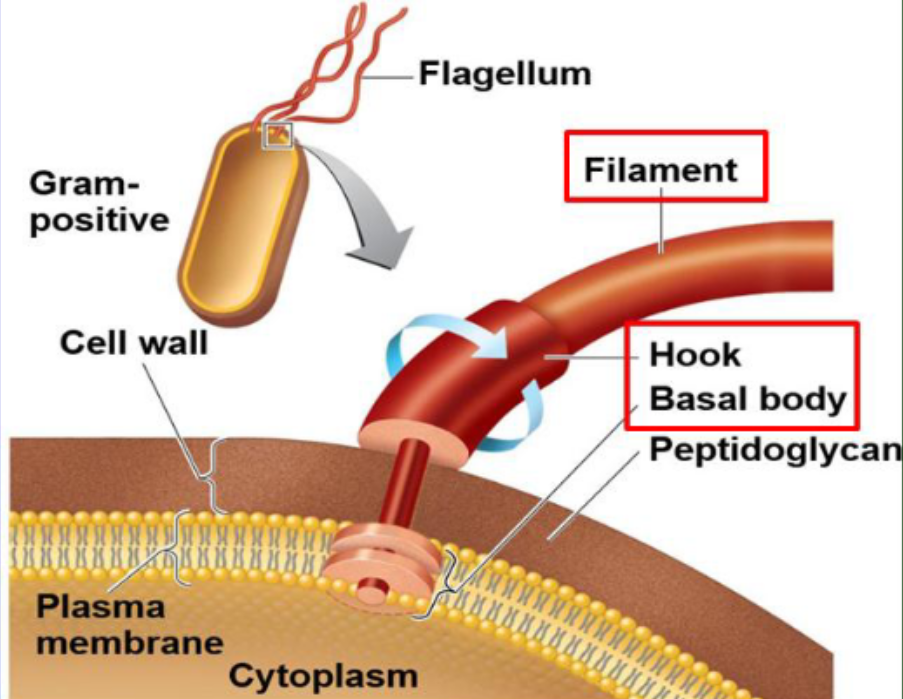
Three parts of a flagellum
Filament (outermost region), Hook (attachment point), and Basal Body (anchors to the cell wall and membrane).
Gas Vesicles
increase buoyancy of bacteria
counterclockwise flagellar motion
results in smooth linear direction - run
clockwise flagellar motion
tumble motion