AP 2 PRAT FINAL QUESTIONS WITH SOME PICS made by angie
1/131
There's no tags or description
Looks like no tags are added yet.
Name | Mastery | Learn | Test | Matching | Spaced | Call with Kai |
|---|
No analytics yet
Send a link to your students to track their progress
132 Terms
lab test that specifically breaks down the types of white blood cells; says what % of each wbc is present in a sample of blood
What is a CBC with differential? (same as complete blood count but specifically looks at what)
A+, A-, B+, B-, AB+, AB-, O+,O-
What are the different blood types?
receive from: A+, A-, O+, O-
donate to: A+, AB+
what can blood type A+ receive and donate to other blood types?
receive from: A-, O-
donate to: A+, A-, AB+, AB-
what can blood type A- receive and donate to other blood types?
receive from: B+, B-, O+, O-
donate to: B+, AB+
what can blood type B+ receive and donate to other blood types?
receive from: B-, O-
donate to: B+, B-, AB+, AB-
what can blood type B- receive and donate to other blood types?
receive from: AB+, AB-, A+, A-, B+, B-, O+, O-
donate to: AB+
what can blood type AB+ receive and donate to other blood types?
receive from: AB-, A-, B-, O-
donate to: AB+, AB-
what can blood type AB-receive and donate to other blood types?
receive from: O+, O-
donate to: A+, B+, AB+, O+
what can blood type O+ receive and donate to other blood types?
receive from: O-
donate to: A+, A-, B+, B-, AB+, AB-, O+, O-
what can blood type O- receive and donate to other blood types?
O-
Which blood type is the universal donor?
AB+
Which blood type is the universal acceptor/recipient?
method to find blood type; determined by antigens (A, B, Rh/D) found on the surface of RBCs; O= A and B absent, Rh=positive; dots=present, without dots= absent, antibodies= what is missing
What is blood typing?
hct; also called packed cell volume (pcv); reports the percentage of RBCs in a sample of whole blood; measured in %
What is hematocrit?
Hb or Hgb; measurement of how much hemoglobin-protein inside RBCs (erthrocytes) that contains iron which binds oxygen and carbon dioxide to transport- is in the blood; measured in g/dl
What is hemoglobin?
neutrophils, eosinophils, basophils, monocytes, lymphocytes
What are the different WBCs?
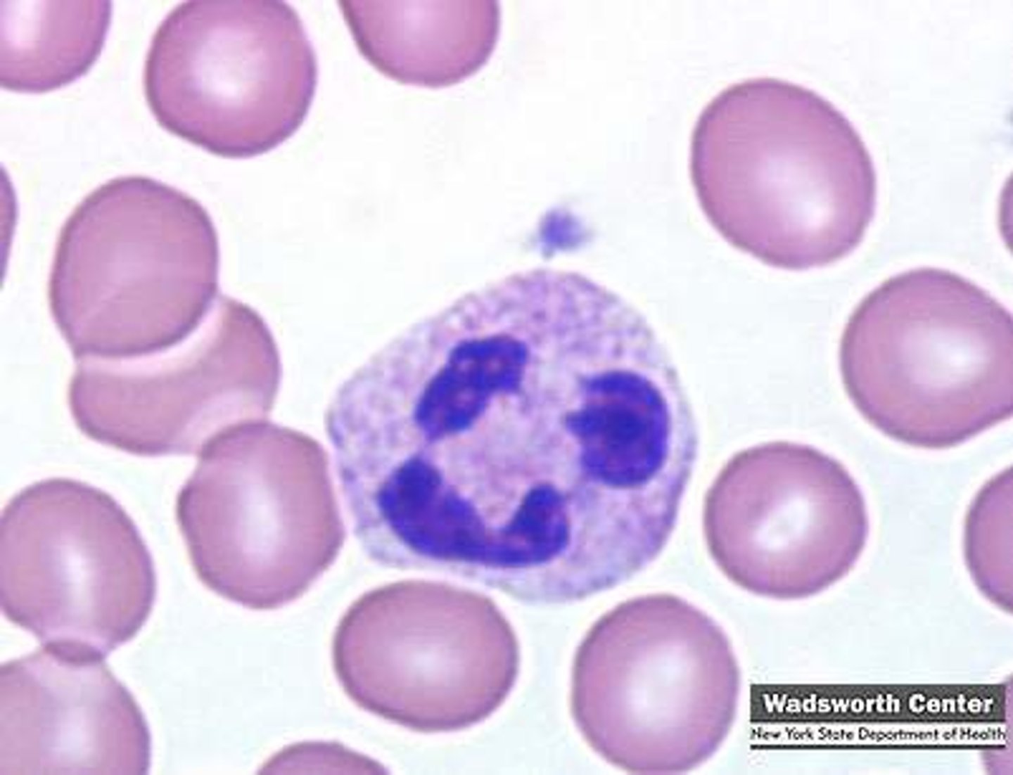
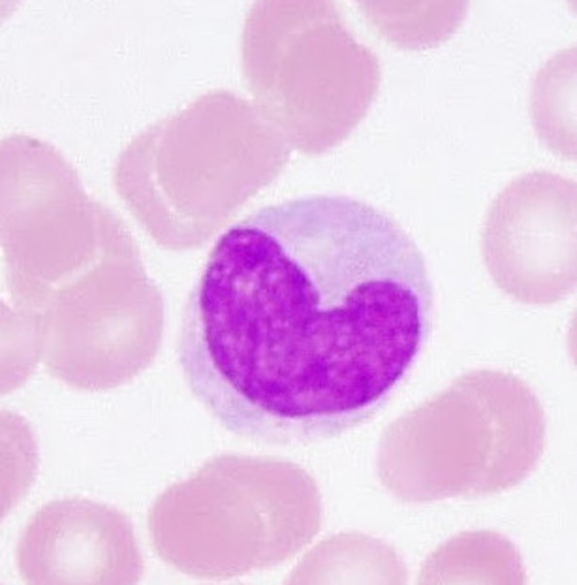
neutrophils
what is this cell?
fights off bacteria pathogens/infection; immunity
what is the function of neutrophils
white blood cell; phagocyte; granulocyte; has lobulated nucleus of 3 or more lobes
what are neutrophils?
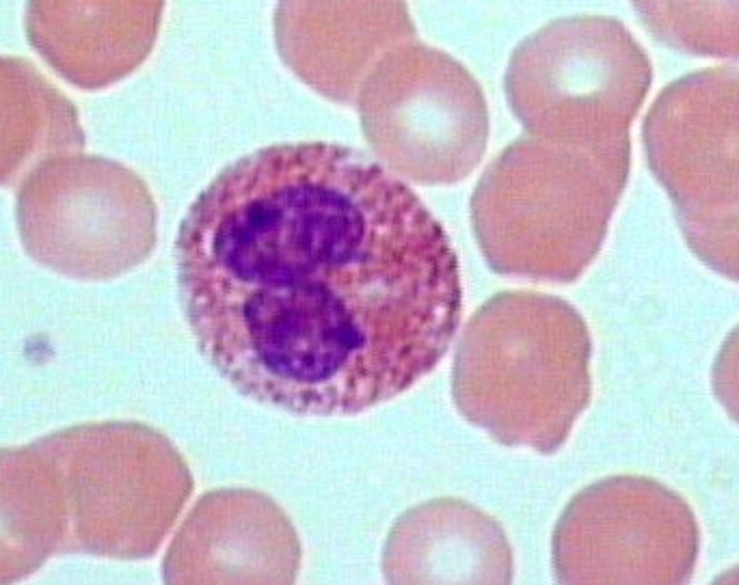
eosinophils
what is this cell?
fights off parasitic infection/allergies; immunity
what is the function of eosinophils
white blood cell; phagocyte; granulocyte; has lobulated nucleus of 2 (bi-lobed) or rarely 3
what are eosinophils
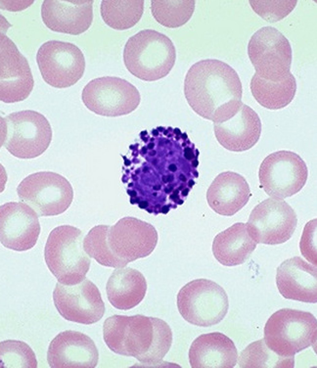
basophils
what is this cell?
releases/secretes histamine and heparin; immunity
what is the function of basophils?
white blood cell; granulocyte; has lobed nucleus that is covered from granules
what are basophils?
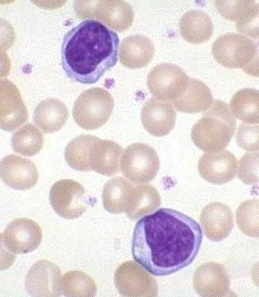
lymphocyte
what is this cell?
fights off viral infection; immunity
what is the function of lymphocytes?
white blood cell; agranulocyte; has no lobes but a huge spherical/indented nucleus; has 3 types- b cells, t cells, natural killer cells
what are lymphocytes?
monocyte
what is this cell?
immunity/phagocytosis
whats the function of a monocyte
white blood cell; agranulocyte; phagocyte; has no lobulated nucleus but kidney bean shaped nucleus; develops into macrophages after leaving the bloodstream and enter the tissue
what is a monocyte
pressure of blood in the aorta (top of the heart) at any given time
What is Blood pressure?
must be an artery that is close to surface of the skin, ex. brachial, radial
where are the proper sites to take BP?
cuff must be above brachial vein, arm must be at heart level (level of aorta)
what is the proper position to take BP?
systolic/diastolic
How is blood pressure reported?
maximum pressure in artery achieved during ventricular contraction
What is systolic pressure?
minimum pressure in artery measured during relaxation
What is diastolic pressure?
throbbing sensation of arteries that can be felt; represents the heart rate (HR)
What is a pulse?
press fingers right above an artery that is close to surface of skin and count bpm
Where is a pulse detected?
simple squamous epithelium
what type of tissue is this?
single layer of flattened cells; found in bowman's capsule, glomerular capillaries, peritubular capillaries and alveoli
what are simple squamous epithelium and where are they found?
simple cuboidal epithelium
what type of tissue is this?
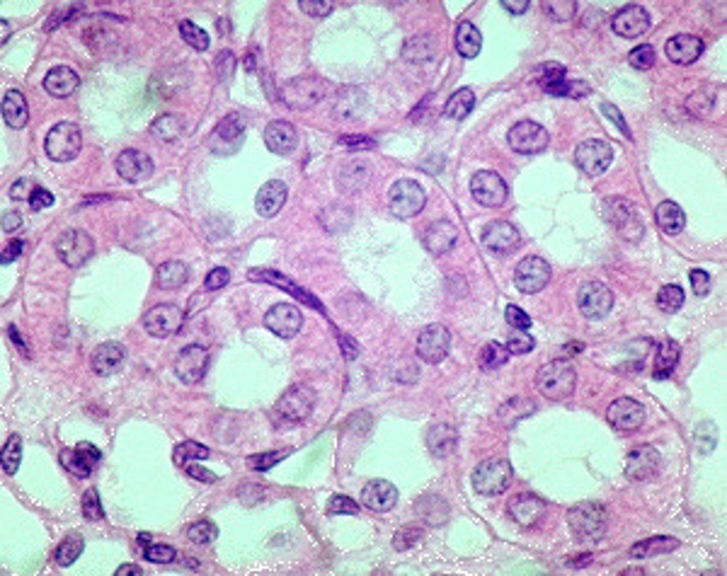
single layer of cube shaped cells; found in nephron tubules and terminal bronchioles
what are simple cuboidal epithelium and where are they found?
transitional epithelium
what type of tissue is this?
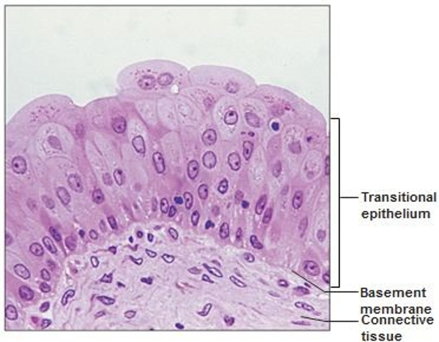
stratified epithelial tissue (stretchy, protective lining); found in ureters, urinary bladder, and part of the urethra
what are transitional epithelium and where are they found?
pseudostratified cilated columnar epithelium
what type of tissue is this?(respiratory)
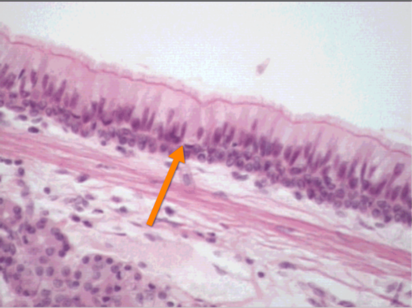
single layer of tall, ciliated cells that fakes looking multi-layered; found in larynx, trachea and primary bronchi which also have c-shaped cartilage rings and goblet cells
what are pseudostratified cilated columnar epithelium and where are they found?
simple columnar epithelium
what type of tissue is this?(respiratory)
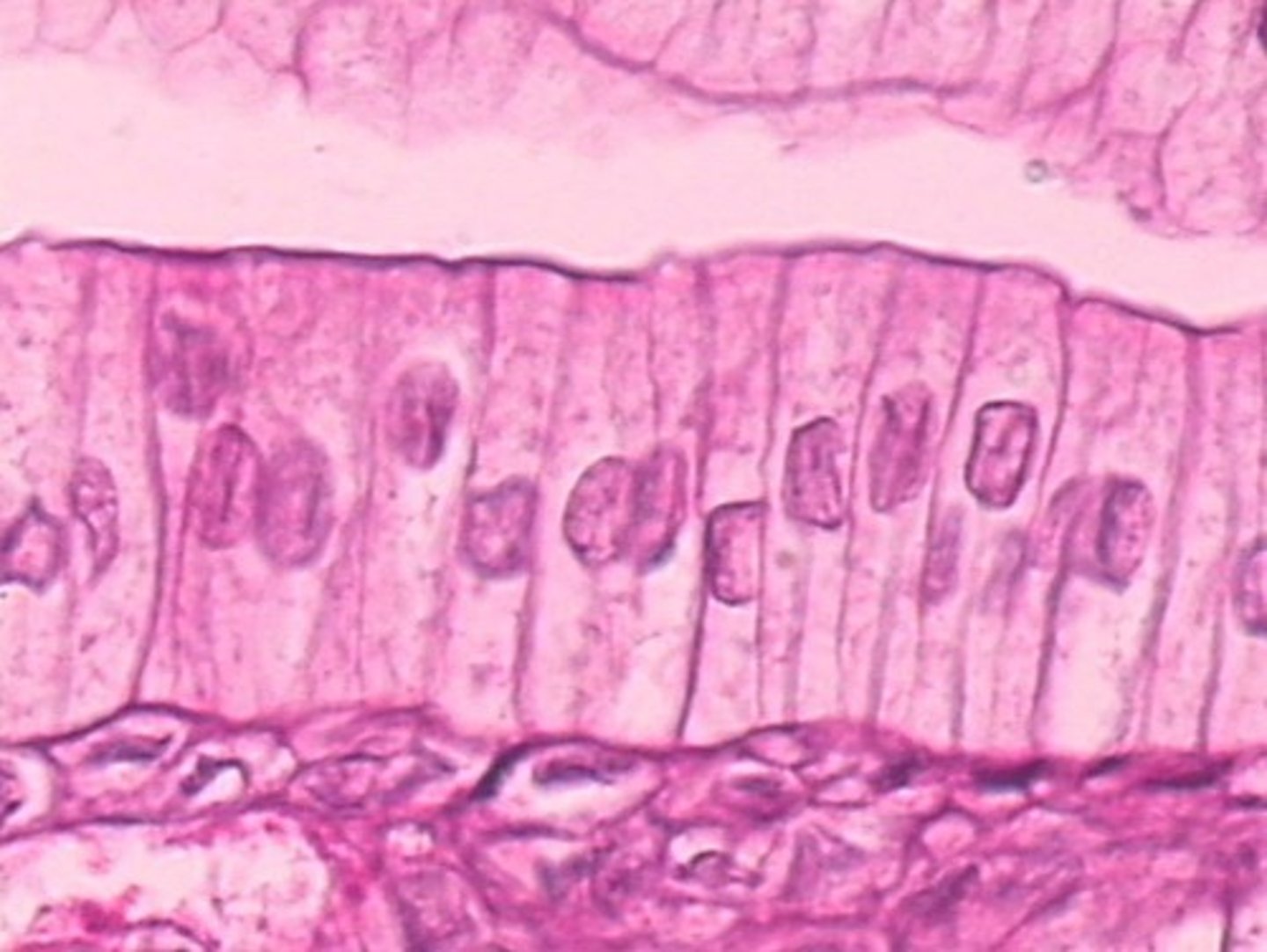
single layer of tall, rectangular (column-shaped) cells; secondary bronchi, tertiary bronchi, and bronchioles (no cartilage)
what is simple columnar epithelium and where are they found?
test to see if the urine system is properly functional
what is a urinalysis
color, odor, and turbidity (cloudiness) of urine, then specific gravity, pH, blood, protein, glucose, ketones, bilirubin, urobilinogen, nitrites, leukocyte esterase
what are the findings of a urinalysis
how concentrated the urine is
what is specific gravity
condition of blood in the urine
what is hematuria
condition of hemoglobin in the urine
what is hemoglobinuria
condition of myoglobin in the urine
what is myoglobinuria
condition protein in the urine
what is proteinuria
uncontrollable diabetes, carbohydrates-free diet, starvation, and pregnancy without diabetes
What can lead to ketones in the body?
low specific gravity (less than 1.005), no glucose, normal urine
what is an indicator of diabetes insipidus?
glucose (higher than 180 mg/dL), ketones, maybe protein
what is an indicator of diabetes mellitus?
blood and protein
what is an indicator of glomerulonephritis?
leukocytes (WBCs) and nitrates
what is an indicator of UTI?
sternocleidomastoid, scalene muscles, serratus anterior
What are the accessory muscles of inspiration?
diaphragm and external intercostals
What are the primary muscles of inspiration?
rectus abdominis, internal intercostals, internal and external obliques
What are the accessory muscles of expiration?
none
What are the primary muscles of expiration?
total lung capacity (TLC), tidal volume (TV), vital capacity (VC), residual volume (RV), inspiratory reserve volume (IRV), expiratory reserve volume (ERV), inspiratory capacity (IC), functional residual cavity (FRC), FEV1, FEV1/FVC
name all the lung volumes and capacities
TLC; volume of air in the lungs upon maximum effort of inspiration; TLC= IRC+TV+ERV+RV
what is total lung capacity
TV; amount of air inhaled and exhaled during normal breathing
what is tidal volume
VC; maximum amount of air that can be exhaled after maximum inhalation; VC=IRC+TV+ERV
what is vital capacity
RV; amount of air remaining in lungs after fully exhaling
what is residual volume
IRV; amount of air that can be inhaled beyond normal tidal volume
what is inspiratory reserve volume
ERV; amount of air that can be exhaled past normal tidal volume
what is expiratory reserve volume
IC; maximum amount of air that can be inhaled including tidal volume ; IC=TV+IRV
what is inspiratory capacity
FRC; volume of air that is left in the lungs after normal passive expiration; FRC=ERC+RV
what is functional residual capacity
amount of air that can forcefully exhaled in 1 sec during a forced vital capacity test
what is FEV1
the ratio of amount of air forcefully exhaled in the first second divided by the total amount that is exhaled; percent of air exhaled in 1 sec divided by total amount of air exhaled (FVC) should be 80% or greater
what is FEV1/FVC
inspiratory reserve volume (IRV)
identify wave 1

vital capacity (VC)
identify wave 2
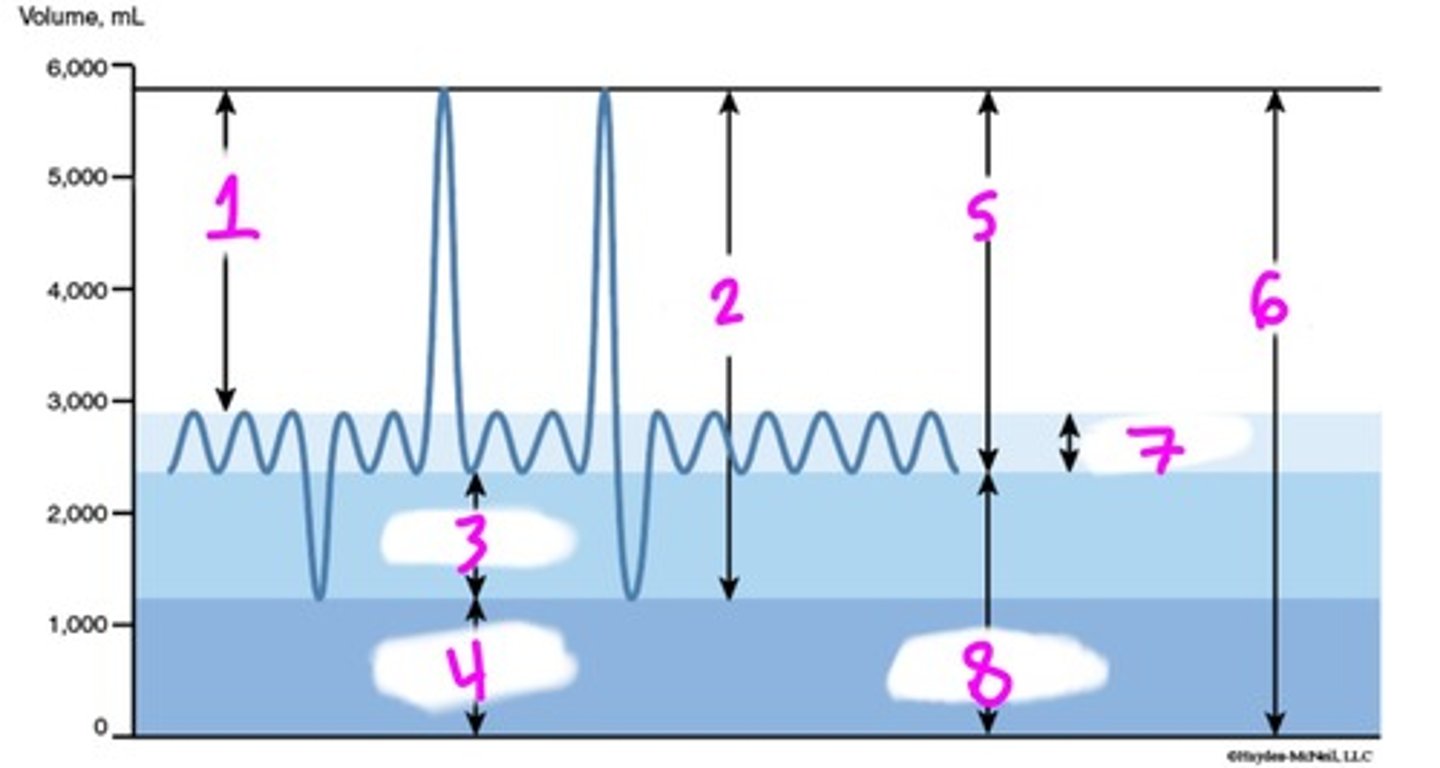
expiratory reserve volume (ERV)
identify wave 3

residual volume (RV)
identify wave 4

inspiratory capacity (IC)
identify wave 5

total lung capacity (TLC)
identify wave 6

tidal volume (TV)
identify wave 7

functional residual capacity (FRC)
identify wave 8

race, height, age, gender
What factors impact spirometry?
a measurement of breathing (or lung volumes); spirometer can measure ventilatory valve
What is spirometry?
test that monitor respiratory function by measuring volumes and capacities of air movement
What is Pulmonary Function Test (PFT)?
ciliated, non-ciliated secretory cells, and basal cells (maybe goblet cells)
What are the cells that are found in the respiratory system?
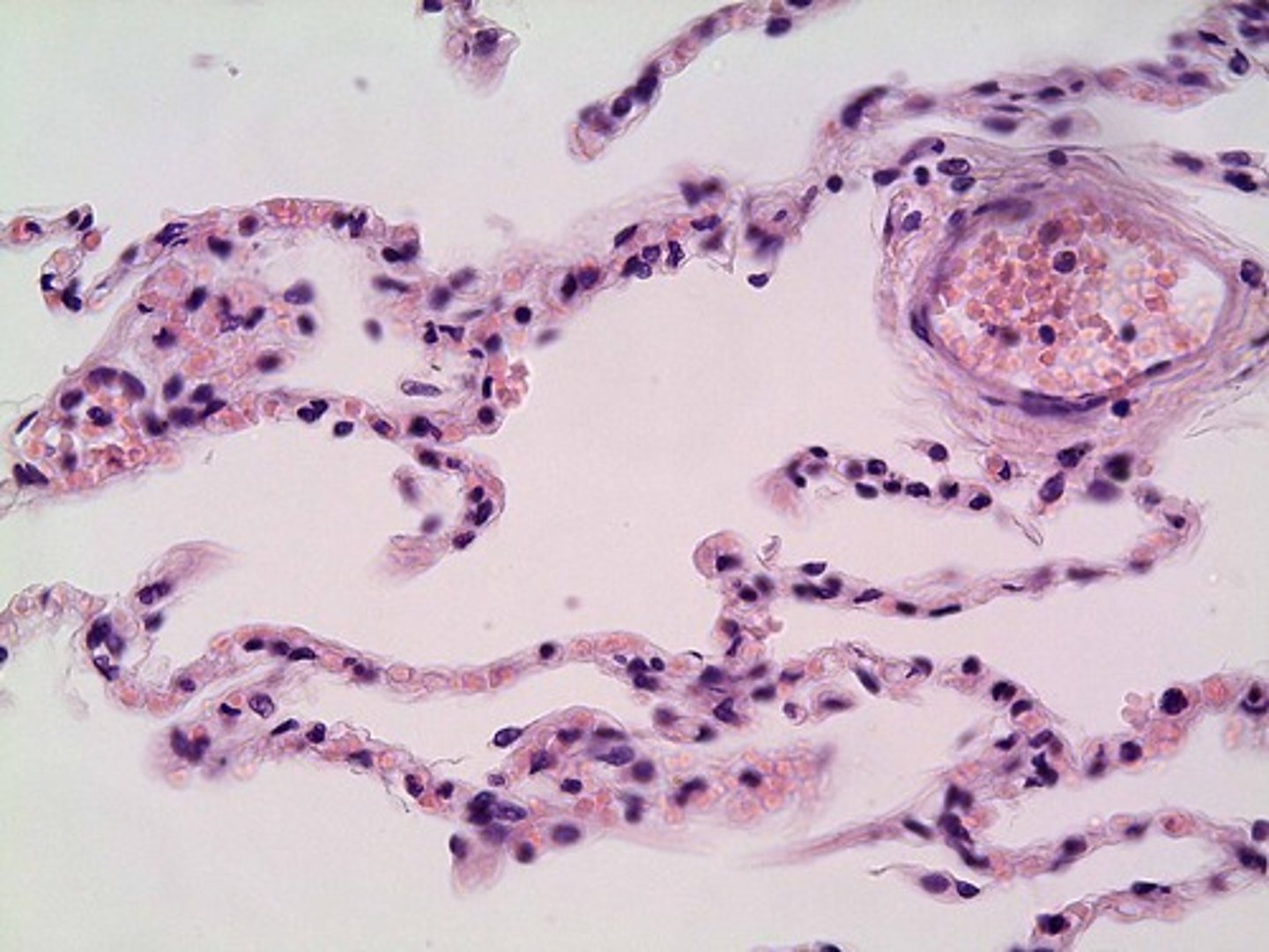
look like clumps of grapes and conduct gas exchange
What do alveoli look like and what is the function?
salivary glands, liver, gallbladder, pancreas
accessory organs of the digestive system
mouth, esophagus, stomach, small/large intestine
major organs of the digestive system
Oral cavity (mouth), pharynx, esophagus, stomach, small intestine, large intestine, rectum, and then anus.
What is the order that food passes the digestive system?
function: produces saliva that contains important enzymes ex. Amylase (helps break down starch); Lipase (helps break down lipids)
structure: salivary gland in the cheek; anterior to the ear;ACCESSORY organ of digestive system
parotid gland function and structure
function: transports food into the stomach; muscles contract to allow peristalsis; glands secrete mucous to moisten food
esophagus function
function: stores and digests food; breaks it up mechanically; chemical digestion of proteins and fat using acid and enzymes; produces chyme
stomach function
function: synthesizes and secretes bile; detoxifies blood and drugs, stores nutrients
structure: upper right quadrant; ACCESSORY organ of digestive system
liver function and structure
function: secretes (concentrates) bile and stores it until needed for digestion
structure: a muscular sac attached to the liver; ACCESSORY organ of digestive system
gallbladder function and structure
function: transports bile to duodenum and empties the bile to aid in digestion
structure: duct formed by common hepatic ducts and cystic ducts
common bile duct function and structure
function: protection, fat storage, infection control
greater omentum function
function: stabilizes stomach
lesser omentum function