LU 5 : Common Eye Problems in Adult
1/28
There's no tags or description
Looks like no tags are added yet.
Name | Mastery | Learn | Test | Matching | Spaced | Call with Kai |
|---|
No analytics yet
Send a link to your students to track their progress
29 Terms
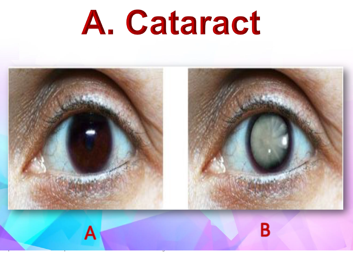
What is cataract ?
A cataract is the opacity of the lens that impair the vision
Explain the pathophysiology of cataract
it occurs due to degenerative opacification of the crystalline lens at any age
it can develop in both eyes and usually one eye is more compromised
visual impairment usually progress at the same rate in both eyes
What are the risk factors for cataract ?
Aging
Myopia
Infection
Aspirin
Cigarette smoking
Chemical burns
Corticosteroids
Diabetes
Poor nutrition
Blunt trauma
What are the types of cataract ?
i) Acquired Cataract
senile cataract - age related
pre-senile cataracts
traumatic cataracts
secondary cataracts
ii) Congenital Cataract
Inborn cataract
Neonates’s genetic defects
What are the clinical manifestations of cataract ?
blurred vision : usually the first symptoms of cataract
glare : refers to the pain felt when the patient looks into the light
halos : vision of light is still there after looking away from bright light
double vision : early symptoms of cataract
poor night vision
What are the complications of cataract ?
increase intraocular pressure (IOP)
blindness
What are the assessment and diagnostic findings for cataract ?
snellen visual acuity test
opthalmoscopy
slit-lamp biomicroscopic examination
What are the treatment for cataract ?
glasses
better lighting
surgery
What are the surgical management for cataract ?
Depends on the maturity of the cataract
For dense cataract : Extracapsular cataract extraction (ECCE) removal of anterior lens and cortex
For immature cataract : Phacoemulsification cataract surgery (PHACO) employs ultrasound energy to emulsify the nucleus
What are the nursing intervention for cataract ? in terms of
preoperative health education
preoperative care
postoperative care
Preoperative health education : i) supine position ii) bring a responsible adult who can drive iii) eye patch will be on affected eye iv) do not touch eyes after surgery
Preoperative care : i) use of anticoagulant is withheld ii) administer dilating drop every 10 minutes for four doses at least 1 hour before surgery iii) antibiotic is given to prevent postoperative infection
Postoperative care : i) eye protection (discharge health education ii) administer medication such as steroid drops iii) recognize signs of complications such as red painful eyes
What are the discharge health education ?
Activities : i) do not do any strenuous activities for a few weeks ii) avoid rigorous exercises iii) do not drive iv) stay away from dusty areas
Protective eye patch : i) to prevent accidental rubbing or poking the eye ii) wear 24 hours after surgery, followed by worn at night for 1-4 weeks iii) wear sunglasses when explore to bright light
Expected side effects : i) slight morning discharge and some redness may be expected for a few days ii) use clean, damp washcloth may be used to remove slight morning eye discharge
Notify the physician if new floaters in vision, decrease in vision or pain
What is Glaucoma ?
is a condition marked by high intraocular pressure (IOP) that damages the optic nerve
normal IOP range is 10-21 mmHg
persistent IOP is above 21 mmHg could be suspicious of glaucoma
Where does fluid forms / location of aqueous formation ?
ciliary body of the eye
Where does fluid exits / location of aqueous drainage ?
trabecular meshwork
What are the types of glaucoma ?
congenital (can manifest anytime from birth)
inherited (cause unknown)
secondary (due to other cause or trauma)
What are the risk factors and complications of glaucoma ?
risk factors : diabetes. hypertension, high myopia, men
complications : progressive optic nerve damage causing loss of vision
What are the types of primary glaucoma ?
Chronic open-angle glaucoma or Primary Open-Angle Glaucoma (POAG)
Acute angle closure glaucoma or Primary Angle-Closure Glaucoma (PACG)
Explain the pathophysiology of Chronic open-angle glaucoma
obstruction to outflow of aqueous humor through the trabecular meshwork into Schlemm’s canal leads to increase IOP
Its usually bilateral.
Increased IOP eventually destroys optic nerves that lead to blindness
Explain the pathophysiology of Acute closed-angle glaucoma
the intraocular pressure rises rapidly due to a sudden blockage of the trabecular meshwork
typically involves sudden, complete, unilateral closure with pupil dilation stimulated by a dark environment, emotional stress
it is an acute, painful condition that can cause permanent eye damage within several hours
What are the sign and symptoms of chronic open-angle and close-angle glaucoma ?
Chronic open-angle glaucoma : i) no early symptoms ii) gradual loss of peripheral vision iii) mildly aching eyes iv) halos around lights
Acute close-angle glaucoma : i) headache ii) pain iii) increased IOP iv) nausea and vomiting v) loss of peripheral vision
What are the laboratory and diagnostic done for glaucoma ?
measure IOP
check visual field
applanation tonometry using tonopen
slit lamp examination
What are the medical management for acute-angle-closure glaucoma ?
Beta-adrenergic blockers : to reduce the formation of aqueous humor in the ciliary body in the eye (eg Timolol Maleate)
Cholinergic agents (topical) : short term management for glaucoma with papillary block (eg Topical pilocarpine)
Alpha-2-adrenergic agonists (topical) : reduce IOP (eg Alphagan or Brimonidine)
Osmotic diuretics : reduce IOP (eg : oral glycerol and IV mannitol)
Carbonic anhydrase inhibitors : reduce IOP (eg Acetazolamide (Diamox)
Prostaglandin : reduce IOP (eg Latanoprost (Xalatan)
What are the surgical management for glaucoma ?
Ophthalmic laser surgery : primary treatment to lower IOP
Trabeculectomy : to allow drainage
What are the patient health education for glaucoma ?
Educate patient on the importance of lifelong medication
adherence
Demonstrate proper eye drop administration technique
Advise on lifestyle modifications to avoid increasing IOP
Schedule and reinforce the need for regular follow-up
appointments
Assess for and address side effects or complications of
treatment
What is diabetic retinopathy ?
high blood sugar levels that may injure the blood vessels of the retina
What are the two stages of diabetic retinopathy ?
NDPR (non-proliferative diabetic retinopathy) : i) early stage of diabetic eye disease ii) tiny blood vessels leak iii) can cause macular ischemia
PDR (proliferative diabetic retinopathy) : i) advanced stage of diabetic eye disease ii) retina start growing new blood vessels iii) very serious and can affect the central and peripheral vision
What are the sign and symptoms of diabetic retinopathy ?
loss of visual acuity
floaters
flashes of light
eye pain
What are the complication of diabetic retinopathy ?
decrease in vision
maculopathy
retinal hemorrhage
vitreous hemorrhage
retinal detachment
IOP
blindess
What are the treatment for diabetic retinopathy ?
laser photocoagulation
surgery