A&P 1 axial skeleton
1/203
There's no tags or description
Looks like no tags are added yet.
Name | Mastery | Learn | Test | Matching | Spaced | Call with Kai |
|---|
No analytics yet
Send a link to your students to track their progress
204 Terms
What is this cranial bone?
Frontal bone is the bone that forms the forehead and the upper parts of the eye sockets.

What is this?
Coronal Suture is the fibrous joint that connects the frontal bone to the parietal bones, running across the top of the skull.

What is this? R/L
Squamous suture is the fibrous joint that connects the temporal bone to the parietal bone, located on the side of the skull.

What is this?
Sagittal suture is the fibrous joint that connects the two parietal bones, running along the midline of the skull from front to back.

What is this?
Lambdoid suture is the fibrous joint that connects the occipital bone to the parietal bones, located at the back of the skull.
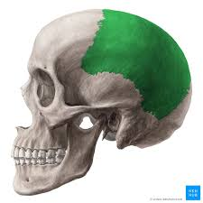
What cranial bones are these? R/L
Parietal bones are two bones that form the top and sides of the skull, playing a crucial role in protecting the brain.

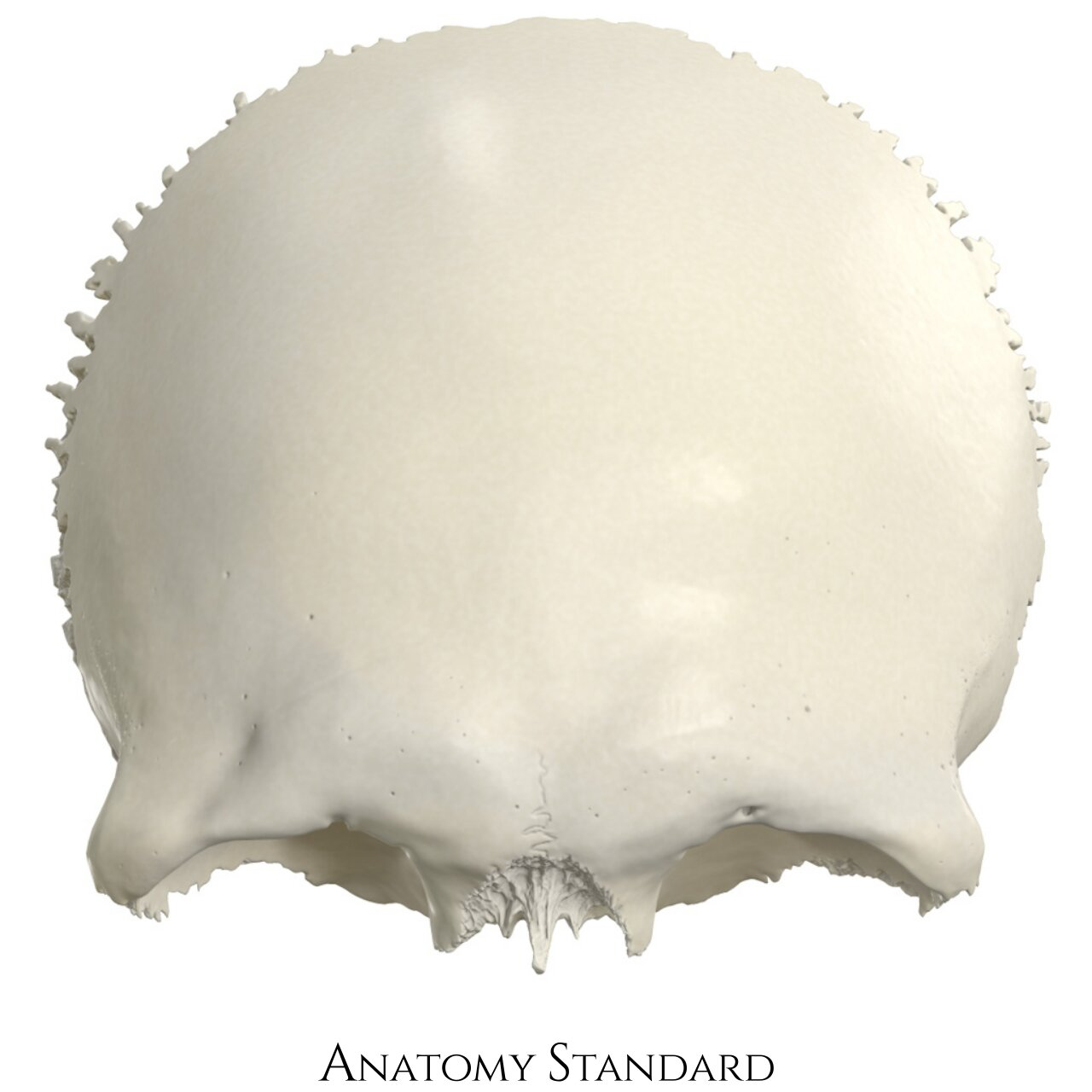
Which cranial bone is this?
Occipital bone is the bone located at the back of the skull, forming the base of the cranium and containing the foramen magnum.

Name these cranial bones R/L
temporal bones (2) located on the sides of the skull

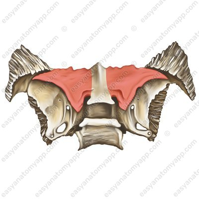
What is this irregular cranial bone?
Sphenoid bone is a complex bone that forms part of the base of the skull and helps to connect the cranial and facial bones.

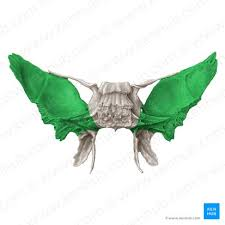
Name this irregular cranial bone
ethmoid bone is a light and spongy bone located between the eyes, forming part of the nasal cavity and the orbits.

What is this bone? R/L
Nasal bone is a small bone that forms the bridge of the nose, contributing to the structure and shape of the face.
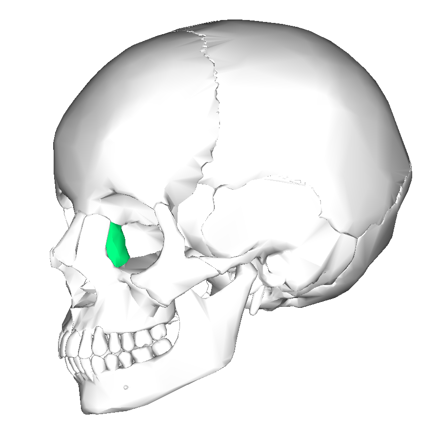
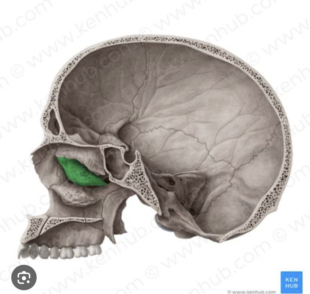
What is this bone? R/L
The lacrimal bone is a small bone located in the medial wall of the orbit, supporting the tear ducts.
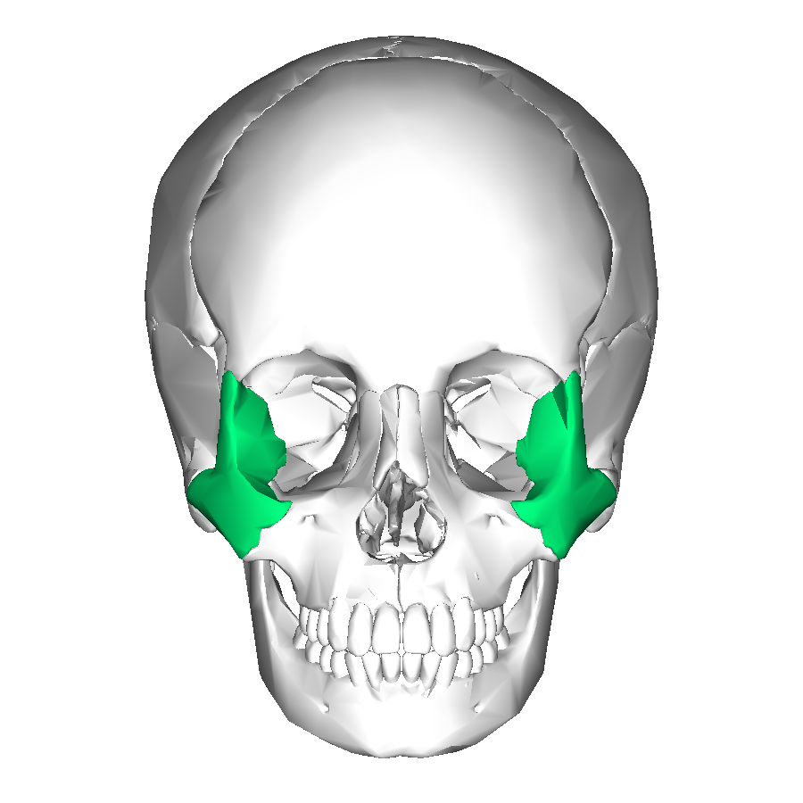
Name this bone R/L
the zygomatic bone is a bone that forms the prominence of the cheek and part of the lateral wall and floor of the orbit.

What is this hole? R/L of what bone
The infraorbital foramen is two openings in the maxilla that allow nerves and blood vessels to pass to the face.

What is this bone?
The maxilla is the upper jaw bone that holds the upper teeth and forms part of the orbits.

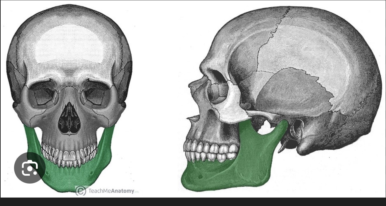
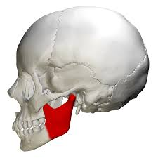
Name this bone
The mandible is the lower jaw bone that holds the lower teeth and is the only movable bone of the skull.
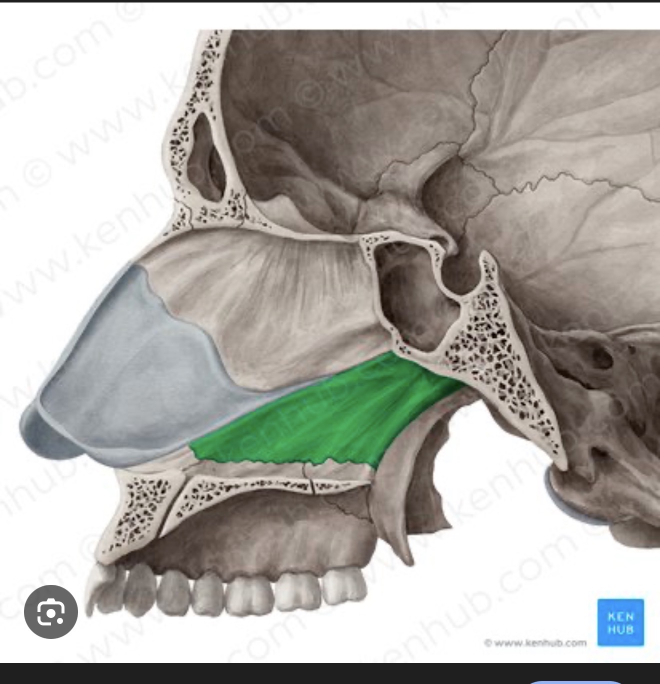
What is this bone?
The vomer is a thin, flat bone that forms part of the nasal septum, separating the nasal cavities.

What is this bone?
Inferior nasal concha is a small, thin bone located in the nasal cavity that helps to filter and humidify the air we breathe.

What is this called?
Inferior orbital fissure is a gap in the skull located between the maxilla and sphenoid bones, serving as a passageway for nerves and blood vessels.

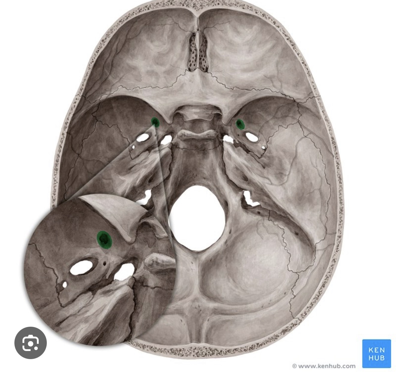
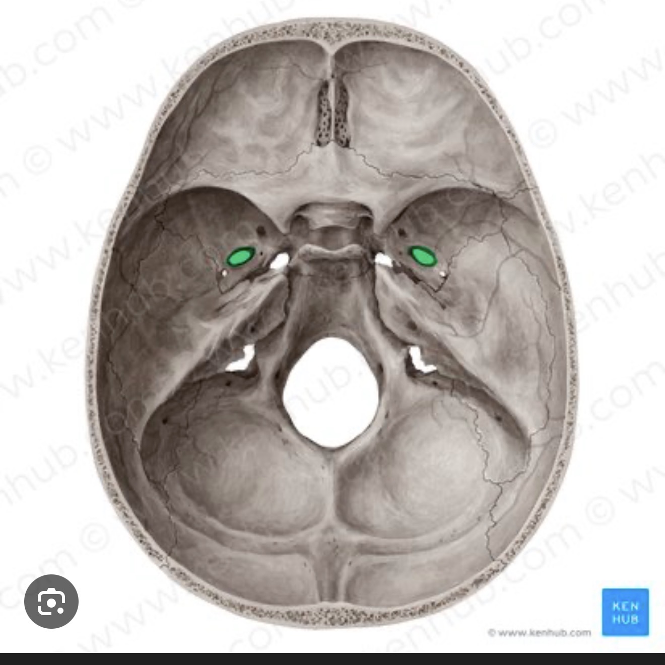
Name this space
Optic canal is a passage in the skull that allows the optic nerve and ophthalmic artery to enter the orbit.

Name this structure
superior orbital fissure is a passage in the skull that connects the middle cranial fossa to the orbit, allowing the passage of cranial nerves and blood vessels.

What is this structure?
Supraorbital margin is the bony ridge located above the orbit, providing support and protecting the eye.
Name this hole? R/L
Supraorbital foramen is an opening in the skull that transmits the supraorbital nerve and artery.
What is this structure?
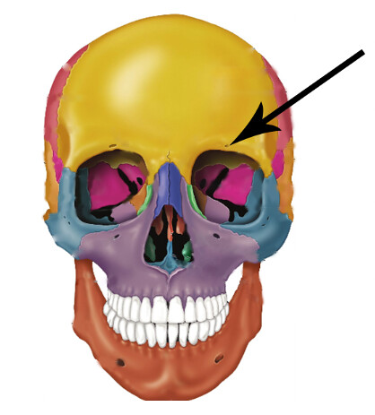
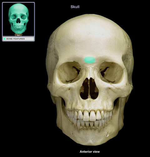
Glabella is the smooth region located between the eyebrows and above the nasal bridge, an important landmark on the frontal bone.

What is this hole?
Mental foramen is an opening in the mandible that allows for the passage of the mental nerve and blood vessels to the chin and lower lip.
What is this structure?
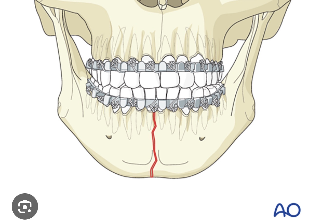
Mandibular symphysis is the midline fusion of the two halves of the mandible, providing structural support and a point of attachment for the lower lip.
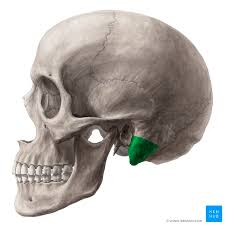
Name this structure R/L?
zygomatic process is a bony projection of the temporal bone that articulates with the zygomatic bone, contributing to the formation of the cheekbone.
What is this?
The frontal sinus is an air-filled cavity located within the frontal bone, aiding in voice resonance and lightening the weight of the skull.
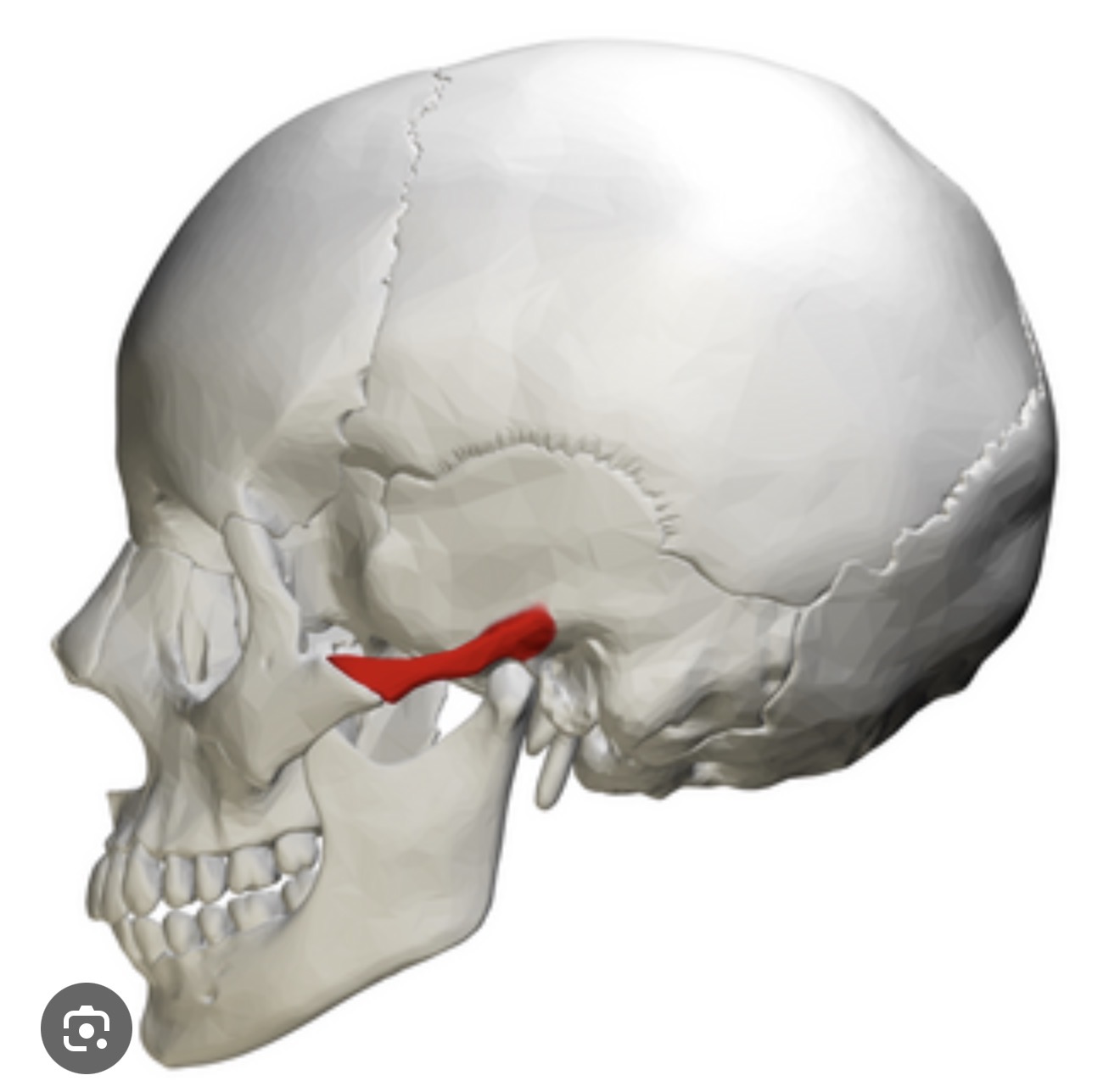
What is this ? R/L
The mastoid process is a bony protuberance located behind the ear, serving as an attachment site for neck muscles and containing air cells that can connect to the middle ear.
what is this? R/L
The external auditory meatus is a canal that leads from the outer ear to the eardrum, allowing sound waves to enter the ear.
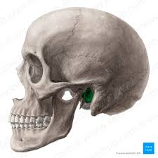
what is this? R/L
mandibular fossa The mandibular fossa is a depression in the temporal bone that articulates with the mandible, forming the temporomandibular joint (TMJ). It is crucial for jaw movement and stability during chewing.
what is this ? R/L
styloid process The styloid process is a slender, pointed projection from the temporal bone, serving as an attachment point for ligaments and muscles associated with the hyoid bone and tongue.

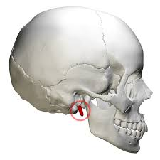
What is this? R/L
stylomoid foramen The stylomastoid foramen is an opening in the temporal bone that allows the facial nerve (cranial nerve VII) to exit the skull, contributing to facial expression and sensation.
what is this ? R/L
internal acoustic meatus The internal acoustic meatus is a canal in the temporal bone that transmits the vestibulocochlear nerve (cranial nerve VIII) and the facial nerve (cranial nerve VII) to the inner ear, playing a key role in hearing and balance.
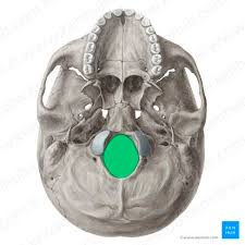
what is this?
foramen magnum The foramen magnum is a large opening at the base of the skull through which the spinal cord connects to the brain, allowing for the passage of nerve connections and blood vessels.
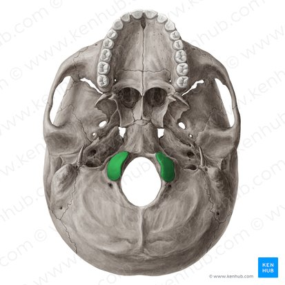
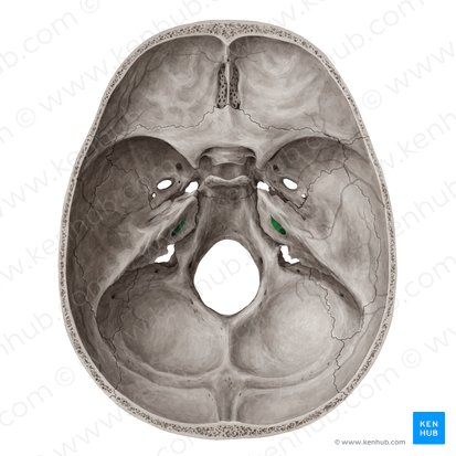
what is this ? R/L
occipital condyle The occipital condyle is a pair of rounded projections located on the occipital bone of the skull. They articulate with the first cervical vertebra (atlas), allowing for nodding movements of the head.
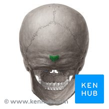
what is this?
external occipital protuberanceThe external occipital protuberance is a prominent bony bump located on the posterior aspect of the occipital bone. It serves as an attachment point for muscles and ligaments of the neck.
what is this ? R/L
greater wingThe greater wing is a large, wing-like structure of the sphenoid bone that extends laterally from the body of the sphenoid and contributes to the formation of the eye socket and the lateral skull.
what is this ? R/L
leeser wing The lesser wing is a small, wing-like structure of the sphenoid bone, located superior to the greater wing. It forms part of the orbit and serves as a boundary between the anterior and middle cranial fossae.

what is this ?
sella turcica The sella turcica is a saddle-shaped depression in the sphenoid bone, located at the base of the skull. It houses the pituitary gland and is bordered by important cranial structures.
what is this? R/ L
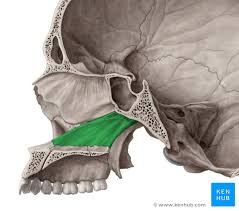
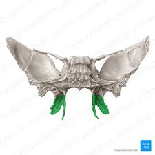
pterygoid process The pterygoid process is a pair of bony projections from the sphenoid bone that extend downward and serve as attachment sites for muscles involved in chewing. They consist of a medial and lateral plate, contributing to the lateral walls of the nasal cavity and the space behind the upper jaw.

what is this?
sphenoidal sinus The sphenoidal sinus is a pair of air-filled spaces located within the body of the sphenoid bone. They are situated posterior to the nasal cavity and drain into the nasal passage, playing a role in reducing the weight of the skull and contributing to the resonance of the voice.

what is this?
crista galli The crista galli is a thin, triangular process of the ethmoid bone that projects upward into the cranial cavity. It serves as an attachment point for the falx cerebri, a membrane that helps stabilize the brain within the skull.
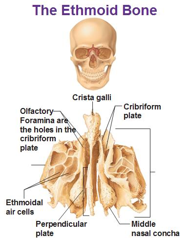
what is this ?
cribriform plate: The facial structure of the ethmoid bone, located in the anterior cranial fossa, that allows passage of olfactory nerves.
what is this?
perpendicular plate A vertical bony structure that divides the nasal cavity into right and left halves, located in the nasal septum.

what is this?
ethmoidal sinus A pair of small air-filled spaces located in the ethmoid bone, contributing to the nasal cavity and involved in the production of mucus.
what is this?
mandible bone Anatomical structure of the jaw.
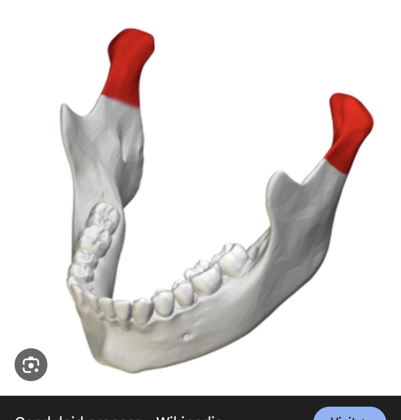
what is this?R/L
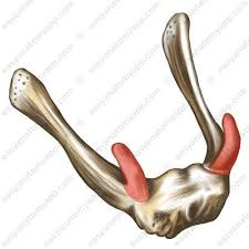
mandibular condyle The rounded end of the mandible that articulates with the temporal bone to form the temporomandibular joint (TMJ).
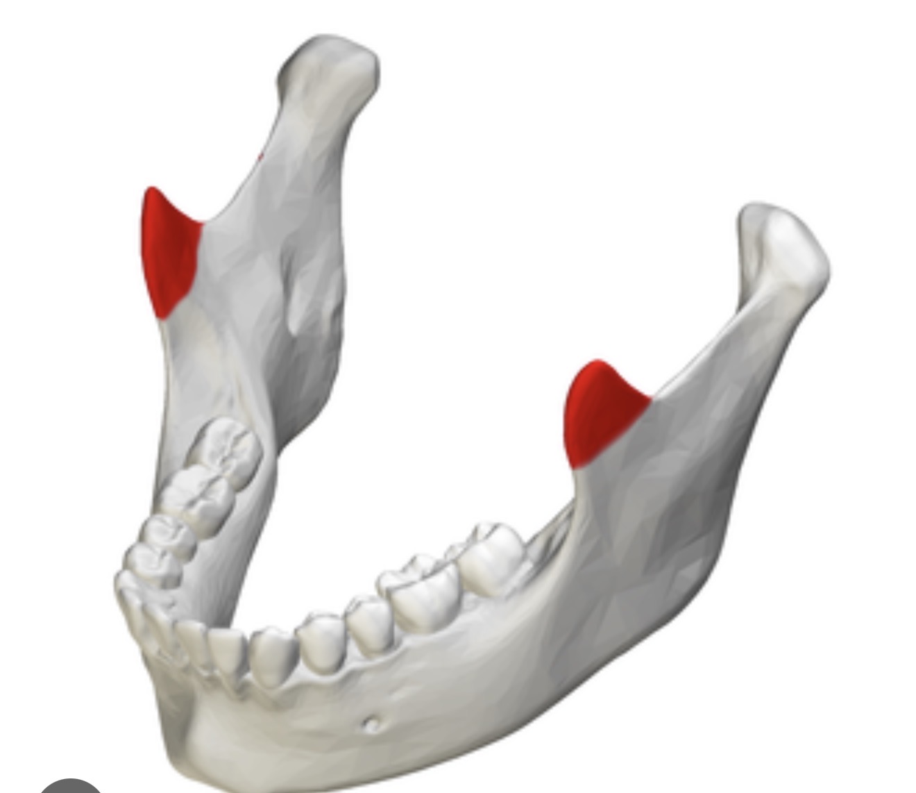
what is this? R/L?
coronoid process A triangular projection on the mandible, serving as an attachment site for the temporalis muscle.
what is this? R/L?
ramus The vertical portion of the mandible that connects the body of the mandible to the skull.

what is this? R/L
mandibular angle The posterior angle formed by the body and ramus of the mandible, providing structural support and muscle attachment.

what is this? R/L
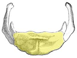
body The horizontal portion of the mandible that holds the lower teeth and provides structure to the jaw.
what is this ?
mental foramen An opening in the anterior mandible for passage of nerves and blood vessels.
what is this? R/L
alveolar margin The upper and lower borders of the mandible that contain the sockets for the teeth.
what is this?
mandibular foramen The ridge on the mandible that contains the sockets for the lower teeth.
what is this?
maxillae bone The two bones that form the upper jaw and hold the upper teeth, also contributing to the formation of the orbits and nasal cavity.

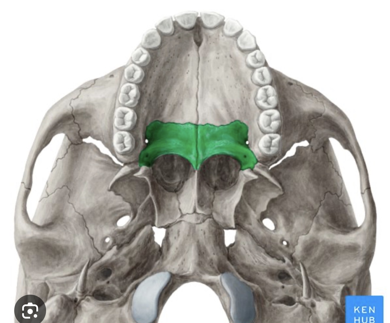
what is this? R/L
palatine process The horizontal extension of the maxillae that forms the anterior part of the hard palate, separating the oral cavity from the nasal cavity.
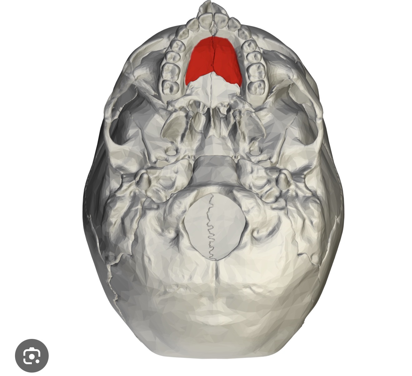
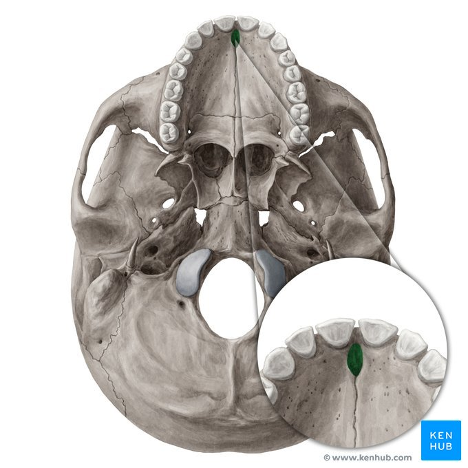
what is this?
incisive fossa A depression in the maxilla, located anterior to the palatine process, that serves as a passage for the nasopalatine nerve and vessels.

what is this ?
maxillary sinus An air-filled space within the maxilla that helps to lighten the weight of the skull, also contributing to the respiratory system.
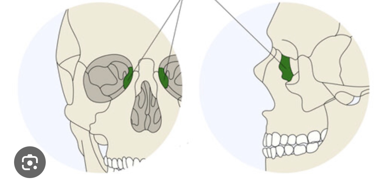
what is this? R/L
lacrimal bone A small bone forming part of the eye socket, located between the frontal process of the maxilla and the ethmoid bone.
what is this? R/L
palatine bone A bone that forms the posterior part of the hard palate and part of the floor of the nasal cavity.
what is this R/L
inferior nasal conchae Thin, curled bone structures located in the nasal cavity that help filter and humidify inhaled air.
what is this ? R/L
vomer bone A thin, flat bone forming the lower part of the nasal septum, separating the left and right nasal cavities.

what is this?
frontal sinus. A singular bone located in the nasal cavity that forms part of the nasal septum, contributing to the separation of the nasal passages.
what is this?
foramen rotundum is a circular opening in the skull that allows the passage of the maxillary nerve.
what is this?
foramen ovale A round opening in the sphenoid bone that allows the passage of the maxillary nerve, a branch of the trigeminal nerve (cranial nerve V).
what is this?
foramen spinosum A small oval opening in the sphenoid bone that allows the passage of the middle meningeal artery and vein.
what is this?
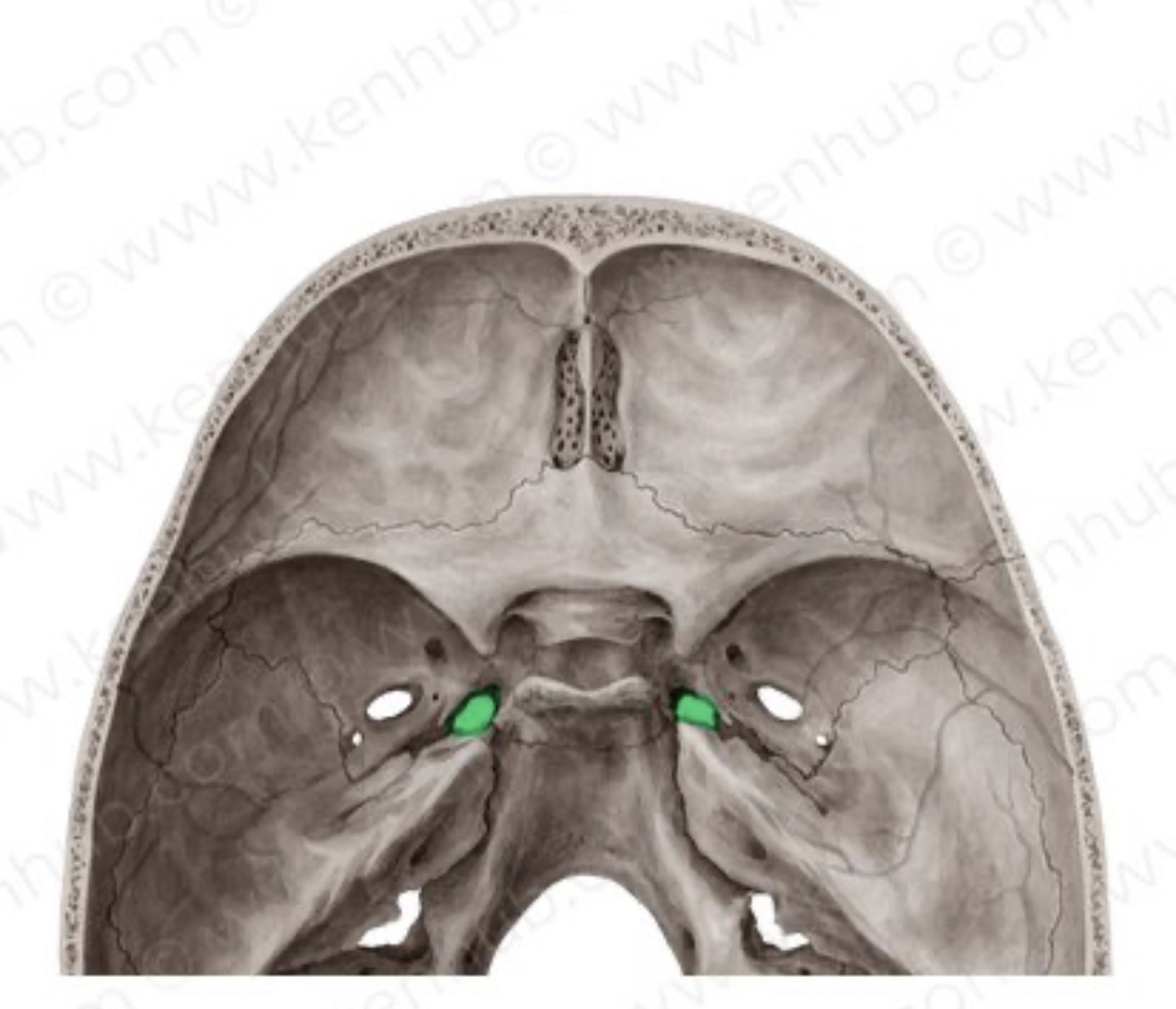
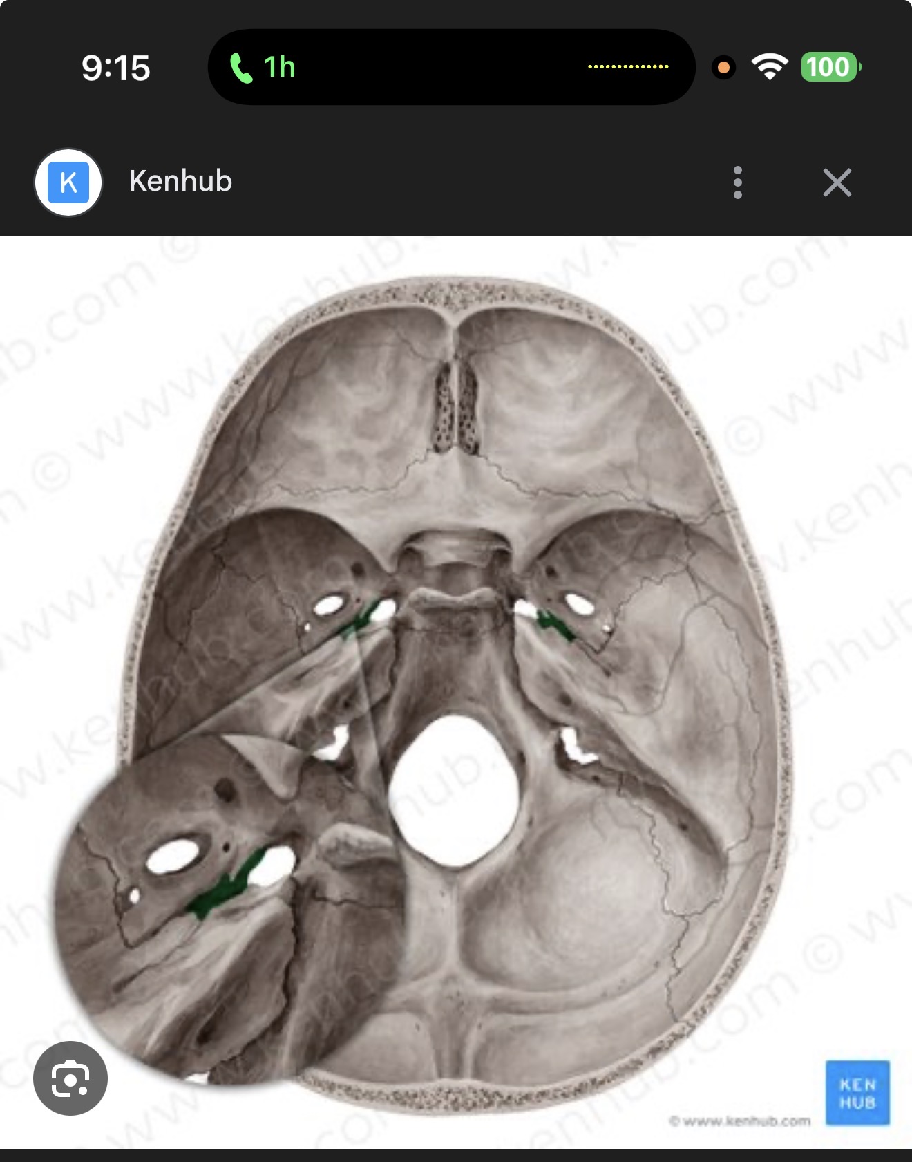
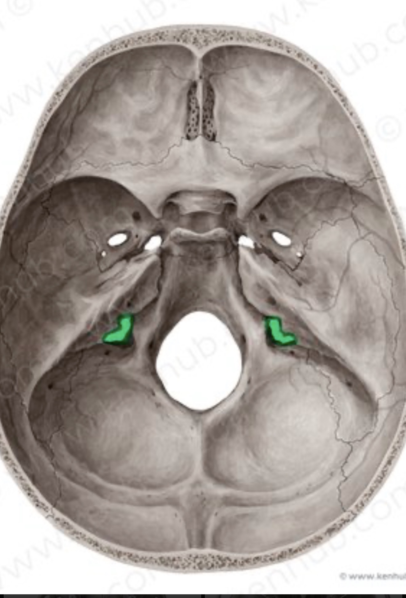
foramen lacerum An irregularly shaped opening situated between the temporal, sphenoid, and occipital bones, providing passage for nerves and vessels.
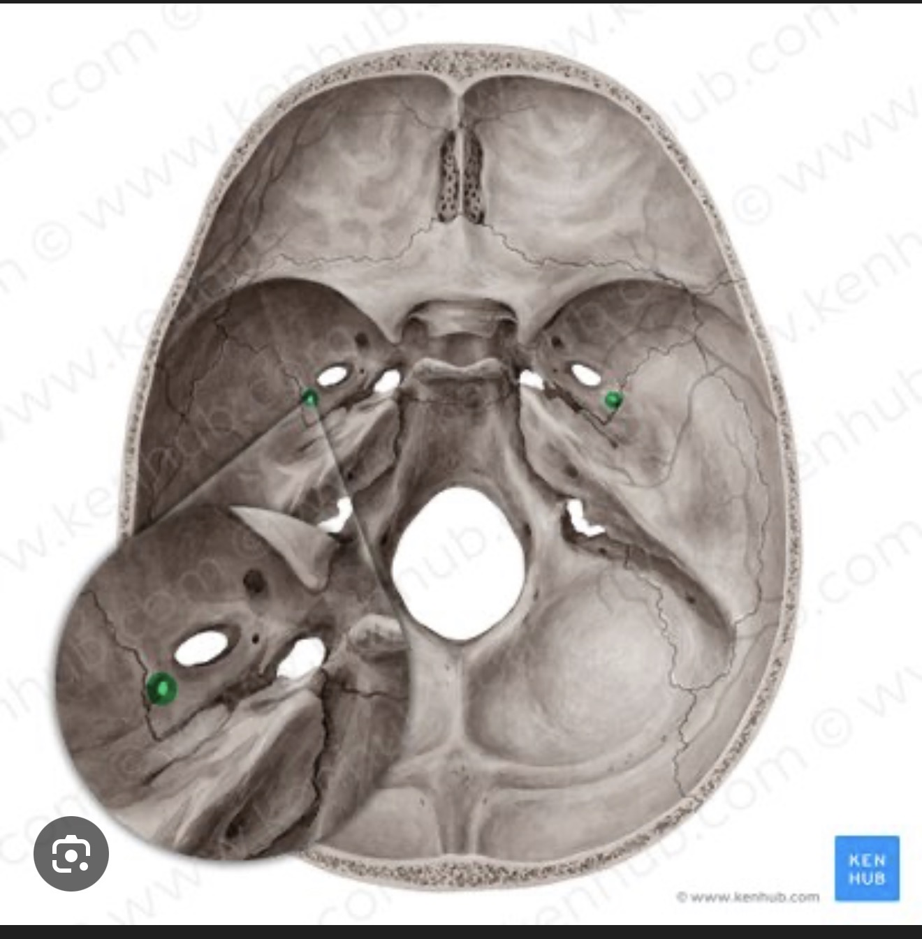
what is this? R/L
carotid canal A passageway in the temporal bone that allows the internal carotid artery and sympathetic nerves to enter the cranial cavity.
what is this? R/L
jugular foramen The jugular foramen is a large opening located between the temporal and occipital bones that allows passage for the internal jugular vein and cranial nerves IX, X, and XI.
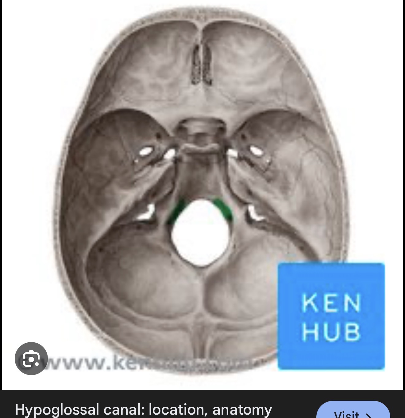
what is this? R/L
hypoglossal canal A passageway in the occipital bone that transmits the hypoglossal nerve (cranial nerve XII) from the brain to the neck.
what is this?
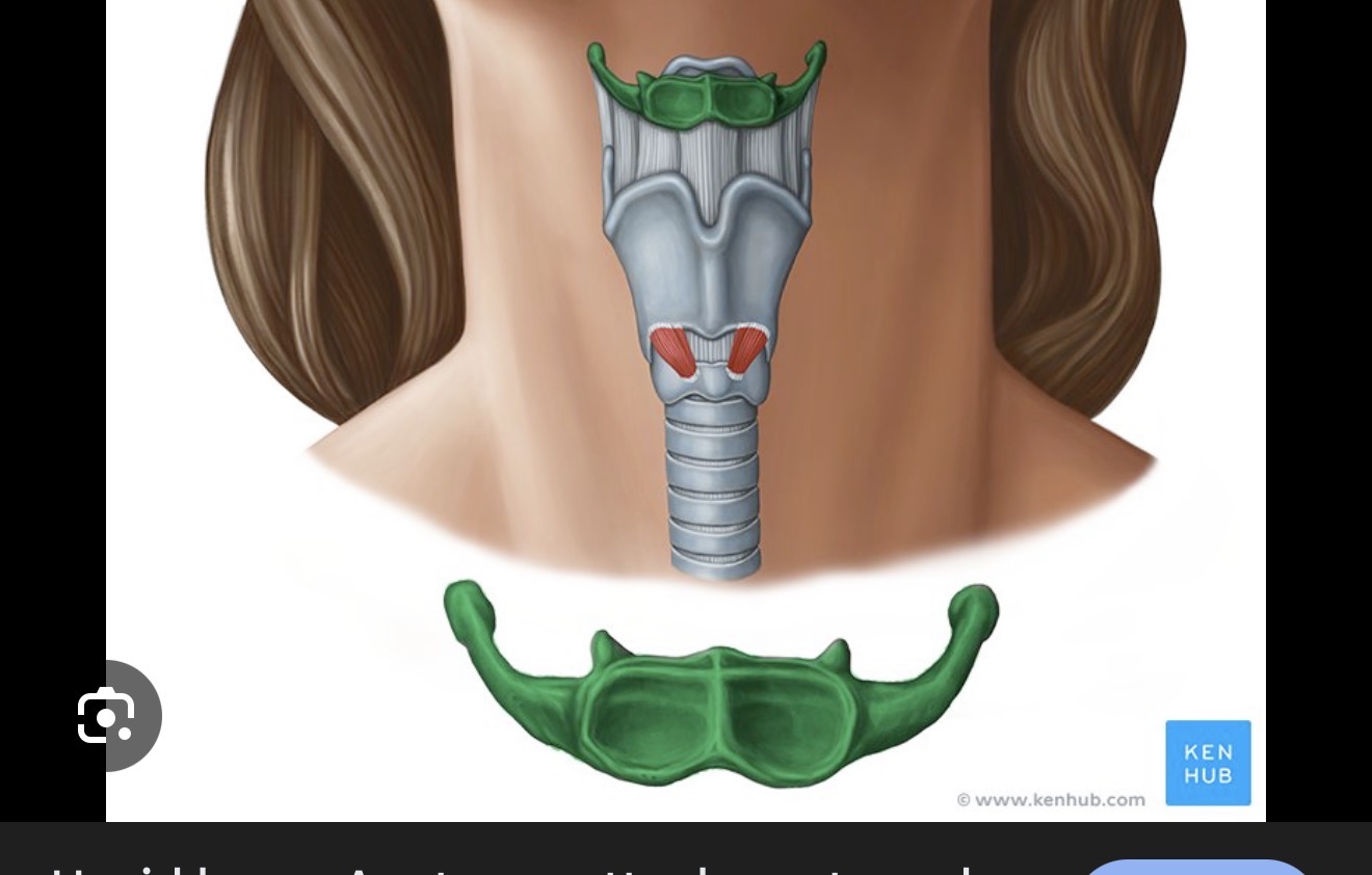
hypoid bone A U-shaped bone in the neck that supports the tongue and serves as an attachment point for muscles.

what is this? R/L
greater horn of hyoid The greater horn of the hyoid is the elongated projection of the hyoid bone that provides attachment for muscles and ligaments of the tongue and floor of the mouth.
what is this? R/L
lesser horn of hyoid bone The lesser horn of the hyoid bone is a small, conical projection on the hyoid that serves as an attachment point for muscles and ligaments, aiding in the movement of the tongue and larynx.
what is this ? R/L
body of hyoid bone The body of the hyoid bone is the central block-like structure that supports the greater and lesser horns and serves as an attachment point for muscles involved in tongue movement and swallowing.
what is this ?
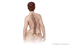
scoliosis is a condition characterized by an abnormal lateral curvature of the spine, often resulting in a C- or S-shaped appearance.

what is this?
kyphois is a condition defined by an excessive outward curvature of the spine, typically in the thoracic region, leading to a hunchback appearance.

what is this ?
lordosis is an excessive anterior curvature of the lumbar spine, creating a swayback appearance.
what is this?
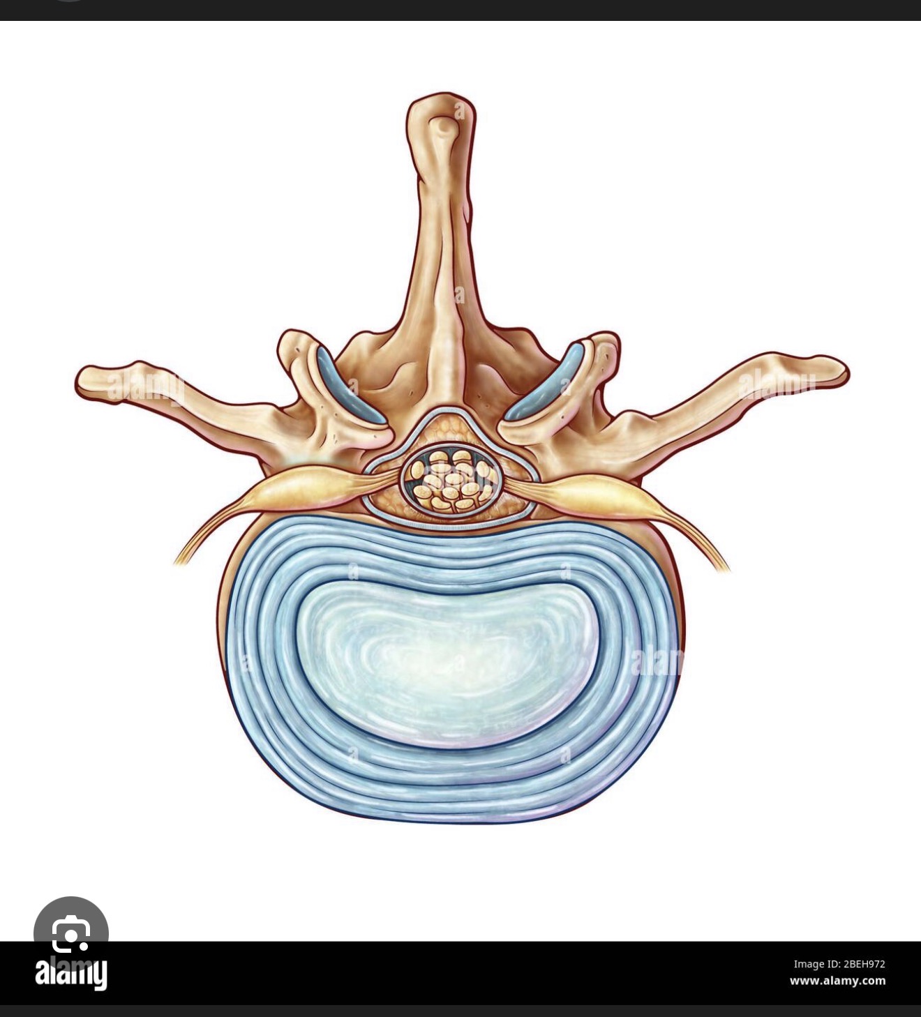
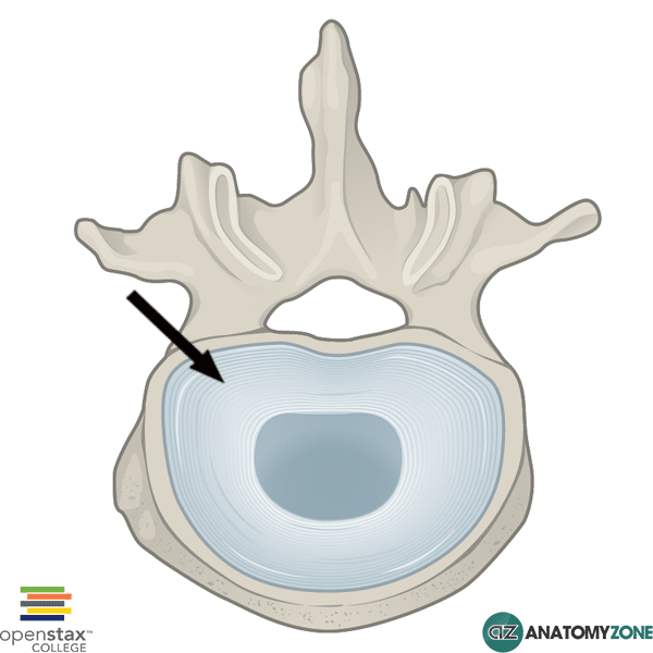
intervertebral disc is a fibrocartilaginous structure located between the vertebrae, serving as a shock absorber and allowing for movement in the spine.

what is this ?
nucleus pulposus of the intervertebral disc consists of a gel-like center surrounded by an outer annulus fibrosus, providing cushioning and support between vertebrae.
what is this?
annulus fibrosus of the intervertebral disc is the tough, outer layer that encases the nucleus pulposus, providing stability and support to the intervertebral disc.
what is this?
herniated disc of the intervertebral disc is the tough outer layer that encases the nucleus pulposus, providing stability and structuring the intervertebral disc.
what is this?
vertebrae that has bulged out of its normal position, typically causing pain and discomfort due to pressure on spinal nerves.

what is this?
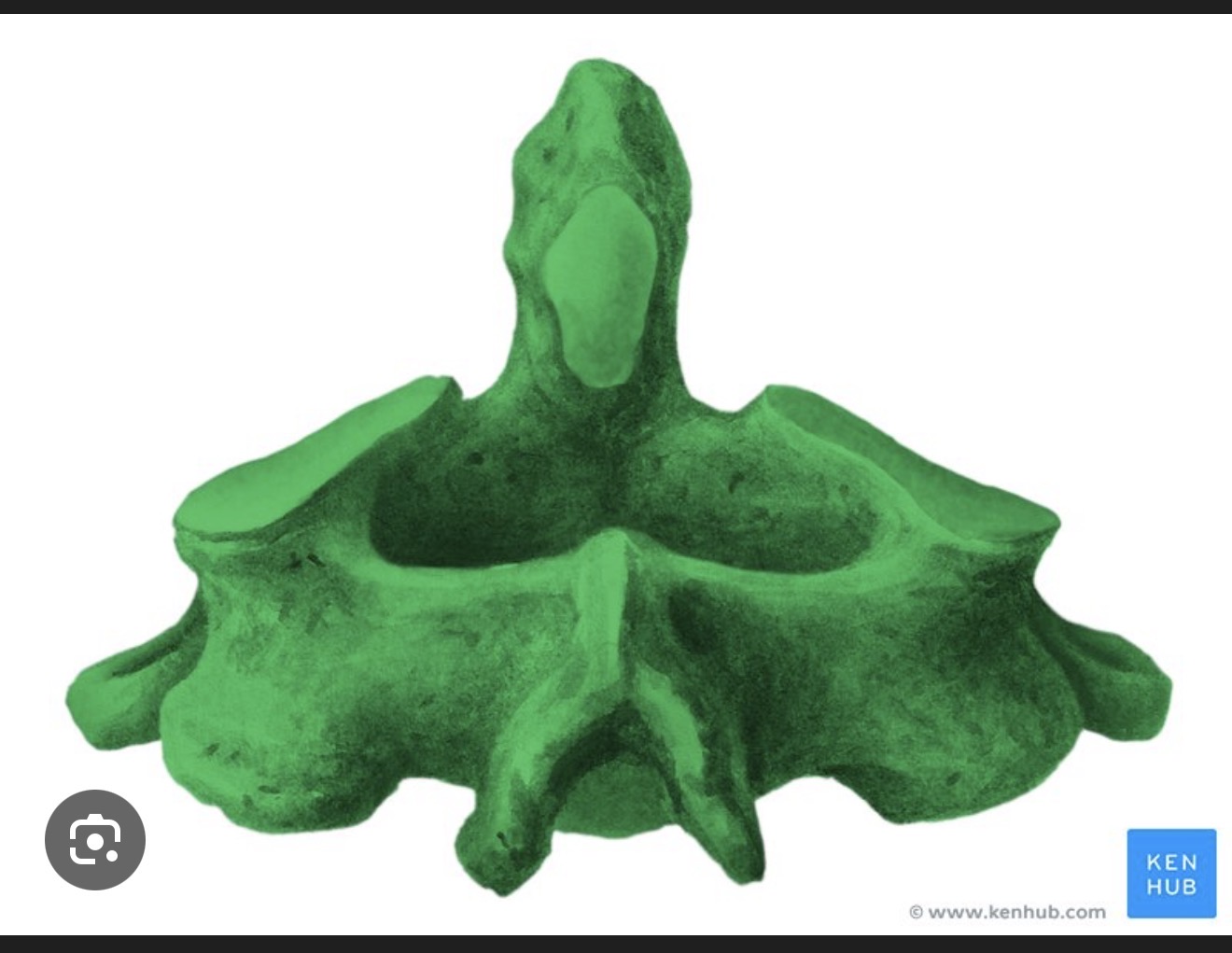
posterior spinous process of a vertebra, projecting posteriorly from the vertebral arch, providing attachment for ligaments and muscles.

what is this? R/L
transverse process is a bony projection on either side of a vertebra, serving as attachment points for muscles and ligaments.

what is this?
body of vertebrae that support the weight of the body and protect the spinal cord, composed of individual vertebrae stacked upon each other.

what is this?
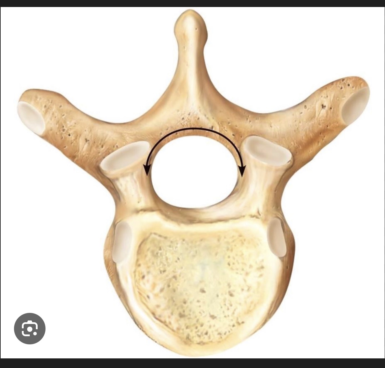
vertebral foramen is the large central opening in a vertebra that allows the passage of the spinal cord.

what is this?
vertebral arch is the bony arch formed from the posterior part of a vertebra, surrounding the vertebral foramen and providing attachment sites for ligaments and muscles.

what is this?
intervertebral foramen is the opening between adjacent vertebrae that allows spinal nerves and blood vessels to exit the spinal column.
what is this?
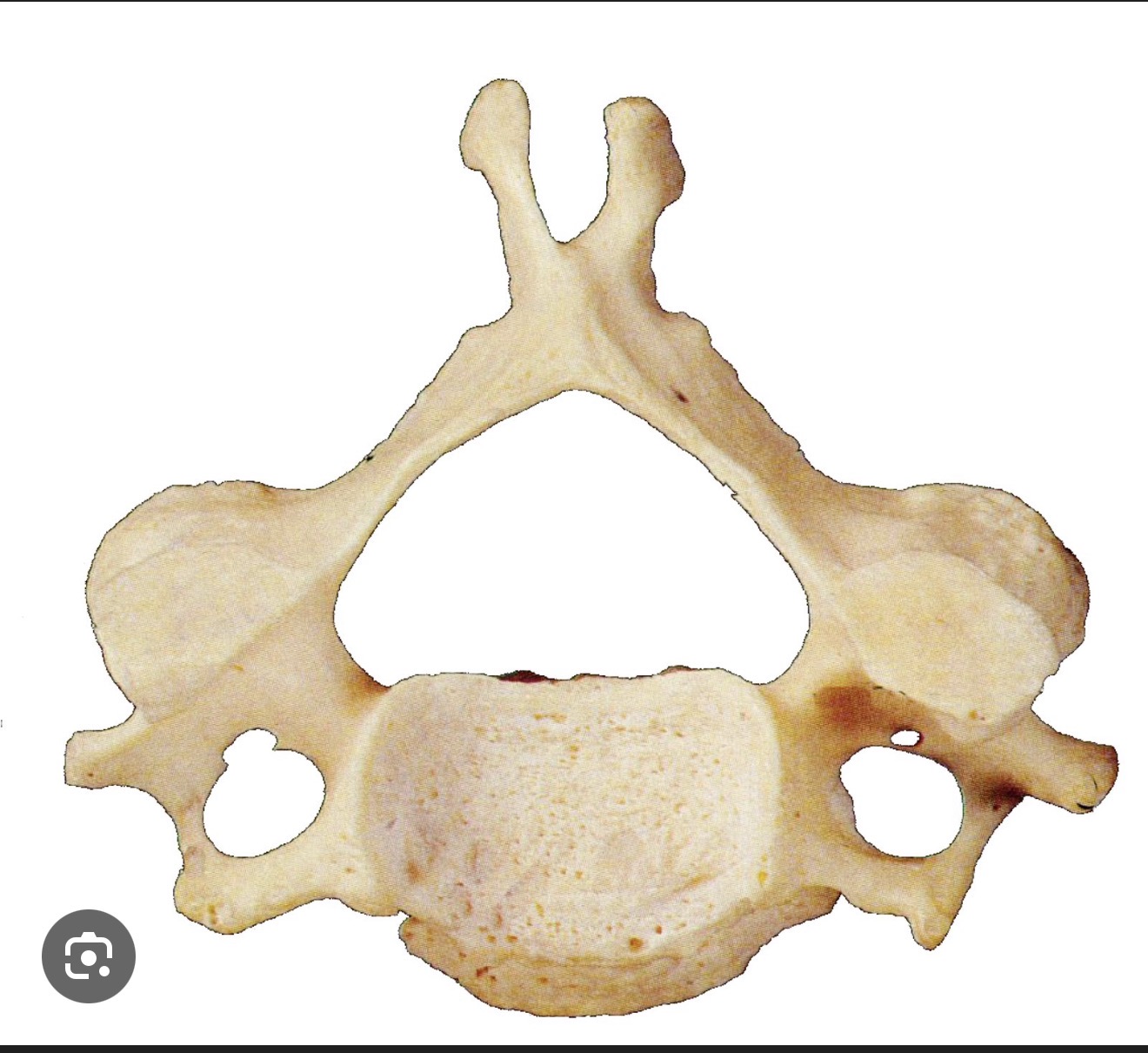
cervical vertebrae are the vertebrae located in the neck region, consisting of seven bones labeled C1 to C7, which support the head and allow for its movement.

what is this?
Transverse foramen of cervical vertebrae is a small opening in each cervical vertebra that allows the passage of the vertebral artery and vein, providing crucial blood supply to the brain.
what is this?
Atlas or C1 of the cervical vertebrae contains openings for vertebral arteries to pass through, providing blood supply to the brain.
what is this?
axis or C2 of the cervical vertebrae is the second cervical vertebra, which allows for rotation of the head and supports the atlas.

what is this?
thoracic vertebrae are the twelve vertebrae located in the upper and mid-back, differentiated by their articulation with the ribs and providing structural support and stability.

what is this?
coastal faucets of the thoracic vertebrae articulate with the ribs and provide support to the rib cage, allowing for flexibility and movement in the thoracic region.

what is this?
lumbarvertebrae are the five vertebrae located in the lower back, known for their robust structure and support for the upper body while allowing for flexibility and movement.

what is this? R/L
clavicles are the two bones that connect the arm to the body, providing structural integrity and support for shoulder movement.

what is this?
sternal end of clavicle

what is this?
The acromial end of clavicle

what is this?
conoid tubercle is a bony prominence located on the inferior surface of the lateral part of the clavicle, serving as an attachment point for the conoid ligament.