BIO (Book 1): Eukaryotic Cell Structure and Function
1/25
There's no tags or description
Looks like no tags are added yet.
Name | Mastery | Learn | Test | Matching | Spaced | Call with Kai |
|---|
No analytics yet
Send a link to your students to track their progress
26 Terms
Cell Theory
The cell theory states that the cell is a fundamental unit of structure, function and organization in all living organisms, and new cells are formed from other existing cells.
Conclusions of cell theory:
All living organisms are composed of one or more cells.
The cell is the most basic unit of structure in all organisms and is the smallest unit of life that are capable of replicating independently
All cells arise only from pre-existing cells.
Cell functions
1. Intake of raw materials, and from these,
2. Extract useful energy, and synthesise its own molecules
3. Grow in an organised manner
4. Reproduce after its own kind
5. Respond and adapt to the external environment
Cell size
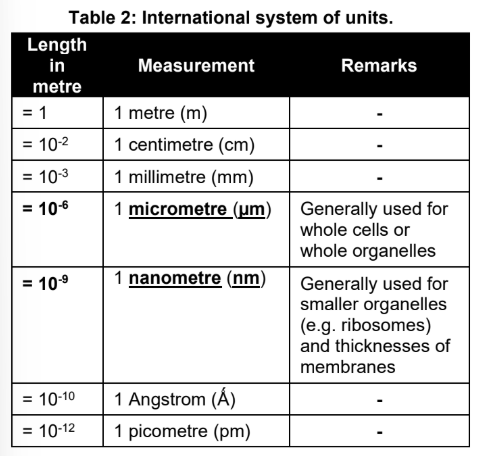
The size (diameter) of cells and subcellular organelles are usually expressed in micrometres or nanometers'
Lower limit of cell size:
It is determined by the minimum amount of space needed to contain the essential elements of its function
Upper limit of cell size:
It is determined by the surface area: volume ratio needed for exchange of materials between the cell and its environment.
As the size of the cell increases, the surface area to volume ratio decreases
The number of chemical exchanges that could be performed with the extracellular environment would be inadequate to maintain the cell, because most of its cytoplasm is relatively far from the outer membrane. Exchange with the extracellular environment is vital as substances like oxygen and nutrients can only enter the cell, and waste products can only leave, in this fashion.
Cell size is thus kept small and hence increase in organism size is accomplished by having a greater number of cells.
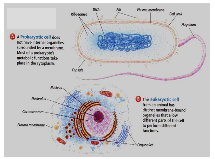
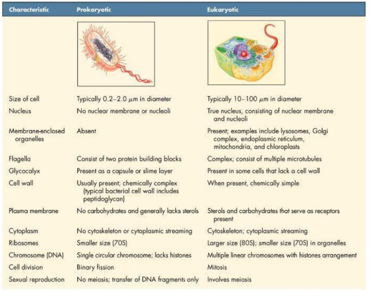
Prokaryotic cells
Prokaryotes do not have:
a) a true nucleus
b) membrane bound organelles
Size:
Prokaryotic cells are generally much smaller than eukaryotic cells
Eukaryotic cell (multicellular)
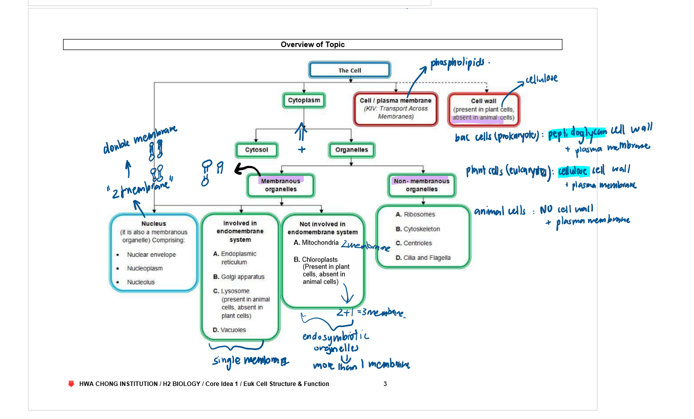
Eukaryotic cells have a plasma membrane on their outer surface, and extensive and elaborately arranged internal membranes that compartmentalise the cell.
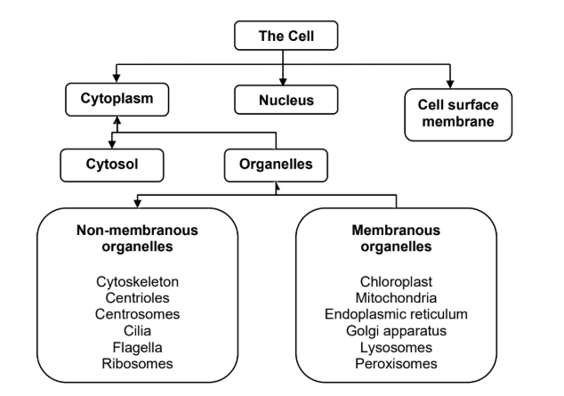
One important compartment is the nucleus, which contains the genetic material of the organism. The contents external to the nucleus are collectively known as the cytoplasm.
The cytoplasm consists of organelles, which are compartments that carry out various functions in the cell, as well as the cytosol, an aqueous matrix in which the organelles and nucleus are suspended.
Organelles can be subdivided into membranous and non-membranous organelles.
In addition to the internal and cell membranes, organisms such as plants and fungi have a cell wall
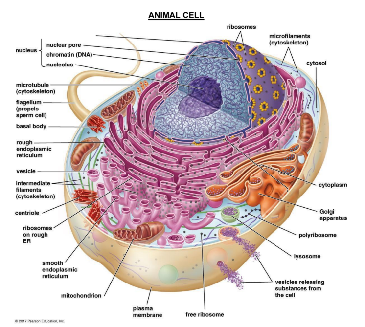
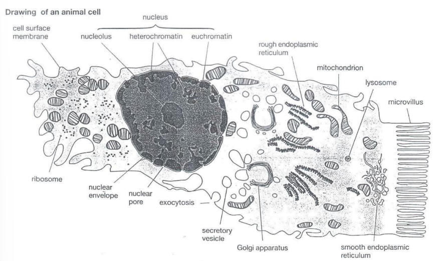
Animal cell
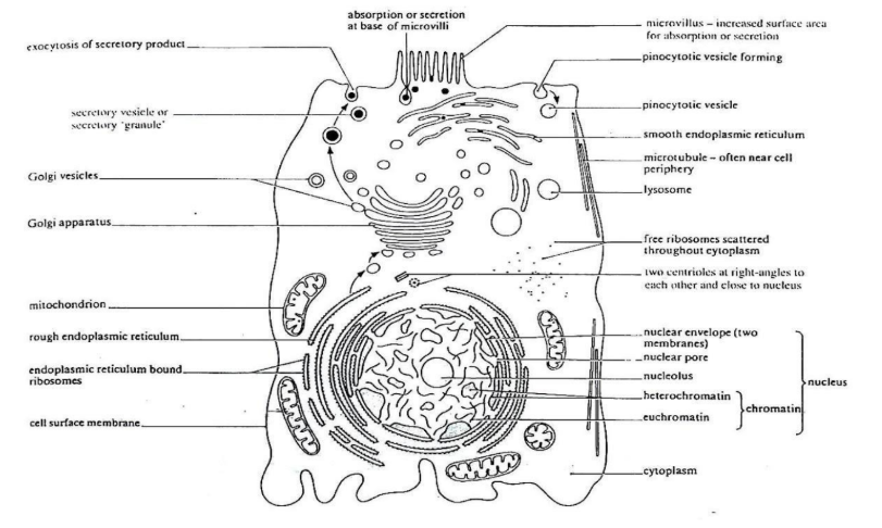
Animal cell
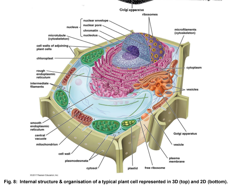
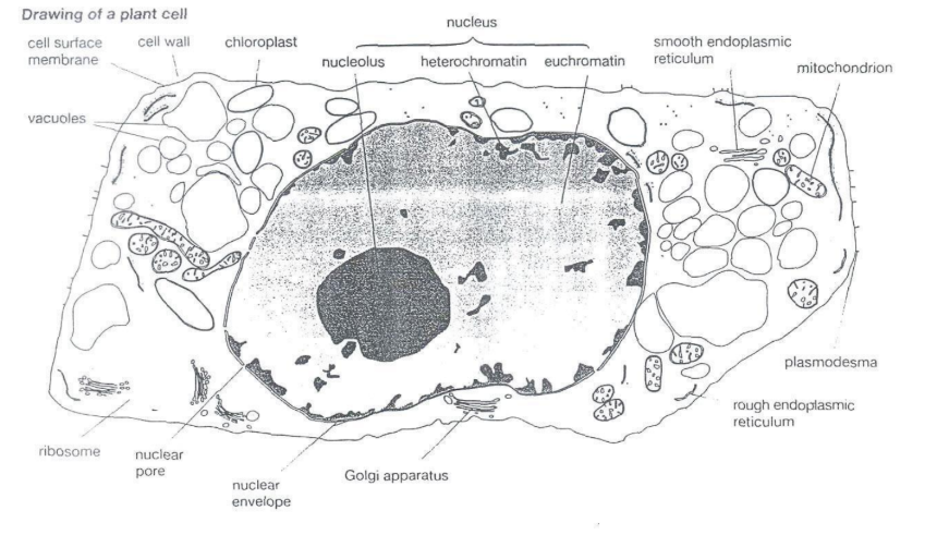
Plant cell
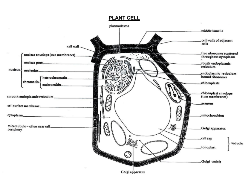
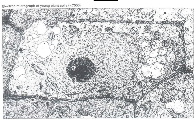
Plant cell
Cytoplasm
Cytoplasm = All organelles + Cytosol within the cell surface membrane, NOT INCLUDING the nucleus
Cytoplasm is bounded by the cell surface membrane, which is composed of phospholipids, proteins and carbohydrates

Cytosol
Cytosol = Aqueous solute rich matrix (the soluble part of the cytoplasm) that appears transparent and lacking structure under the LM
It contains about 90% water
Substances dissolved in cytosol:
Various essential ions and soluble organic molecules such as sugars and amino acids
Soluble proteins including enzymes
Cytoskeleton – a network of fine strands of globular and fibrous proteins, which provides infrastructure and support to the cell.
Membranous organelles
Membranous organelles are compartmentalised spaces within the cytoplasm and are surrounded by membranes which are structurally and biochemically similar to the plasma membrane.
Advantages to have Membranous organelles
The presence of membranes surrounding the organelles allows the maintenance of characteristic differences between the contents of each organelle and the cytosol.
The compartmentalisation of specific reactions provide different local environments for which incompatible processes can occur simultaneously.
The presence of membranes also helps to increase membrane surface area.
These internal membranes also allow for the embedding of enzymes and proteins that mediate many cellular reactions.
The greater the membrane surface area, the larger the number of enzyme complexes that can be embedded, and thus increasing efficiency of many reactions by providing optimal enzyme concentration for reactions to occur.
Nucleus
Outline:
It is the largest organelle in the animal cell
Easily seen with the light microscope
Present in every cell with a few exceptions (eg: red blood cell)
Usually spherical or oval
Size between 5-20 micrometres
DOUBLE-MEMBRANED organelle
Function:
Nucleus encloses genetic material and protects DNA from metabolically active cytoplasm.
The double membrane is perforated with pores to enable exchange of substances between the nucleus and cytoplasm.
Nucleus — Components — Nuclear envelope
It is a double membrane and it separates the contents of the nucleus from the cytoplasm.
Each of the two membranes is a lipid bilayer
The outer membrane of the nucleus is continuous with the membrane of the endoplasmic reticulum
The inner and outer membranes are continuous with each other and the region between them is known as the perinuclear space, which is continuous with the ER lumen.
The nuclear envelope is perforated by nuclear pores, made up of a large protein complex, which allow macromolecules such as mRNA and rRNA to exit the nucleus, and proteins (eg: enzymes) to enter and exit the nucleus.
Nucleus — Components — Nucleoplasm
Nucleoplasm is an aqueous matrix within the nucleus containing proteins, metabolites, ions, RNA and chromatin (the genetic material of the cell).
Chromatin is composed of coils of DNA wound around basic protein known as histones which exist in two forms:
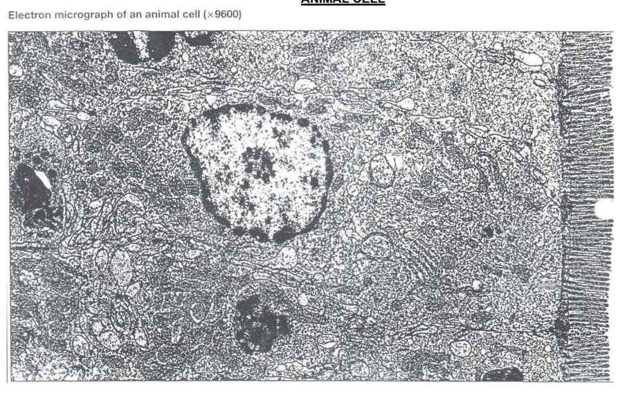
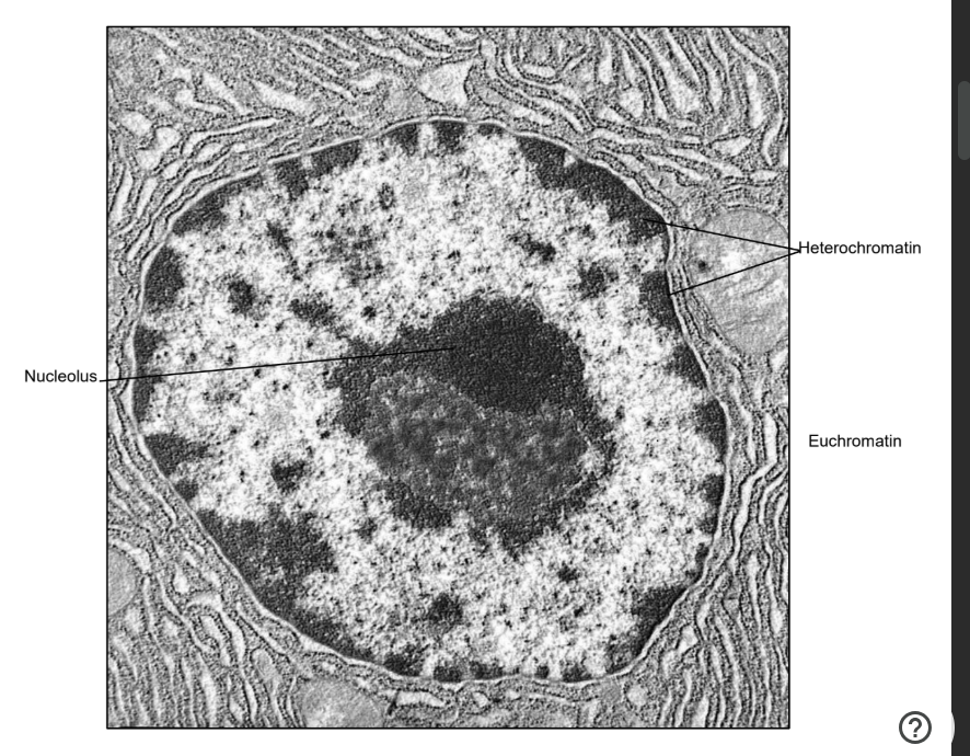
Loosely coiled chromatin known as euchromatin, which appears as light-coloured patches in the electron micrograph of the nucleus
Tightly coiled chromatin known as heterochromatin, which appears as dark-coloured patches in the electron micrograph of the nucleus
Nucleus — Components — Nucleolus
The nucleolus appears as a dense mass in the nucleus when viewed under the electron microscope
It is composed of DNA carrying rRNA genes, RNA and protein, which functions to synthesize a specific type of RNA known as ribosomal RNA (rRNA) that forms a component of ribosomes
Endomembrane System
The endomembrane system is composed of a number of inter-related membrane sacs within the cytoplasm of the cell. These membranes are related either by direct physical continuity or by the transfer of membrane segments known as vesicles
The system functions in part to manufacture proteins and lipids. Individual components of the endomembrane system each have a unique structure and function.
Components of the endomembrane system include:
1. Rough & Smooth endoplasmic reticulum
2. Golgi apparatus / Body
3. Lysosomes
4. Vacuoles
Endoplasmic reticulum
(ER) consists of an extensive network of hollow, membranous tubules, sacs or sheets called cisternae (singular: cisterna).
The internal space of the ER is known as the cisternal space, or lumen, which is continuous with the perinuclear space.
It is distinguishable from the Golgi apparatus as it has a flatter, more compact packing that is sheet-like.
Specialised structures for functions:
The extensive network of cisternae increases membrane surface area for synthesis
For rER, it allows for more ribosomes to be embedded on it for synthesis of more polypeptides
For sER, the membrane allows for enzymes to be embedded, so that steroid and phospholipid can be synthesised
Hollow cisternae accommodate newly synthesised substances and allow for packaging of contents into vesicles for transport to the GA
There are two distinct but connected regions of the ER: the smooth and rough ER, which differ in structure and function.
Endoplasmic Reticulum — Rough Endoplasmic Reticulum
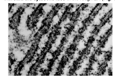
Structure:
Rough ER has a sheet-like appearance which appear rough due to the presence of ribosomes that stud the cytosolic face of the rough ER
Function:
These rough ER-bound ribosomes are sites of protein synthesis where a polypeptide chain is synthesised at the bound ribosome
The polypeptide chain then enters the ER lumen, which is the site of protein folding, through a protein channel in the rER membrane where the polypeptide chain folds into its native conformation.
These proteins are either destined for export, or are targeted to various cellular organelles (In contrast to proteins synthesized by free ribosomes in the cytosol which remain in the cytosol).
Significance:
Cells that are active in protein secretion usually have abundant rough ER.
Some proteins synthesised in the rough ER can also directly enter the membrane of the ER to form ER membrane proteins.
Proteins that leave the rER are enclosed in vesicles known as transport vesicles.
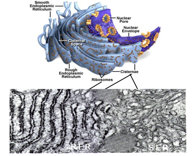
Endoplasmic Reticulum — Smooth Endoplasmic Reticulum
Structure:
The smooth ER is a network of tubules which lack ribosomes, resulting in its smooth appearance
Function (all processes require ATP):
Synthesis of lipids
Oils, phospholipids and steroids like sex hormones
Cells that are active in hormone secretion usually have abundant sER.
Metabolism of carbohydrates
Detoxification of drugs and poisons
Storage of calcium ions for use in muscle contraction, as well as cell signalling
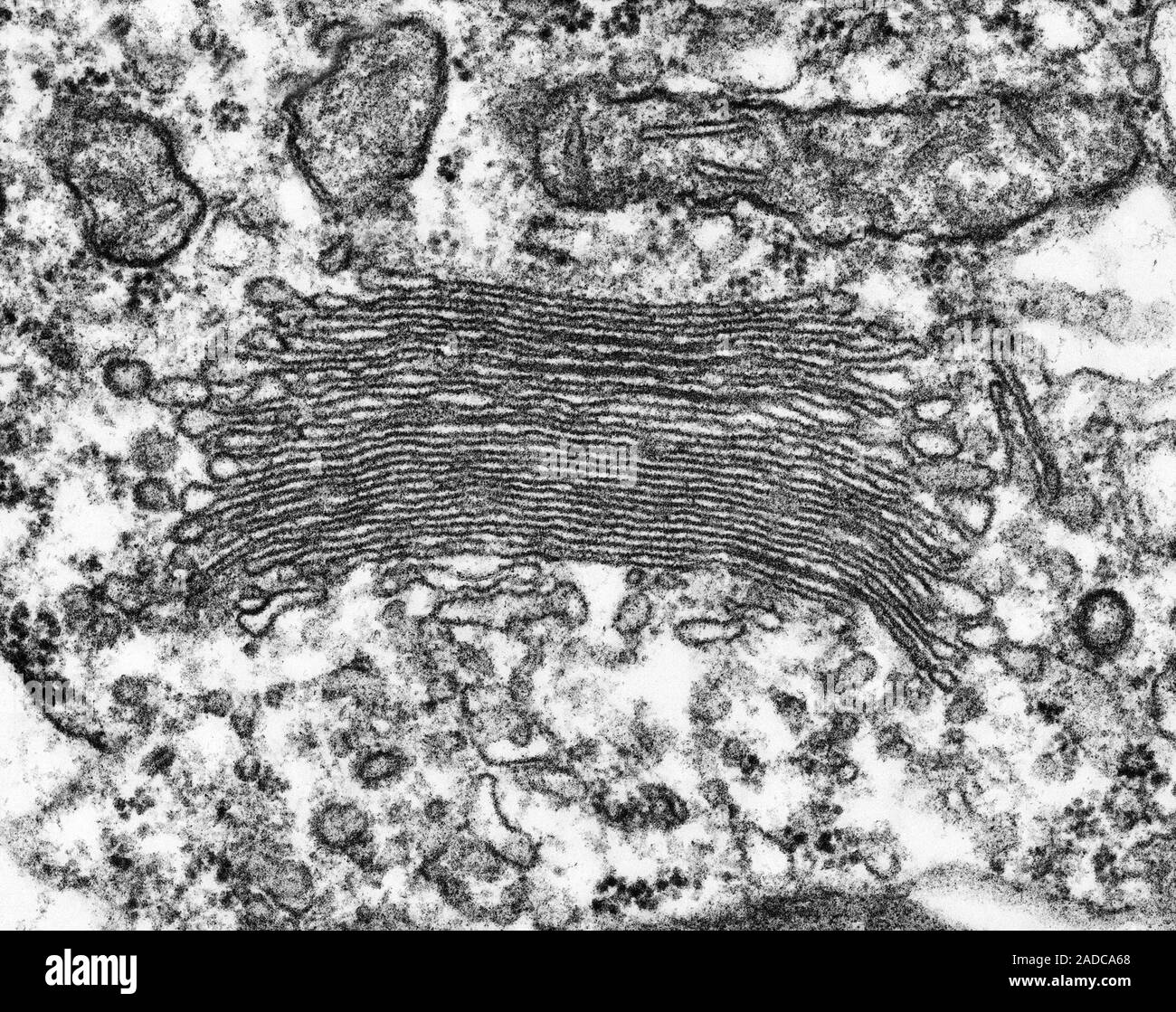
Golgi Apparatus / Golgi Body
Structure:
It consists of a stack of flattened, membrane-bound sacs called cisternae (singular: cisterna), which separates its internal space from the cytosol.
Each stack differs in thickness and molecular composition.
A distinct polarity exists in the stack of GA:
→ A cis face (forming face)
→ A trans face (maturing face)
New cisternae are constantly being formed at the cis face by receiving transport vesicles from the ER. The membranes of transport vesicles from the ER fuses with the cis face membrane and deposit their contents into Golgi cisternal space.
At the trans face, membranes bud off to form secretory vesicles, which contain materials to be transported to the extracellular matrix.
Membranes can also bud off from the Golgi trans face to form lysosomes.
Between the Golgi sacs, Golgi vesicles are responsible for transferring materials between the parts of the Golgi.
Some Golgi vesicles also bud off from the trans face to transport substances to other organelles in the cell.
Function:
It is where some modifications occur:
→ Glycosylation (addition of sugar groups)
→ Trimming (removal of excess monomers)
Different Golgi cisternae contain different enzymes for modification, and hence ER products are progressively modified as they move through the stacks of the Golgi complex from the cis face to the trans face.
The processed and packaged contents are then passed on to other components of the cell by vesicles that bud off the GA complex.
Significance:
Cells that are active in any form of secretion usually have abundant GA, as the abundant flattened cisternae provide increased surface area for vesicle reception and budding.
Multiple cisternae also allow for different modification processes to occur simultaneously.
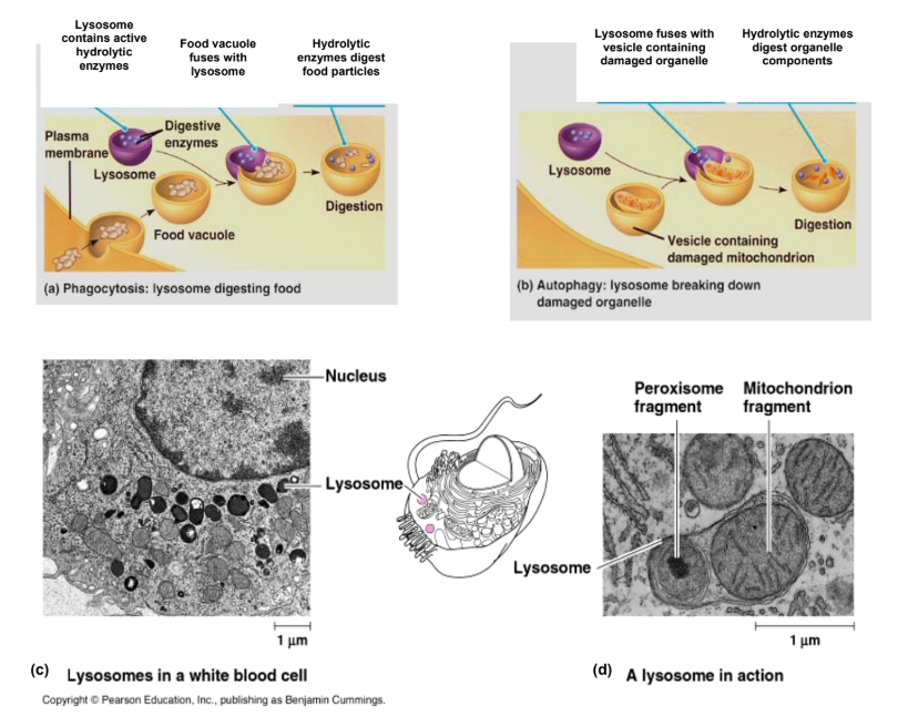
Lysosomes
Structure:
The lysosome is a membranous organelle that appears homogenously electron-dense under the EM
It contains hydrolytic enzymes (proteases, nucleases, lipases and acid phosphatases) that can digest most biological macromolecules.
Enzyme contents are synthesised on rER and transported to GA for further processing.
Due to the acidic (pH 5) nature of lysosome contents and hydrolytic activity of enclosed enzymes, lysosomal contents must be prevented from spilling into the cytoplasm under normal cell conditions.
Therefore, segregation of contents within the membrane provides optimal pH for hydrolytic reactions and protects cellular contents from hydrolysis.
Functions:
Digestion of materials taken into cells:
Food particles are engulfed by endocytosis to form food vacuoles, which fuse with lysosomes to form endosomes.
The enzymes then digest the endosome’s contents which later end up in the cytosol to be used as food for the cell.
It can also be a defence mechanism against bacteria in certain cell types; in such cases, the result of fusion is known as a phagocytic vacuole.
Autophagy (‘removal’) of worn-out organelles
Unwanted structures within the cell are enclosed by a membrane of unknown origin, forming a vesicle.
This vesicle then fuses with the lysosome to form an autophagic vacuole.
Autolysis:
Cells can self-destruct in a controlled manner when programmed to, in a process known as apoptosis.
It is a normal event in development and differentiation such as destruction of tadpole tail to become a frog.
It can also occur when a cell senses that it has become a threat to its environment. For autolysis to occur, there must be a mass release of lysosomal contents in the whole cell.
Vacuoles
Vacuole membranes in both animal and plant cells originate from the ER and GA, and thus vacuoles are considered part of the endomembrane system.
In animal cells:
Vacuoles are small, mobile organelles that serve to house and transport substances
Eg: Food vacuoles and phagocytic vacuoles.
Present in larger amounts than plant cells
In plant cells:
Generally a large central vacuole surrounded by a single membrane known as a tonoplast
The solution within the tonoplast is known as cell sap, and differs in composition from the cytoplasm.
The plant cell vacuole is a versatile component that has the ability to regulate the substances it concentrates.
Functions of the plant vacuole include:
1. Storage of organic compounds (e.g. proteins) and inorganic ions (e.g. K+ and Cl-
)
2. Disposal site for toxic metabolic by-products
3. Contains pigments (e.g. red and blue pigments that colour the petals)
4. Plant protection by accumulating compounds that are toxic or unpalatable to consumers
5. Cell growth and elongation as water accumulates in the vacuole; plant cells can therefore
increase in size with minimal investment in cytoplasm synthesis and without sacrificing surface
area to volume ratio, as cytoplasmic contents are pushed to the periphery of the cell.
Non-membranous organelles
Non-membranous organelles are not enclosed in a membrane.
Summary