PE, DVT, Pneumothorax, & Atelectasis
1/140
There's no tags or description
Looks like no tags are added yet.
Name | Mastery | Learn | Test | Matching | Spaced | Call with Kai |
|---|
No analytics yet
Send a link to your students to track their progress
141 Terms
True or False: there are a lot of PEs that can break off and become DVTs
true
What are PEs/DVTs?
complication of thrombus formation in deep vein circulation
What will 50-60% of proximal DVTs do?
embolize to pulmonary circulation
What are the PE statistics?
third leading cause of death among hospitalized patients
Who is most likely to have a DVT?
>40 years old, caucasians and black people with more complications
What is the pathophysiology of DVTs?
virchow’s triad
formation of fibrin clot
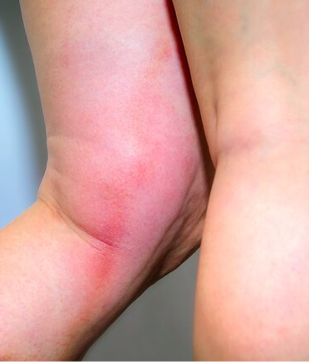
What are the s/sxs of DVTs?
edema
erythema/warmth
pain
homan’s sign may be positive
palpable indurated cord-like vein (rope-like structure)
Can deep veins usually be palpated?
no, but in DVTs it can get to a point where it becomes a palpable indurated cord-like vein
What is virchow’s triad?
endothelial injury, venous stasis, and hypercoagulability
How is homan’s sign performed?
provider dorsiflexes the foot ont he affected side, and if pain is produced, that is a positive homan’s sign, but it is well-known to be inaccurate
What are the potential endothelial injuries that can contribute to 1/3 of Virchow’s triad?
surgery
IV drug users
trauma
What are the potential causes of venous stasis that can contribute to 1/3 of Virchow’s triad?
imobility
traveling
hyper-viscosity
increased central venous pressure
What are the potential causes of hypercoagulability that can contribute to 1/3 of Virchow’s triad?
medications
disease
inherited genetic defects
What are medications that can lead to hypercoagulability?
oral contraceptives or estrogen replacements
What are inherited genetic defects that can lead to hypercoagulability?
clotting deficiencies or abnormalities (ex. Factor V, antithrombin 3 defieincey, Protein C or S deficiencies)
What are the reasons someone may have increased central venous pressure, leading to venous stasis?
pregnant people and people with truncal obesity will have downward pressure on the femoral veins and can lead to a DVT because it blocks the blood trying to get up and out of the legs
True or False: hyperlipidemia leads to hypercoagulability, but one of the things that doesn’t is malignancies
false; both can lead to hypercoagulability states
What is used for DVT risk stratification?
well’s scoring
What is the first imaging test done in a suspected DVT?
US of affected limb with doppler
Even though US is the preferred test, what is the gold standard?
venography
What labs can we get when we suspect a DVT?
D-dimer
coagulation studies
anti-thrombin III
CRP
ESR
Why is venography gold standard but not always preferred?
it takes longer, is more invasive, and there’s the potential of a rxn to the dye
True or False: Everyone has a little D-dimer in their body because of their natural degradation processes
True
Why can D-dimers support the diagnosis of a clot?
it will be more elevated than usual in cases of large or multiple small clots
When can’t you trust a D-dimer
When people have smaller, insignificant clots that are not creating oclusions (no edema, erythema, etc), then it’s unclear; if someone has less RFs and the levels are elevated, it’s much more likely to be DVT (even if not symptomatic)
What are CRP and ESR?
non-specific inflammatory markers
What is the only part of the well’s score that isn’t given a +1 if present?
Alternative diagnosis to DVT is more likely to get a -2
How does Well’s scoring work in cases where DVTs are unlikely (<2)?
a D-dimer is done; negative → no DVT, positive → do US, positive US → anti-coagulate, negative → no DVT
How does Well’s scoring work in cases where DVTs are unlikely (>2)?
First, get a D-dimer; if negative or positive → US, if no DVT, you could still do venography, or repeat US in 1 wk, then no DVT again confirms, if positive → anticoagulate
When do we automatically do an US for DVTs?
If they have 3 or more criteria from well’s scoring
What score of Well’s criteria do you take action, and what do you do?
What is the goal of DVT treatment?
prevent embolus, prevent recurrent DVT, prevent post-thrombotic syndrome that can lead to permanent venous valve damage
What are the treatment types of DVTs?
anticoagulation, thrombolytic therapy, surgical extraction
What are the DVT treatments in order of preference?
direct oral anticoagulant → warfarin → heparin
What are the direct oral anticoagulants?
apixaban (eliquis)
rivaroxaban (Xarelto)
edoxaban (savaysa)
dabigatran (pradax)
What is the MOA of apixaban (eliquis)?
inhibits factor Xa
What is the MOA of rivaroxaban (Xarelto)?
inhibits factor Xa
What is the MOA of edoxaban (Savaysa)?
inhibits factor Xa
What is the MOA of dabigatran (pradax)?
direct thrombin inhibitor
What is Warfarin (Coumadin)?
a vitamin K antiagnoist
What does warfarin (Coumadin) do?
inhibits clotting factors II, VII, IX, X
What should be monitored while patients are on warfarin (coumadin), and what should the results be?
monitor INR (2.0-3.0 levels)
How would you reverse the effects of Warfarin (Coumadin)?
vitamin K
What is the MOA of LMWH?
binds to and accelerates antithrombin III
What are the types of LMWH?
dalteparin (Fragmin)
enoxaparin (lovenox)
When is LMWH CI?
in renal insufficiency
What are the 2 types of heparin?
LMWH
unfractioned heparin
How is unfractionated heparin different from LMWH?
more specific for inactivation of thrombin
What will the aPTT of patients on unfractioned heparin be?
1.5-3x the upper limit of the normal range
What are the potential complications of unfractionated heparin?
hemorrhage, HIT
What is the MOA of fondaparinux?
indirectly inhibits factor Xa
What is the most preferred DVT anticoagulantion regimen in low-risk patients?
direct oral anticoagulation alone
What is a PE?
thrombus that has broken off and travels to the pulmonary vasculature
How does a air emboli occur?
air introduced into the venous system, then the air creates a bubble and the bubble itself travels, lodges somewrhere and cuts off everything
How does an embolus form due to amniotic fluid?
amniotic fluid has a different density, so it can rise and move and has the opportunity to block something
How does a fat emboli occur?
usually trauma related, a blood vessel could be opened and teared, and little particles can be introduced to the vein
How can foreign body emboli occur?
common with trauma and IV drug users
How can a septic emboli form?
usually from vegetative plaque of endocarditis that originates in the heart
What are the types of emboli?
thrombus
air
amniotic fluid
fat
foreign body
septic
What is the pathophysiology of PEs?
clot travels and lodges in part of the pulmonary arterial circulation or systemic artery
What are respiratory consequences of PEs?
increased alveolar dead space
hyperventiliation
hypoxemia (ventiliation-perfusion mismatch)
decreased regional surfactant
What is surfactant needed for?
needed for proper lung tissue function, for it to contract and inhale with inhalaltion and exhalation
What are the hemodynamic consequences of PEs?
reduces cross sectional area of pulmonary vascular bed
increases right ventricular afterload
may results in right-sided HF or ventricular failure
potential pulmonary arterial vasoconstriction from neurohormonal reflexes
What are the signs of PEs?
dyspnea
cough
pleuritic chest pain
What may be found on PE in patients with a pulmonary embolus?
tachypnea
hypoxia
rales
fever
accentuated S2
tachycardia
signs related to underlying cause (thrombophlebitis, pregnnacy, drug use)
What is the gold standard test to diagnose a PE?
pulmonary angiography, specifiically helical CT pulmonary angiography
What lab is taken for suspected PEs?
D-dimer is used to exclude PE in a low-probability patient
What test can be done if a CT is not avaliable or CI to diagnose a suspected DVT?
ventilation-perfusion lung scan (VQ)
What are the findings of an ECG in a patient with a PE?
sinus tachycardia and nonspecific ST segment, and wave changes
What are the wave changes seen on ECG in potential PE patients?
Deep S wave in lead 1, significant Q wave and T wave inversion in lead 3
What are the treatments for PE?
anticoagulation (IN STABLE PATIENTS)
thrombolytic therapy
inferior vena cava filter
surgical extraction (embolectomy)
What is the last resort treatment in a PE?
surigcal extraction (embolectomy) due to high risk of mortality
What are the types of anticoagulation used in PE treatment?
direct oral anticoagulants (preferred agent)
warfarin
heparin
fondaparinux
What type of heparin is preferred in PE treatment?
LMWH over unfractionated heparin
True or False: direct oral anticoagulants should be used with another anticoagulant in PE treatment
false; direct oral anticoagulants can be used as monotherapy
What is a CI of a inferior vena cava filler in PE treatment?
anticoagulants
When is an inferior vena cava filter in PE treatment indicated?
recurrent thromboembolism despite adequeate anticoagulation
chronic recurrent pulmonary emboli with pulmonary hypertension
What are the ways to prevent PEs?
ambulation
hydration
anticoagulation
compression stockings (30-40 mmHg)
What is a pneumothorax?
accumulation of air in the pleural space (potential space between the visceral and parietal pleurae)
How can a pneumothorax present?
spontaneous or traumatic
How does a primary spontaneous pneumothorax occur?
no underlying lung disease or trauma
How does a secondary spontaneous pneumothorax occur?
complication of preexisting lung disease that alters normal lung structure
How does a traumatic pneumothorax occur?
penetrating or blunt trauma
How does a iatrogenic pneumothorax occur?
during/following a medical procedure
How does a tension pneumothorax occur?
penetrating trauma, infection, CPR, mechanical ventilation; most dangerous kind
Who can have a primary spontaneous pneumothorax?
tall, thin male
age 10-30 y/o, younger people
What are the potential RFs of a primary spontaneous pneumothorax?
± history of smoking or FH of pneumothorax
What causes a primary spontaneous pneumothorax?
subpleural apical blebs/bullae rupture
What is the pathophysiology of a pneumothorax?
pleural space fills with gas from a ruptured bleb → gas pressure outside lung overcomes gas pressure inside lung → lung collapses until the rupture is sealed
What are the s/sxs of a pneumothorax?
sudden chest pain on affected side
sudden dyspnea
tachycardia
What may we find on PE when checking the affected side?
decreased breath sounds on auscultation
hyperresonance on percussion
decreased tactile fremitus
decreased movement of chest
How do people with a tension pneumothorax present?
hypotension and mediastinal/trachel shift
hyperressoance on affected side
hpyoxia
respiratory failure
What is the best way to diagnose a pneumothorax?
chest radiograph
What may be seen on a chest radiograph in a patient with a pneumothorax?
visceral pleural line with no lung markings beyond affected lung portion (“companion line”)
pleural effusion
“deep sulcus” sign (supine)
What might be seen in a tension pneumothorax on chest radiograph?
large amount of air in affected side
shift of mediastinumm toward unaffected side
What is a complication of a pneumonthorax?
pneumomediastinum
subcutaneous emphysema
Where would we expect air to leak out in a pneumomediastinum?
air leaks out of skin overlying the chest wall
What is the treatment for an asymptomatic pneumothorax patient?
supportive care and treat depending on risk of recurrence
How is a tension pneumothorax treated?
needle aspiration in 2nd intercostal space at MCL
chest tube placement