2. cells
1/248
There's no tags or description
Looks like no tags are added yet.
Name | Mastery | Learn | Test | Matching | Spaced | Call with Kai |
|---|
No analytics yet
Send a link to your students to track their progress
249 Terms
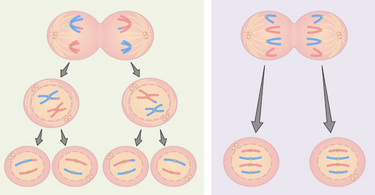
summarise the difference (in products) between mitosis and meiosis
mitosis: produces 2 daughter nuclei each w/ the same number of chromosomes as the parent cell and each other
meiosis: produces 4 daughter nuclei w/ half the number of chromosomes as the parent cell
name the 3 stages of interphase:
G1 (growth stage 1)
S (synthesis)
G2 (growth stage 2)

what occurs in G1 of interphase?
cell grows
proteins synthesised
organelles replicate → prepares for DNA replication
what occurs in S of interphase?
DNA is replicated
what occurs in G2 of interphase?
cell finishes growing
protein synthesised → prepares for cell division
what is mitosis significant for?
growth
repair
differentiation
what are the stages of the cell cycle?
interphase (made up of G1, S and G2)
Prophase
Metaphase
Anaphase
Telophase
cytokinesis
what is the centrosome? what are some of its key features?
where spindle fibres grow from
located near the nucleus
not present in plants/fungi
in cytoplasm?
1 centrosome contains 2 centrioles

what is the centriole?
1 centriole = 9 groups of microtubules
spherical shape
what happens (generally) during interphase?
DNA replication
Organelle (including centrosomes) duplication
Cell growth
Transcription/translation
Obtain nutrients
Respiration

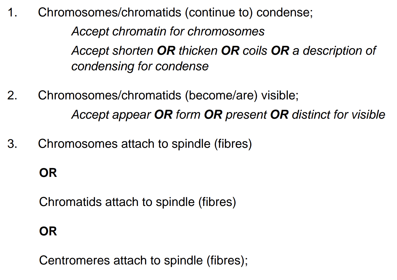
what happens during prophase?
chromosomes condense so they become visible
chromosomes attach to spindle fibres (/metaphase)
nucleolus disappears and nuclear envelope breaks down
what happens during metaphase?
chromosomes arrange along middle of cell:
spindle fibres form from centrioles
chromosomes line up along spindle fibres across middle of cell
what happens during anaphase?
centromeres divide
sister chromatids pulled to opposite poles of cell
spindle fibres shorten (/prophase/metaphase)
what happens during telophase?
nuclear envelope reforms:
spindle fibres disintegrate
nucleolus reforms
nuclear membrane reforms
chromosomes decondense
what happens during cytokinesis?
division of the cytoplasm and cell membrane into 2 genetically identical daughter cells
what is the proteome?
all of the proteins in an organism (essentially like the genome but for proteins)
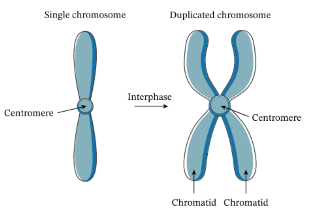
what is the centromere?
centre of a chromosome, where spindle fibres attach

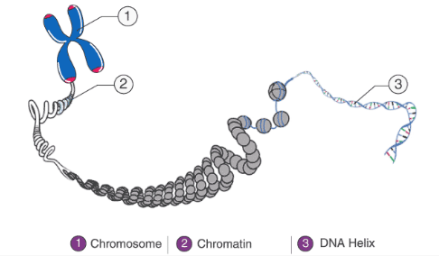
explain the difference between a chromosome and a chromatid
chromosome = DNA molecule that contains part of the genetic material of an organism
chromatid = one arm of a duplicated chromosome - one copy of a newly copied chromosome
(confusingly, chromosomes can look like I or X)
what is a cell?
smallest building block in an organism and are capable of carrying out specific functions
how are cells able to carry out different functions?
differentiation
what is ultrastructure?
the internal structure of a cell as not visible on a light microscope
what are the three domains of all living things?
archaea
bacteria
eukaryota
what is a cell made of (main groups)?
proteins
polysaccharides
lipids
nucleic acids
what is the significance of the proteins w/in a cell?
enzymes in the cytoplasm and inside organelles
associated w/ the membrane
associated w/ DNA in the nucleus
associated w/ RNA in ribosomes
cytoskeleton
what is the significance of the polysaccharides w/ in a cell?
associated w/ the membrane
makes up cell walls
stored for energy reserve
what is the significance of the lipids w/in a cell?
stored for energy reserve
phospholipids make up cell membranes
cholesterol associated w/ membrane and controls fluidity
where are the nucleic acids w/in the cell found?
DNA: associated w/ proteins in the nucleus
RNA: mRNA, rRNA, tRNA - different types found in nucleus or cytoplasm or associated w/ proteins in ribosomes
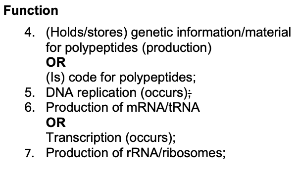
what is the function of the nucleus?
contains genetic material for polypeptides
DNA replication
production of mRNA/tRNA/transcription
production of rRNA/ribosomes
what is the structure of the nucleus?
nuclear envelope/double membrane and pores
chromosomes/chromatin/DNA w/ histones
nucleolus/nucleoli
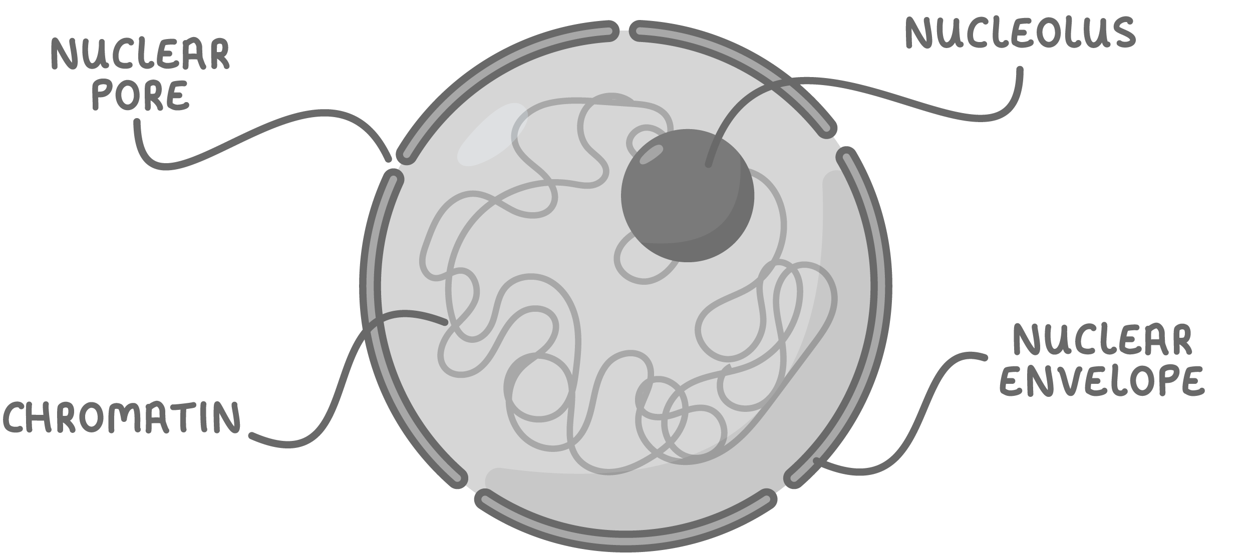
what is the difference between chromosomes and chromatin?
chromosomes:
linear molecule of DNA tightly wrapped around histone proteins
DNA is only in this form during cell division
chromatin:
DNA more loosely associated w/ histones
enclosed in the nucleus
what are the two types of endoplasmic reticulum (ER)?
rough endoplasmic reticulum (RER)?
smooth endoplasmic reticulum (SER)?
what is the structure of the RER?
continuous w/ the nuclear membrane, membrane bound
has ribosomes on cisternae
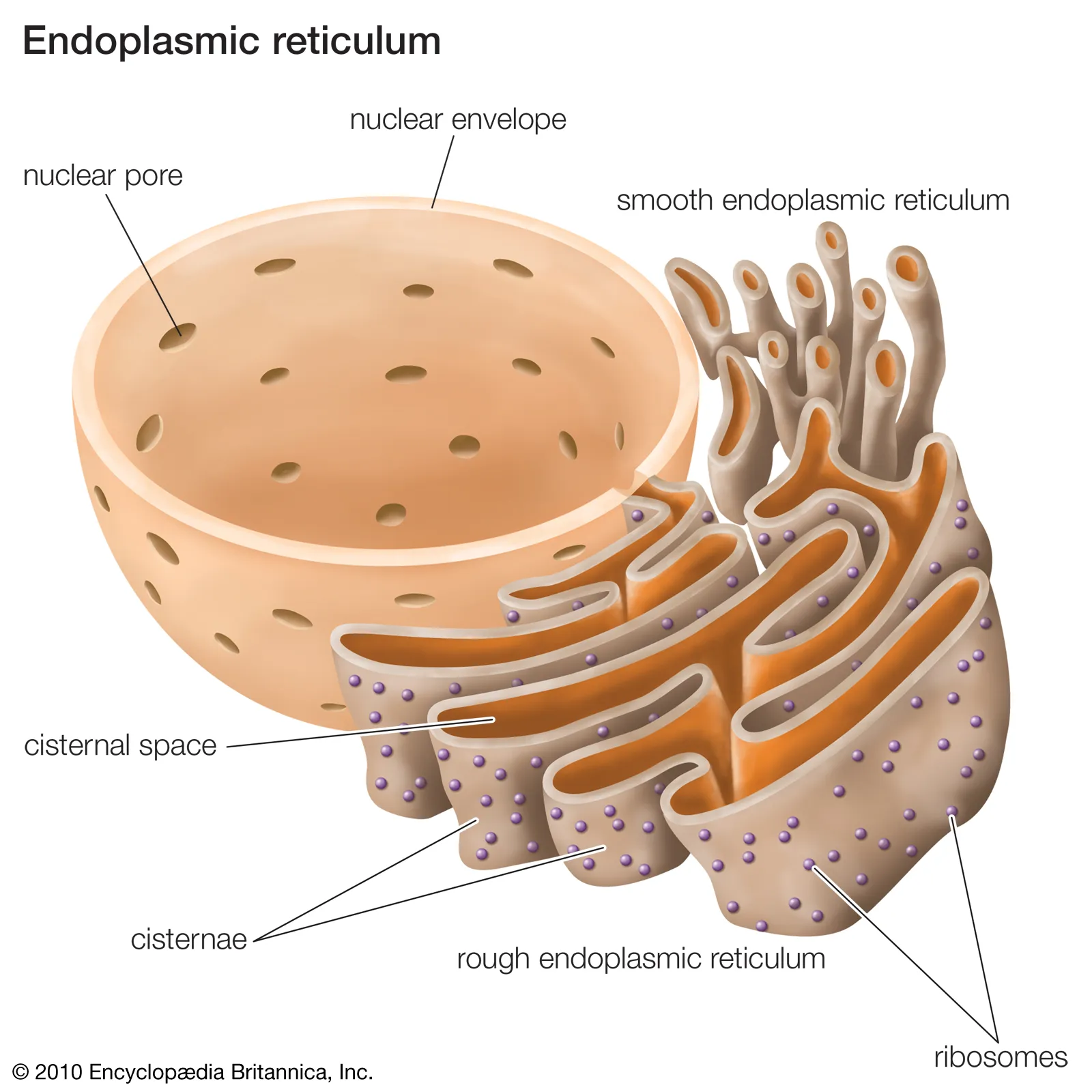
what is the function of the RER?
cisternae provide large SA for protein synthesis
protein collects inside RER and transported throughout cell
what is the structure of the SER?
membrane bound
no ribosomes (hence smooth)
more tubular than RER

what is the function of the SER?
synthesises, stores and transports lipids, steroids and carbohydrates
what is the function of the ribosomes?
site of protein synthesis
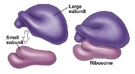
what is the structure of the ribosomes?
made up of ribosomal RNA (rRNA) and protein
consists of a large and small subunit
two types: 80S (found in eukaryotic cells) and 70S (found in prokaryotic cells, mitochondria and chloroplasts)
what is the structure of the Golgi apparatus?
membrane bound
stacks of cisternae (flattened, membrane bound sacs)
vesicles are continuously pinched off from the ends
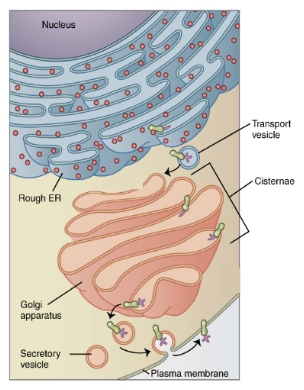
what is the function of the Golgi apparatus?
modifying/packaging/transporting proteins/glycoproteins
modifying/packaging/transporting lipids/glycolipids
form vesicles and lysosomes (as they are Golgi vesicles)
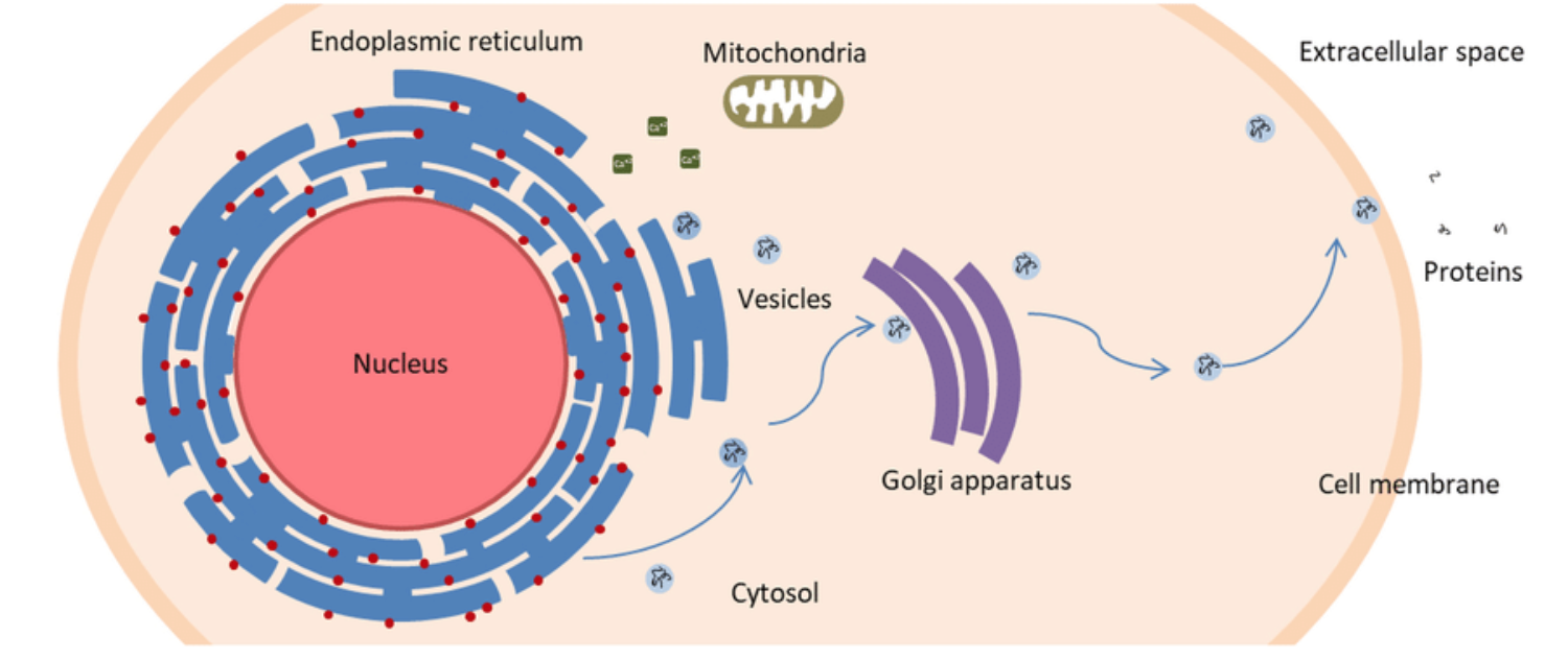
what events take place when a protein is made and sent to the extracellular space?
a gene is transcribed into mRNA in the nucleus
the mRNA leaves through a nuclear pore
the mRNA reaches a ribosome on the RER
the ribosome synthesises a protein into the RER
the protein is packed in a vesicle that leaves the RER
the vesicle reaches the golgi and fuses w/ its cisternae
the protein is released outside
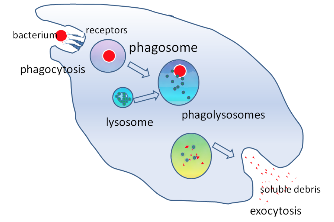
what are lysosomes?
type of Golgi vesicle containing hydrolytic enzymes (proteases, lipases, lysozymes)
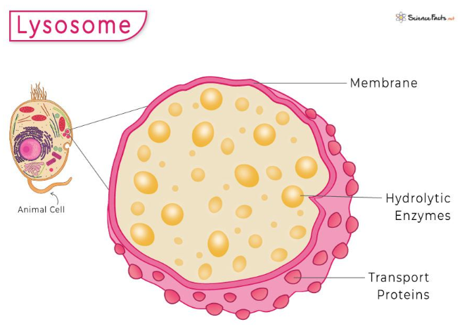
what is the function of lysosomes?
digests unwanted material in the cell:
hydrolyses material ingested by phagocytic cell
exocytosis - releases enzymes to outside of cell to destroy material
digests worn out organelles
autolysis - complete breakdown of cells after they have died
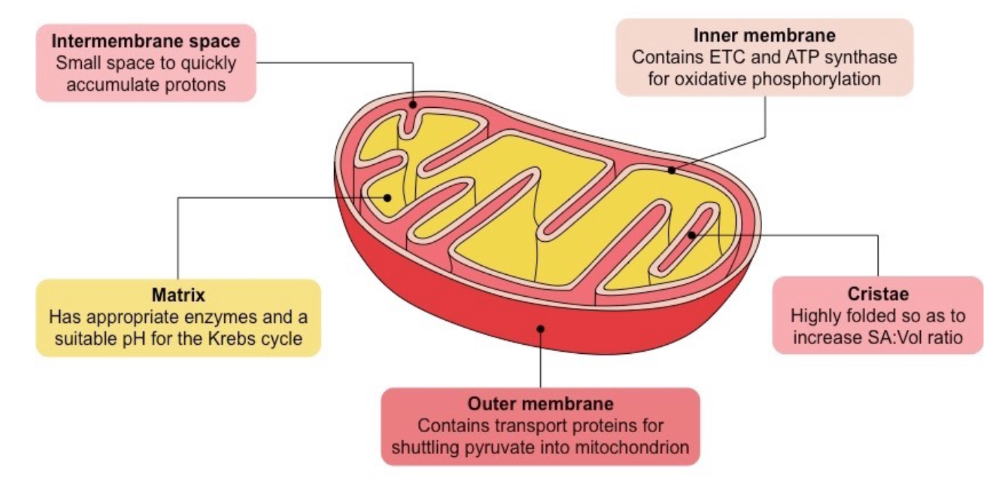
what is the function of the mitochondria?
site of aerobic respiration
releases ATP during respiration - source of energy for cell activities
what is the structure of the mitochondria?
double membraned (membrane bound) - inner membrane folds to form cristae, where respiratory enzymes are embedded
fluid centre = mitochondrial matrix - also contains respiratory enzymes as well as loop of mitochondrial DNA, proteins, lipids and ribosomes
give the 4 main steps of cell fractionation:
sample preparation
homogenisation
filtration
ultracentrifugation
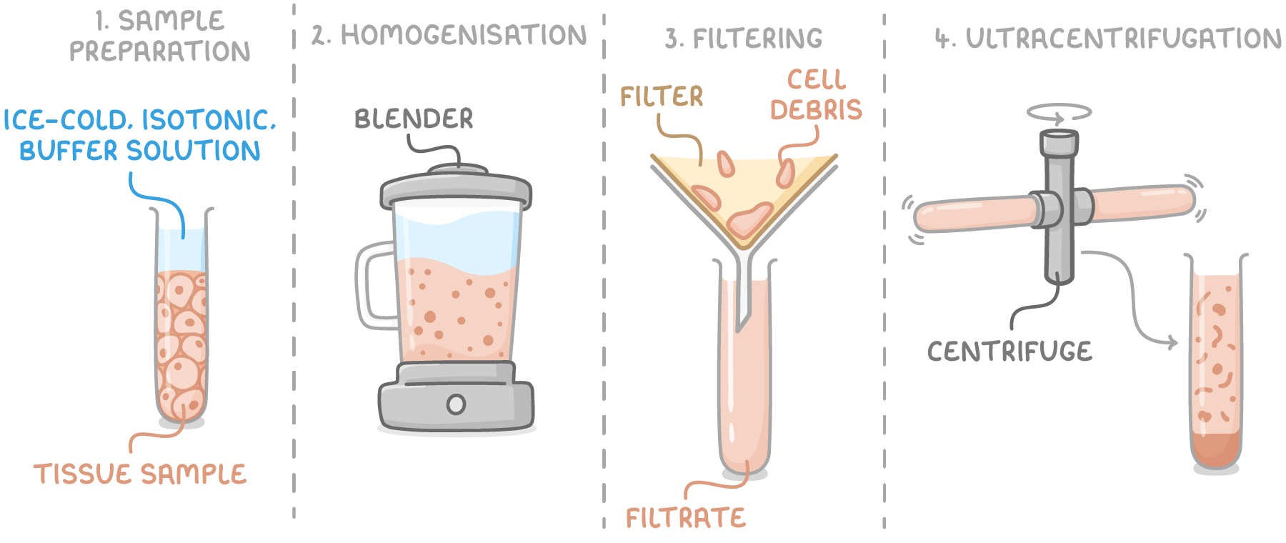
describe the sample preparation stage:
sample is placed in an ice cold, isotonic, buffered solution
why must the solution used in sample preparation be ice-cold?
reduce enzyme activity that might otherwise digest organelles
why must the solution used in sample preparation be isotonic?
ensures ψ inside and outside organelles is the same, so they do not burst as a result of osmosis
why must the solution used in sample preparation be buffered?
keeps pH constant so that organelle structures are not damaged and enzymes do not denature
describe and explain the homogenisation stage:
cells are broken open
this disrupts the plasma membrane, allowing the organelles to be released into the solution
describe the filtration stage:
mixture filtered to remove cellular debris and tissue fragments
describe the ultracentrifugation stage:
filtrate centrifuged at a low speed → dense organelles (nuclei) form pellet at bottom of tube, less dense organelles remain suspended in supernatant
supernatant transferred to new tube and centrifuged at a higher speed → next heaviest organelles (chloroplasts/mitochondria) settle out
transfer supernatant into new tube and repeat entire process, increasing speed each time until all organelles have been separated into distinct layers
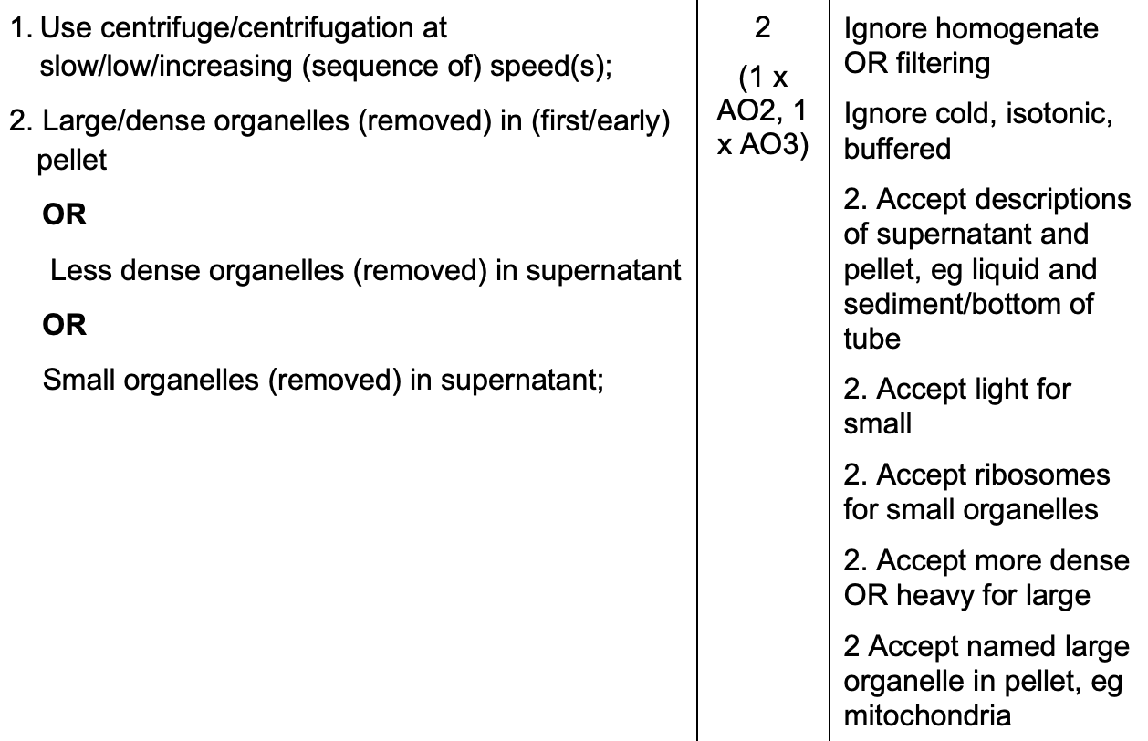
give the order of organelles from heaviest to lightest:
nuclei
chloroplasts
mitochondria
lysosomes
ER
ribosomes
what is the pellet?
sediment at bottom of tube - contains heavier organelles
what is the supernatant?
liquid remaining above pellet - contains lighter organelles
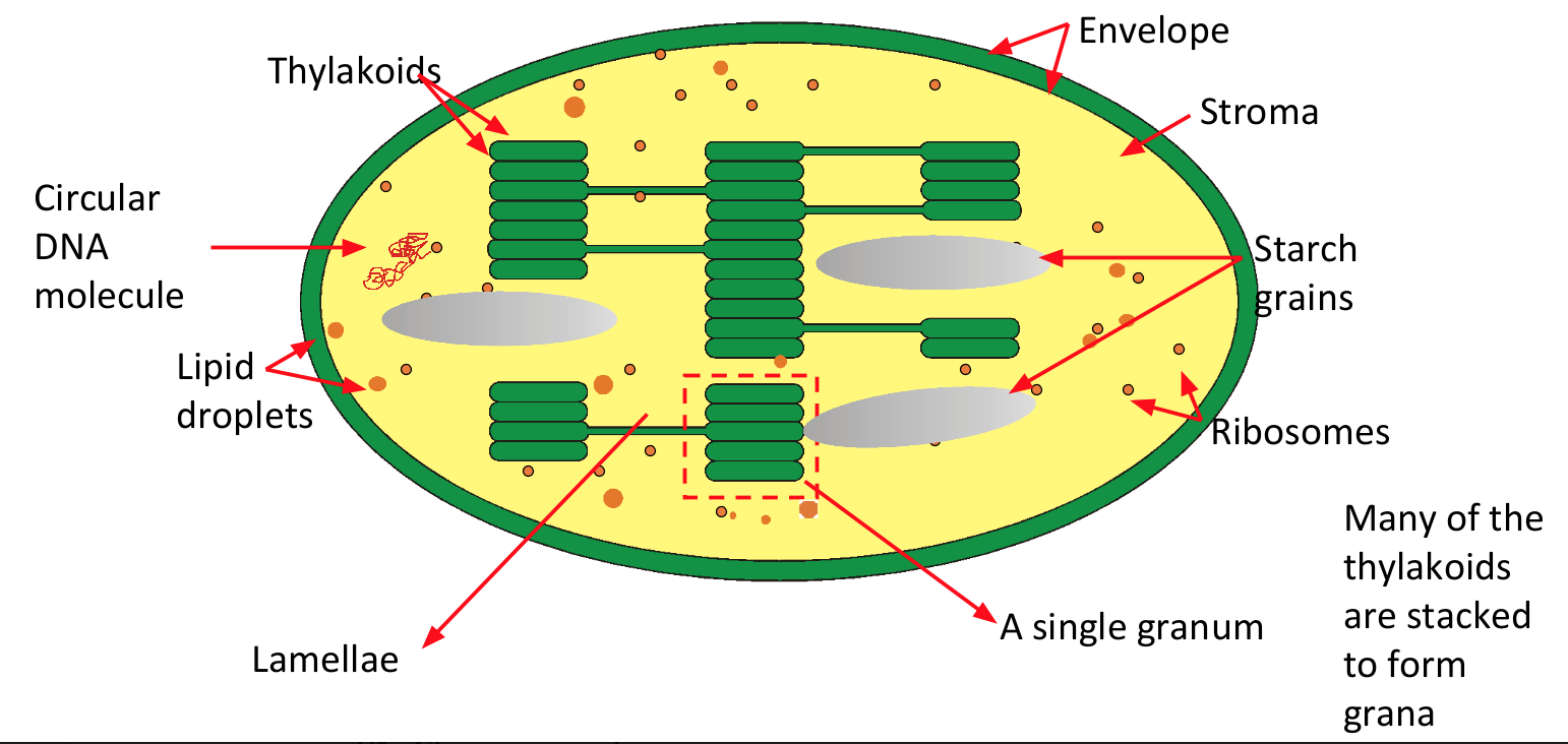
what is the function of the chloroplasts?
site of photosynthesis
what is the structure of the chloroplasts?
double membraned
grana - sticks of disc like structures called thylakoids (membranes that contain photosynthetic pigment - chlorophyll)
stroma - fluid filled matrix containing enzymes for photosynthesis
contain both DNA and ribosomes
what is the structure of the vacuole?
fluid filled sac surrounded by a membrane called tonoplast
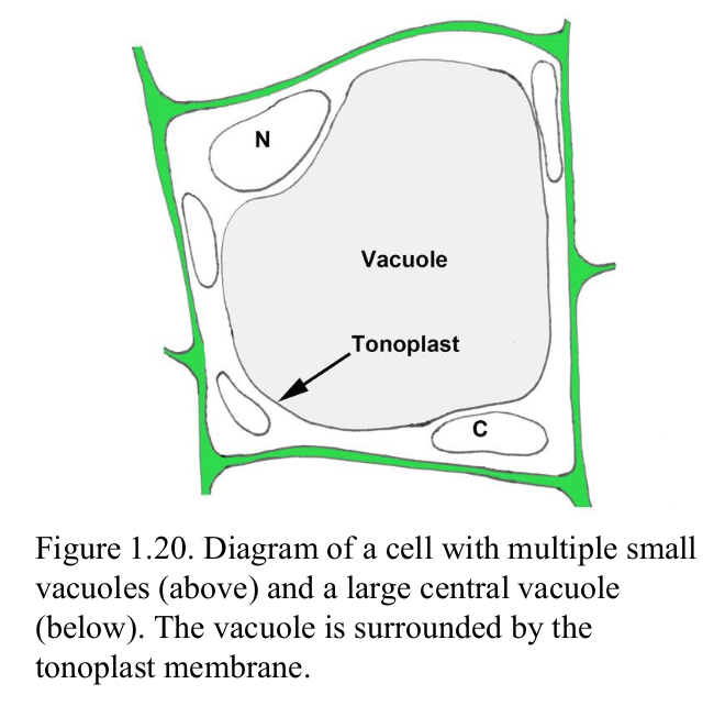
what is the function of the vacuole?
makes cells turgid by pushing cytoplasm against cell wall → provides support
temporary store of sugars and amino acids
stores and excretes waste products
pigments may give cell colour
what is the function of the cell wall?
provides structural support and strength to the cell
prevents cell from bursting under osmotic pressure
what is a plant cell wall made up of?
cellulose microfibrals
what is an algal cell wall made up of?
cellulose and/or glycoproteins
what is a fungal cell wall made up of?
chitin, glycan and glycoproteins
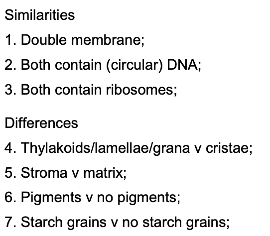
outline the similarities in and the differences between the structures of chloroplasts and mitochondria (4):
accept chloroplasts have chlorophyll whereas mitochondria do not
what are the 3 key types of microscope?
optical/light microscope
transmission electron microscope
scanning electron microscope
what is magnification?
how many times larger an image is compared to the object
what is resolution?
minimum distance between 2 objects in which they can still be viewed as separate
how do you convert from mm → µm?
x1000
how do you convert from µm → nm?
x1000
what is the equation for magnification?
magnification = image size/object size
what is an eyepiece graticule?
circular piece of glass/plastic that sits on a ledge in the eyepiece, w/ a scale engraved into it
will always appear the same no matter the magnification

what is a stage micrometer?
slide w/ a very accurate scale engraved into the glass, placed on microscope stage
distance between lines remains same no matter the magnification
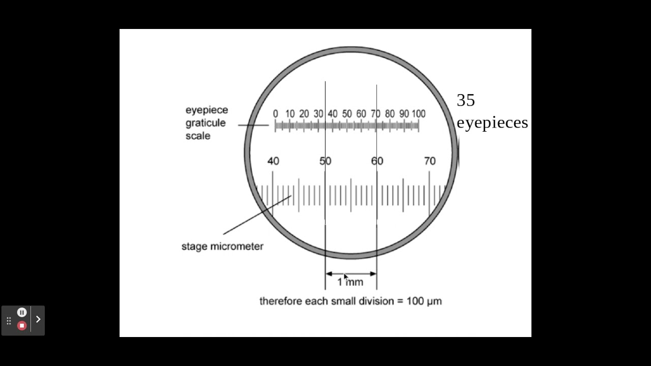
why must we calibrate the eyepiece graticule?
graticule scale is arbitrary (represents different lengths for different magnifications), so needs to be calibrated for any new microscope or new magnification
how does a light microscope work? what is its max magnification and resolution?
visible light is bent through the lens system to enable the user to see the specimen
max resolving power = 0.2 µm
max magnification = x1500
evaluate the use of a light microscope:
strengths:
living specimens can be viewed
limitations:
much lower resolving power and magnification compared to electron microscopes
stains must be used, as cells transparent and components not distinguishable, which usually kills cells
specimen must be extremely thin for light to pass through
a scientists prepared alveolar tissue to view using optical microscope. the scientist cut very thin slices of the alveolar tissue.
explain why the scientist used very thin slices of alveolar tissue with the optical microscope (2)
what is the maximum magnification and resolution of a transmission electron microscope and scanning electron microscope?
max magnification = x1,500,000
max resolving power = 0.0002 µm (TEM), 0.02 µm (SEM)
how does a transmission electron microscope work?
electron beam penetrates the cell, providing details of cell’s internal structure
denser parts of specimen absorb more electrons, making them look darker - this is how an image is formed
evaluate the use of a transmission electron microscope:
strengths:
higher resolving power and magnification than light microscopes
limitations:
can only be used on thin specimens
complex staining process
b&w image
can only be used on dead specimens as system must be in a vacuum
may contain artefacts as a result of how the specimen is prepared
how does a scanning electron microscope work?
beam of electrons moves across specimen, knocking electrons off the specimen
electrons gathered in a cathode ray tube - this is how an image is formed
evaluate the use of a scanning electron microscope:
strengths:
higher resolving power and magnification than light microscopes
produces 3D images
limitations:
complex staining process
b&w image
can only be used on dead specimens as system must be in a vacuum
may contain artefacts as a result of how the specimen is prepared
lower resolving power than transmission electron microscope
what is a bacterial cell wall made up of?
murein (peptidoglycan)
give 2 features of all prokaryotic cells that are not features of eukaryotic cells (1)
reject plasmids - smaller versions of circular DNA which are not necessary for survival
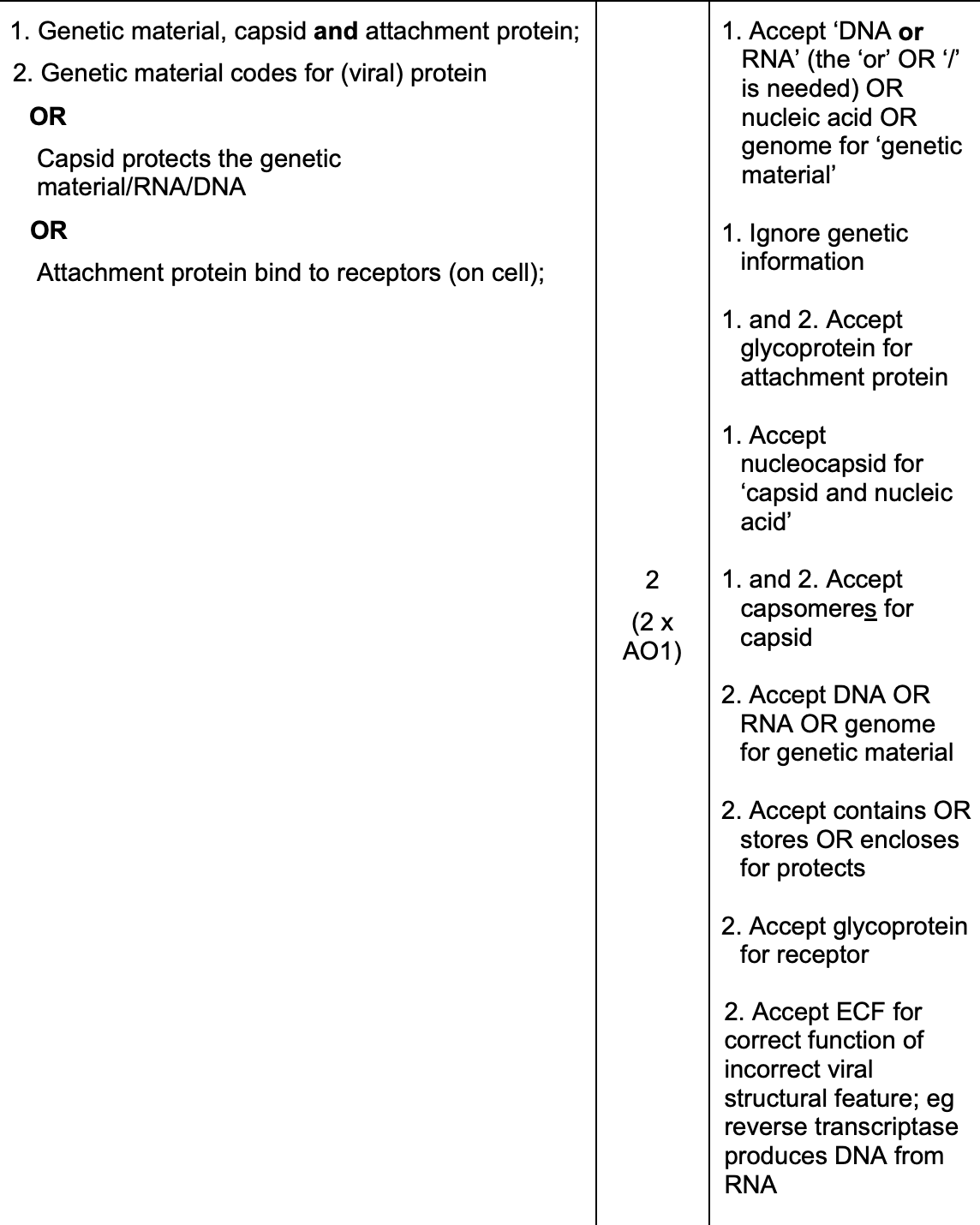
give 3 structural features found in all virus particles and describe their function:
capsid - protects genetic material
genetic material - codes for viral protein
attachment protein - to bind to receptors on cell
name the 7 key structural components of a general bacterial cell

flagellum
genetic material (circular and plasmids)
cytoplasm
ribosomes
capsule
murein cell wall
cell membrane
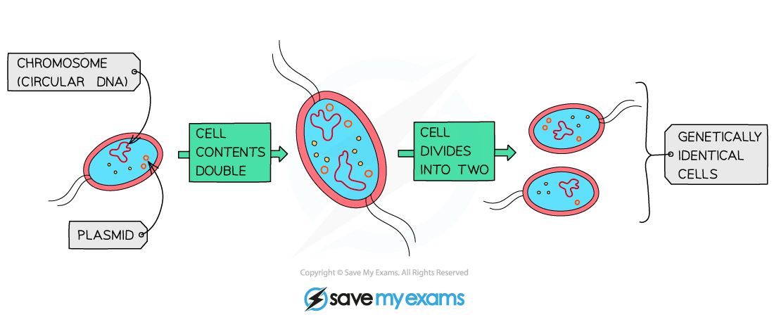
describe the process of binary fission:
circular DNA and plasmids replicate (main DNA loop only replicated once but plasmids can be replicated many times)
cell grows and DNA loops move to opposite poles of cell
cytoplasm begins to divide and new cell walls begin to form
cytoplasm divides and 2 daughter cells are produced w/ 1 copy of circular DNA but a variable no. of copies of plasmids
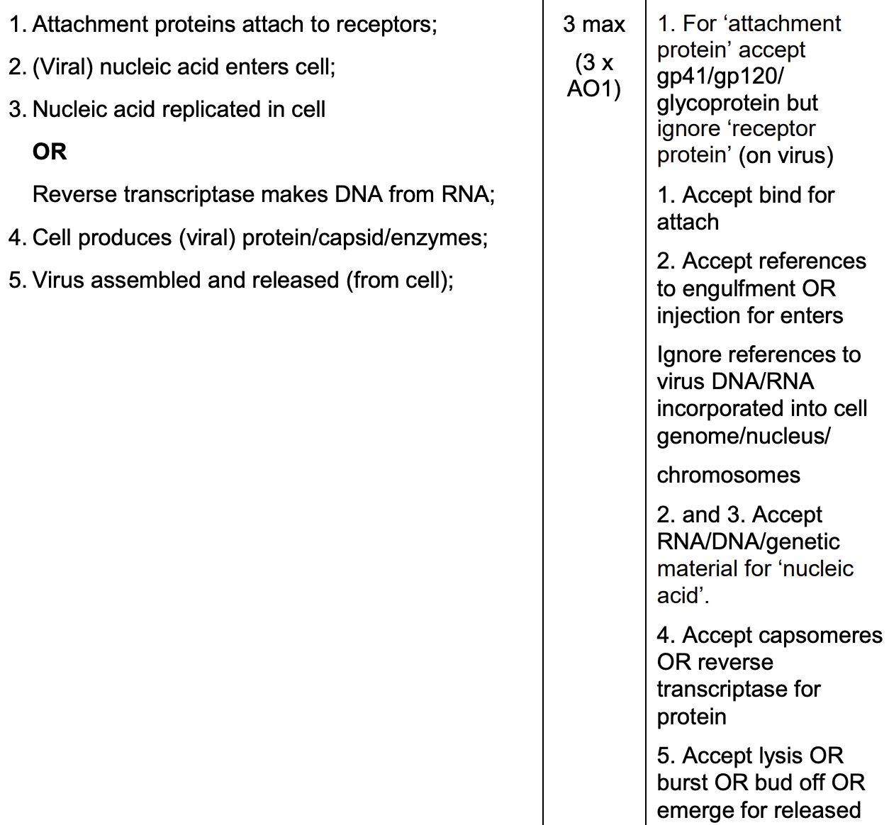
describe viral replication (3)
attachment proteins attach to receptors
nucleic acid/DNA/RNA enters cell
nucleic acid replicated in cell/reverse transcriptase makes DNA from RNA
cell produces protein/capsid/enzymes
virus assembled and released from cell
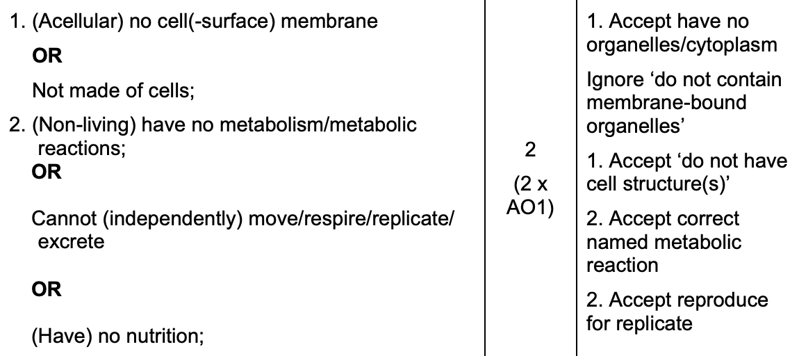
why are viruses described as acellular and non living? (2)
not made of cells/no cell surface membrane
no metabolism/nutrition
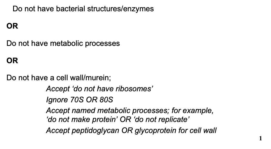
give one reason why antibiotics are ineffective against viruses:
any 1 from:
do not have bacterial structure/enzymes
do not have metabolism
do not have cell wall/murein
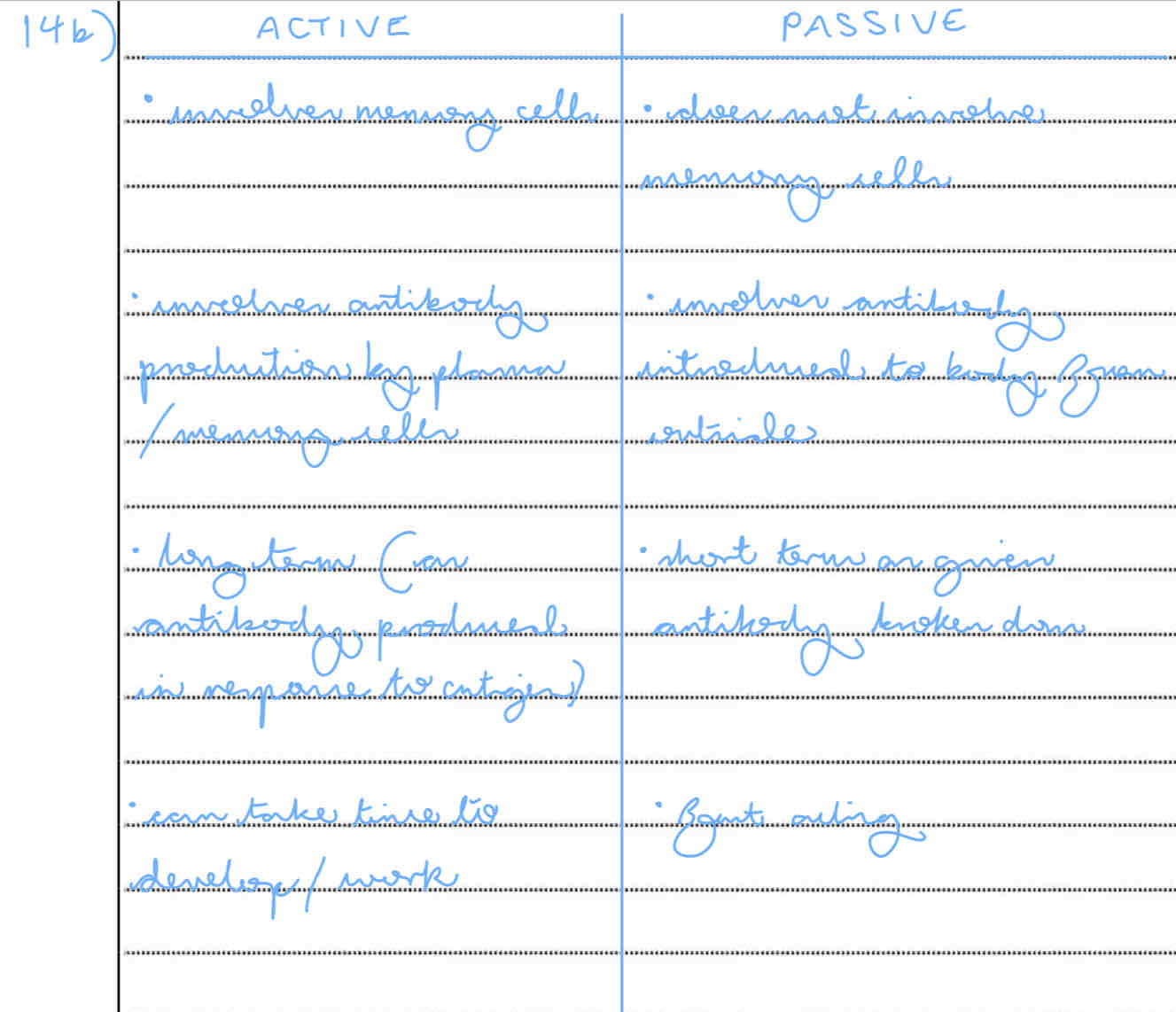
describe the differences between active and passive immunity (5)
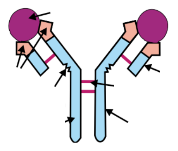
can you label this antibody?
yes
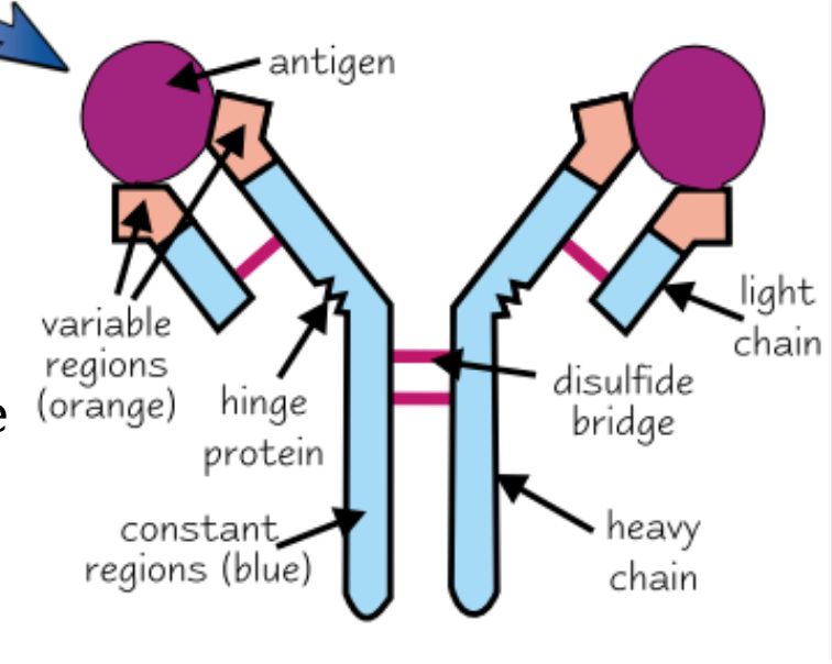
how do antibodies help to destroy pathogens?
bind and neutralise toxins
agglutination
what is agglutination? why is it significant?
antibodies are flexible, which cause pathogens to clump together
→ this makes the pathogens easier to locate and destroy by phagocytes
draw the shape of the primary/secondary immune response graph:

explain the shape of the primary/secondary immune response graph:
primary immune response:
takes time for clonal selection and expansion of specific T and B cells
antibodies do not begin to appear in the blood for several days after the foreign antigen enters the body (this is when symptoms occur)
some B cells differentiate during clonal expansion into plasma cells and memory cells but plasma cells are short lived :(
secondary immune response:
B memory cells recognise antigen and quickly / and differentiate into plasma cells and more memory cells
antibodies made more quickly and in a higher conc → pathogens killed before symptoms develop
what is active immunity?
immunity developed after immune system makes its own antibodies
what is natural active immunity?
immunity developed after immune system makes its own antibodies following infection
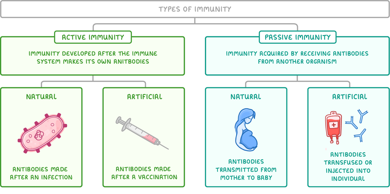
what is artificial active immunity?
immunity developed after immune system makes its own antibodies following vaccination

what is passive immunity?
immunity acquired by receiving antibodies from another organism
what is natural passive immunity?
immunity acquired by transmission of antibodies from mother to baby
