Microbiology Lab Final
1/68
There's no tags or description
Looks like no tags are added yet.
Name | Mastery | Learn | Test | Matching | Spaced | Call with Kai |
|---|
No analytics yet
Send a link to your students to track their progress
69 Terms
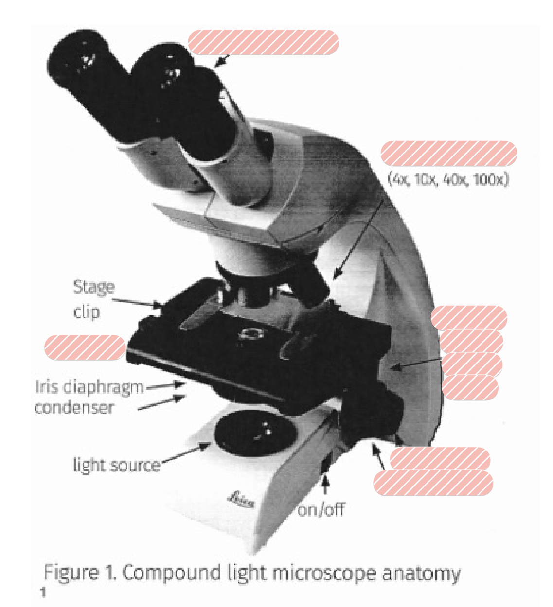
Label the:
Ocular lenses
Objective Lenses
Stage
Fine focus
Coarse focus
Stage adjustment knobs
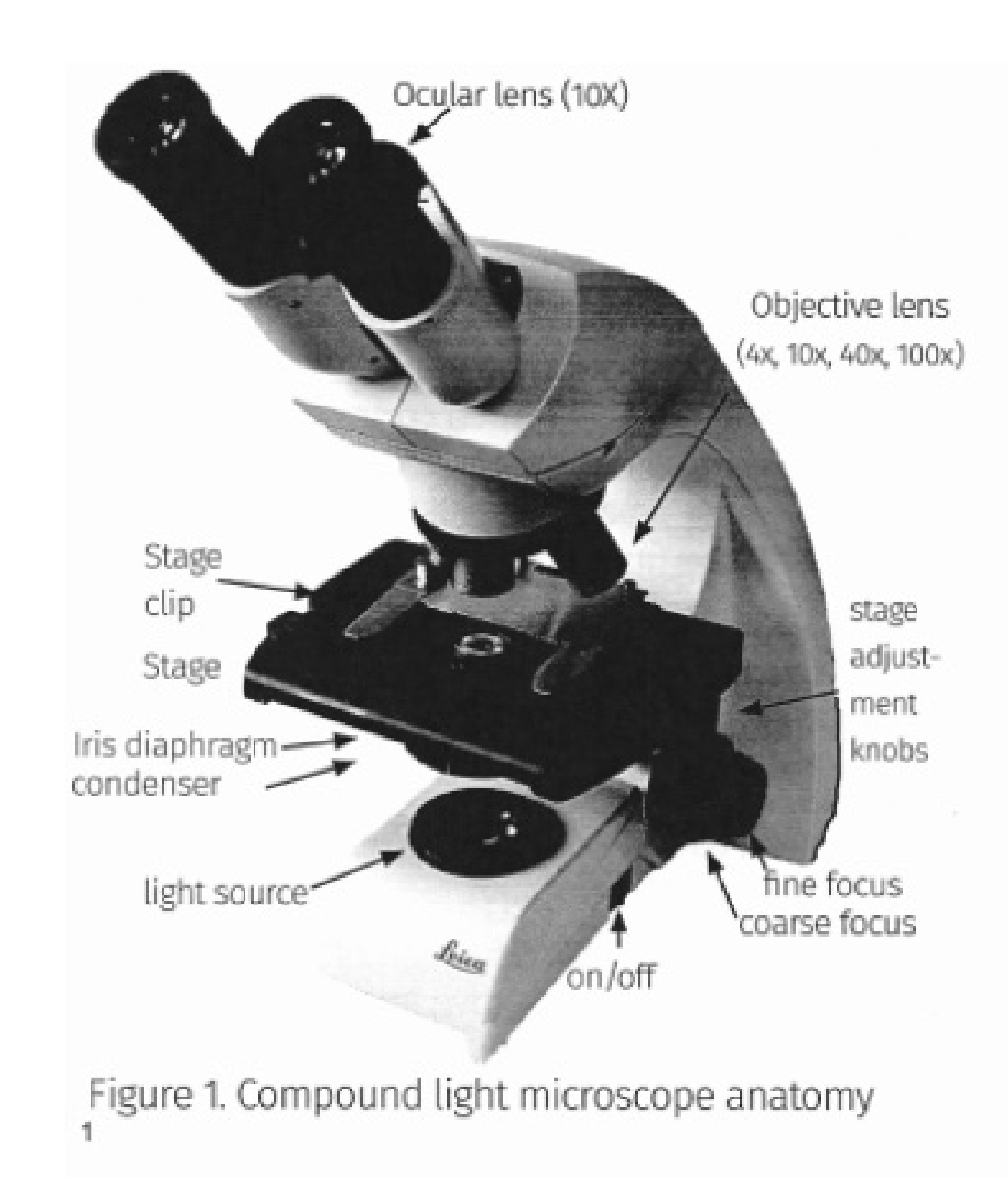
What is the magnification of the 4 objective lenses?
Scanning (4x)
Low power (10x)
High power (40x)
Oil Immersion (100x)
Which objective lens do we use to view bacterial specimens and what is it called?
The 100x (Oil Immersion) lens is used to view bacterial specimens
If you are focussing the microscope you start with
the 4x or 10x objective using the coarse focus
Once focused on the 4x/10x objectives you will switch to higher objectives and only use the
fine focus
Purpose of Isolation Streak
To separate individual bacteria from a mixture to obtain a pure culture (single species)
How do you know if an isolation streak is successful?
Characterized by visible, individual colonies separated from each other, typically appearing in the 3rd or 4th quadrant
When/what is a Gram stain used for?
differentiates gram positive (purple/violet) vs gram negative (pink/red) bacteria, based on cell wall thickness
Process for a Gram stain?
1) Crystal violet (Primary stain; both cells will be colored) →
2) Iodine (Mordant; sets the dye) →
3) Ethyl alcohol (decolorizer; Gram - cells colorless and Gram + remain purple) →
4) Safranin (counterstain; Gram - cells red/pink, and Gram + remain purple)
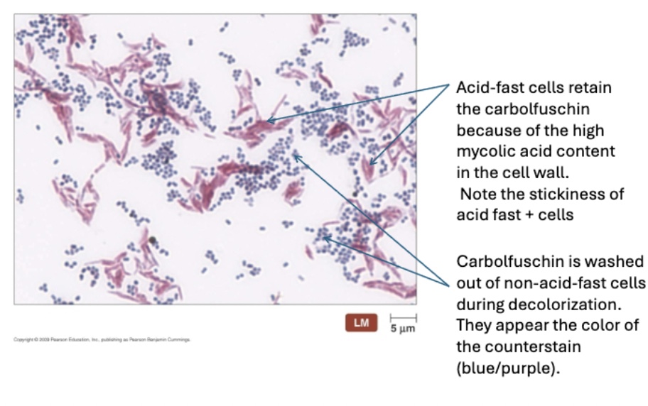
When/what is a Acid-fast stain used for?
Identifies Mycobacterium species; acid-fast bacteria retain carbol fuchsin (red), while non-acid-fast bacteria take up methylene blue.
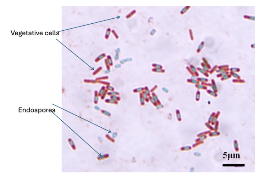
When/what is a Endospore stain used for?
Identifies bacterial spores (green) from vegetative cells (red/pink)
When/what is a Capsule stain used for?
Highlights bacterial capsules as halos surrounding a cell, often using a negative stain background
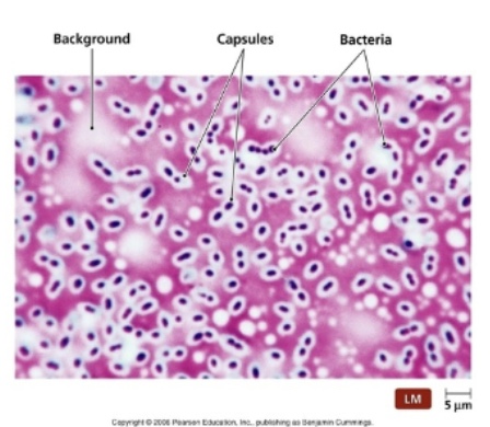
It is a negative staining method that uses an acidic stain (congo red) for the background and a basic stain (crystal violet) for the bacterial cell
Capsule staining
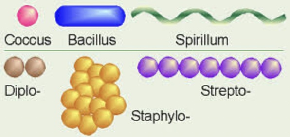
Recognize/draw the following cellular morphologies: cocci, bacilli
Cocci (spherical/round)
Bacilli (rod-shaped)
Be able to recognize/draw the following cellular arrangements: singular, staphylo-, strepto-
Singular (individual)
Staphylo (clusters)
Strepto (chains)
If you are performing a Gram Stain and forget to add the crystal violet stain what will happen?
All cells will eventually look pink/red from the counterstain (safrinin)
If you are performing a Gram Stain over decolorize it (add too much ethyl alcohol or leave too long), what will happen?
Gram-positive cells (purple/violet cells) will lose their purple color and appear pink (false negative)
If you are performing a Gram Stain over undercolorize it (don’t leave on for too long or don’t add enough), what will happen?
Gram-negative cells retain purple and do not look pink/red after adding the counter stain (false positive)
Bacterial cell walls are _ charged and attract_(positive) dyes
negatively, basic
For capsule stains, _(negative) dyes are used because they are
acidic, repelled by the cell, staining only the background
Encapsulated bacteria in a simple or Gram stain will show a
clear, unstained area (the capsule) around the cell
An Acid-fast bacteria may stain inconsistently or appear
“ghost like” in a standard Gram stain due to their waxy mycolic acid walls
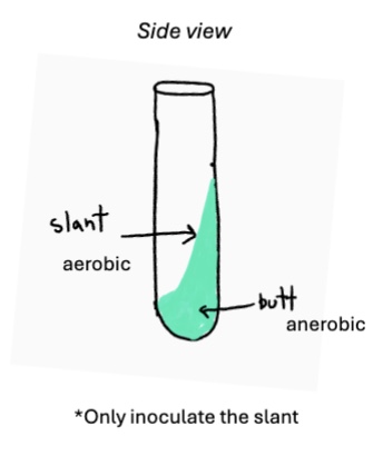
Is a differential medium used primarily to determine the oxygen requirements of microorganisms
Fluid Thioglycolate (FTG)
FTG result where growth is only present at the top
Obligate Aerobe
FTG result where growth is only present at the bottom
Obligate Anaerobe
FTG result where growth is only present throughout, but much thicker at the top
Facultative Anaerobe
FTG result where growth is uniform throughout
Aerotolerant Anaerobe
What does it mean if there is a red turbid area extending from the stab line?
The test is positive, the bacteria is motile
What does it mean if there is red growth only on the stab line?
The test is negative, the bacteria is not motile
What are two qualities of motility media that help us determine motility (general)*?
We used a semi-solid agar and a TTC indicator that turns red where bacteria grow
What is the selective ingredient in the MSA (Mannitol Salt Agar)?
7.5% Sodium Chloride (Salt)
Only bacteria that can tolerate _ will grow on an MSA plate
salt; Gram negative will not grow, some Gram positive will
What is the differential ingredient in the MSA (Mannitol Salt Agar)?
Mannitol (sugar) and Phenol Red (pH indicator); if the bacteria can perform Mannitol fermentation media color will change
MSA selects against
non-staphylococci
What does it mean if there is growth on an MSA plate? What does MSA select for? What does it mean if an MSA plate stays red?
If there is growth the bacteria is salt-tolerant (Gram positive), but does not ferment mannitol
What does it mean if an MSA plate turns yellow around the growth?
The bacteria is salt-tolerant (Gram positive) and ferments mannitol
What are the (general) selective ingredients in an EMB (Eosin Methylene Blue) plate?
Eosin Y and Methylene Blue dyes
Eosin Y and Methylene Blue dyes select against
Gram-positive bacteria
What is the main differential ingredient in an EMB plate?
Lactose
What does it mean if there is no growth on an EMB plate? What does EMB select against?
If there is no growth the bacteria is Gram-positive and EMB selects against Gram-positive bacteria
What does it mean if there is growth on an EMB plate? What does EMB select for?
Growth means that this is a Gram-negative bacteria as EMB selects for Gram-negative bacteria
What does it mean if growth on an EMB plate does not change color?
The bacteria is Gram-negative, but does not ferment lactose
What does it mean if growth on an EMB plate turns green/metallic/dark purple?
The bacteria is Gram-negative and ferments lactose (Green = E.coli)
What are coliform bacteria and why do we monitor environmental samples for coliform growth?
Gram-negative bacteria that are lactose fermenting bacilli used as indicators of fecal contamination
For MSA and EMB, what happens when you forget the selective ingredient? What happens if you forget the differential ingredient?
Forgetting the selective ingredient causes a false positive because non-target bacteria (like Gram-negatives on MSA or Gram-positives on EMB) are no longer inhibited and will grow freely, while forgetting the differential ingredient causes a false negative because the bacteria will grow without producing the characteristic color changes or sheens needed to identify their metabolic traits.
What is the name of the enzyme that breaks down tryptophan?
Tryptophanase
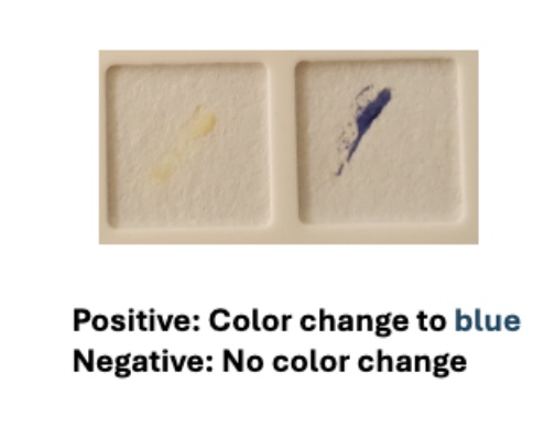
What does a positive indole test look like? What does this tell you about the organism?
A positive indole test has a red/pink ring at the top which indicates that the bacteria produces indole (organism has tryptophanase enzyme)
What does a negative indole test look like? What does this tell you about the organism?
A negative indole test has a yellow/greenish ring at the top which indicates that the bacteria does not produce indole (organism doesn’t have a tryptophanase enzyme)
What does a positive MR test tell you about the bacteria?
Red = positive test → The bacterium performs mixed acid fermentation producing stable acids (pH of media is low)
What does a positive VP test tell you about the bacteria (general)?
Red = positive test → The bacterium performs butanediol fermentation producing unstable acids (acetoin) (pH of media is neutral)
How are these two tests similar? (MR-VP)
They are performed as a combination test, using the same MR-VP broth medium. The broth contains glucose that the bacteria use for fermentation. Both tests determine how a microorganism metabolizes glucose. Differ in which specific end products they detect (stable acids for MR vs acetoin for VP)
What 2 results indicate a positive citrate test? What does it tell you about the organism?
A blue color or growth on the slant, which tells us that the bacterium uses citrate as a sole (not sure if sole) carbon source. If citrate is used as a carbon source → it will produce alkaline compounds that increase media pH and change color of pH indicator to blue
What does a negative citrate test look like? What does it tell you about the organism?
The media stays that green color with no growth present → bacterium does not use citrate as a carbon source

What is the enzyme that breaks down urea?
Urease
What does a positive urease test look like? What does this tell you about the organism?
The media will turn pink (due to pH increase) indicating that the bacteria produces urease enzyme → breaks down urea into ammonia (indicating pH increase from ammonia, buffer in media requires LOTS of ammonia to be produced)
What does a negative urease test look like? What does this tell you about the organism?
The resulting media will be a yellow/orange color indicating the bacteria does not have a urease enzyme
What does a positive and negative catalase test look like? What does each result tell you about the organism?
Positive test: Look for the immediate formation of bubbles. This tells you the organism produces the enzyme catalase.
Negative test: Look for no bubbles. This tells you the organism does not produce catalase.
What is the reagent added in the catalase test? In the presence of catalase, what two products does the reagent get broken down into?
Reagent added: Hydrogen Peroxide (H2O2).
Two products formed: Catalase breaks the reagent down into water (H2O) and oxygen
What does a positive and negative oxidase test look like? What does each result tell you about the organism?
Positive test: The bacteria or swab turns dark blue or purple within 20 seconds. This indicates the organism produces the enzyme cytochrome c oxidase.
Negative test: There is no color change within 20 seconds. This tells you the organism does not produce cytochrome c oxidase.
Be able to do and interpret an oxidase test.
Procedure: Use a wooden applicator stick (avoid metal loops as they can cause a false positive) to pick up a small amount of bacterial growth and rub it onto an oxidase reagent slide or a swab infused with the reagent.
Looking at TSI test, the result is a yellow slant/yellow butt, what do you interpret?
A/A results (Acid/Acid) → Bacteria fermented Glucose (mainly) and lactose and/or sucrose fermenter after → Glucose is used first, followed by the higher concentrations of lactose and sucrose. This produces enough acid to keep the entire tube yellow.
Looking at TSI test, the result is a red slant/yellow butt, what do you interpret?
K/A (Alkaline/Acid) → Bacteria fermented glucose only → Because glucose is in low concentration (0.1%), it is used up quickly, turning the whole tube yellow initially. Once the glucose is gone, the bacteria on the aerobic slant switch to using peptones, which raises the pH and turns the slant red
Looking at TSI test, the result is a red slant/red butt, what do you interpret?
K/K (Alkaline/Alkaline) → bacteria fermented no sugars and used proteins (peptones) instead → No acid is produced because no sugars are fermented. The bacteria use peptones aerobically (turning the slant red) or both aerobically and anaerobically (turning both slant and butt red)
Looking at TSI test, the result is a black precipatate, what do you interpret?
Sulfur reduction requires an acidic environment; therefore, if you see a black butt, you assume it is acidic (Yellow/A) even if the black color masks the yellow.
K/A + H₂S (if the slant is red and the butt is black)
If an organism produces gas while it ferments, how will you know?
Yes we can tell if there are cracks in agar, or agar is pushed up from bottom of tube
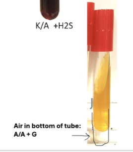
If an organism reduces sulfur how will you know?
If we see a black precipitate form at the butt of the tube. Sulfur reduction requires an acidic environment; therefore, if you see a black butt, you assume it is acidic (Yellow/A) even if the black color masks the yellow.
β (Beta) hemolysis
Zone clearing of the media → Total/complete destruction of red blood cells
α (alpha) partial hemolysis
Media cloudy/greenish → partial destruction of red blood cells
γ