GA Exam 2 - Wrist + Hand
1/40
There's no tags or description
Looks like no tags are added yet.
Name | Mastery | Learn | Test | Matching | Spaced | Call with Kai |
|---|
No analytics yet
Send a link to your students to track their progress
41 Terms
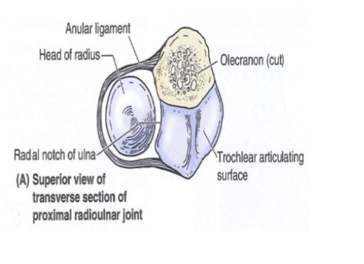
Proximal radioulnar joint
Synovial joint
Head of the radius
articulates with the radial
notch of the ulnaRadius is held in place by
the anular ligament
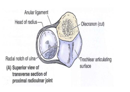
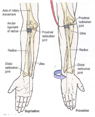
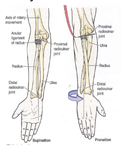
Movements of radioulnar joint
During pronation and supination the radial head rotates within the anular ligament
pronator teres
look at lab practical quizlet
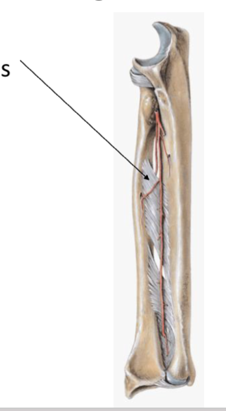
interosseous membrane
connects radius and ulna along their diaphysis
Forms the radioulnar syndesmosis joint
Distal radioulnar joint
Head of Ulna articulates
with the ulnar notch on
the radiusFibrocartilagenous articular disc (a.k.a. triangular ligament)
connects
Volar
palkm
Thenar
base of thumb
Hypothenar
base of pinkie
Muscles of Pronation and Supination
Supination: supinator and biceps brachii (esp during resistance)
Pronation: pronator quadratus and pronator teres
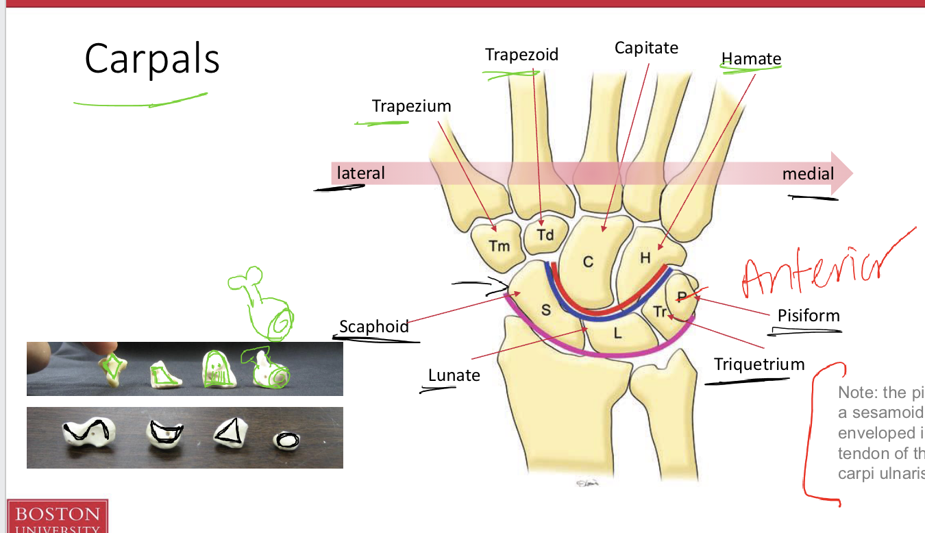
Pisiform is a _____ bone
sesamoid bone
Carpals Lateral to medial
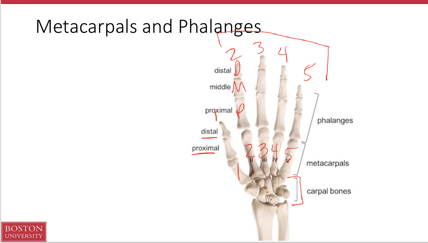
Metacarpals and Phalanges
Wrist complex contains 2 proximal-distal joints:
Radiocarpal
Midcarpal
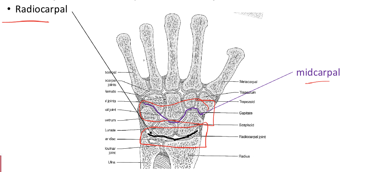
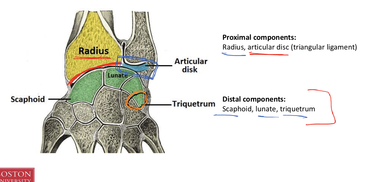
Proximal and Distal components of the Radiocarpal joint
Proximal components:
Radius, articular disc (triangular ligament)
Distal components:
Scaphoid, lunate, triquetrum
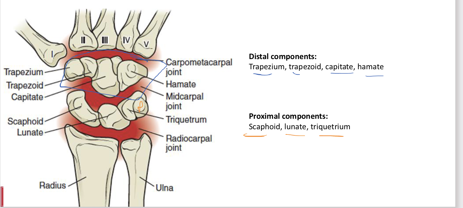
Proximal and Distal components of the Midcarpal joint
Distal components:
Trapezium, trapezoid, capitate, hamate
Proximal components:
Scaphoid, lunate, triquetrium
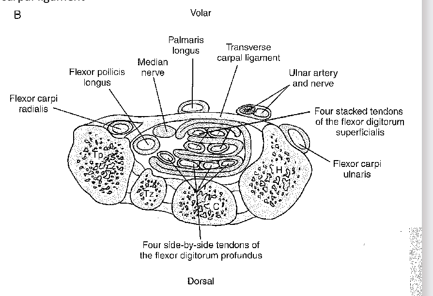
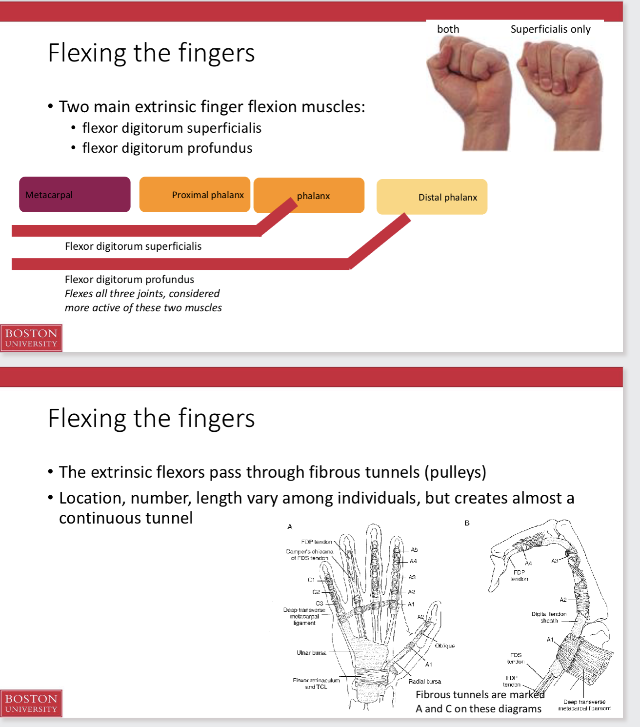
The carpal tunnel
A large, fibrous ligament stretches over the volar side of the wrist—transverse carpal ligament or flexor retinaculum
Forms a tunnel with the carpals (carpal tunnel) through which tendons pass
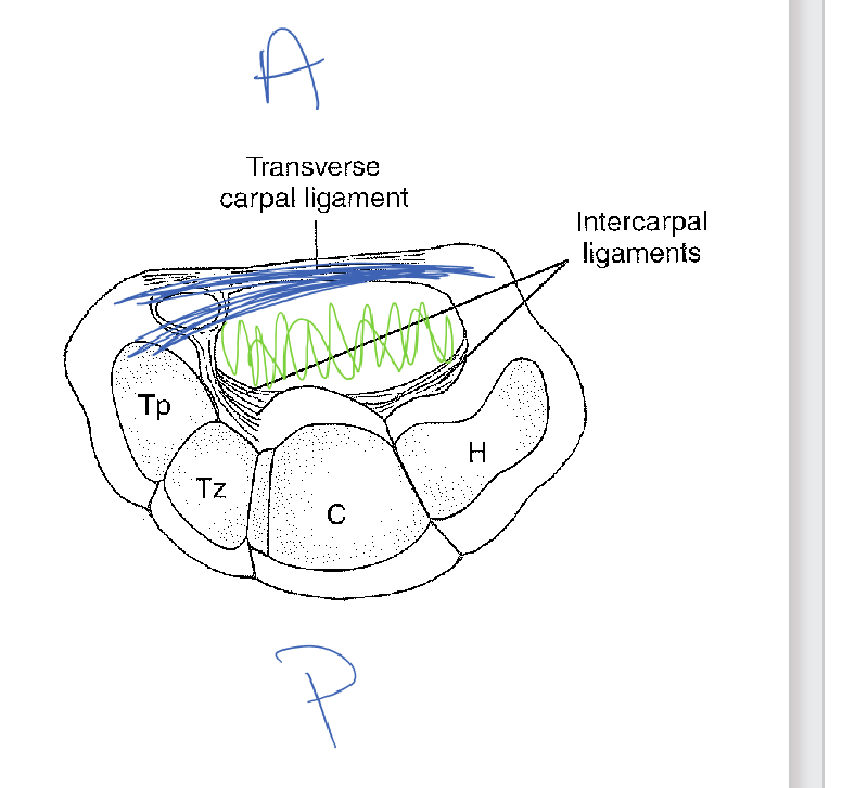
Intercapal joints
Joints between carpals
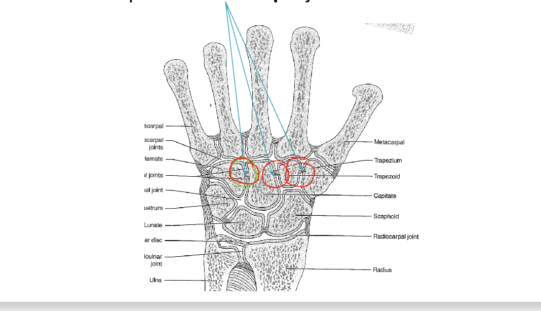
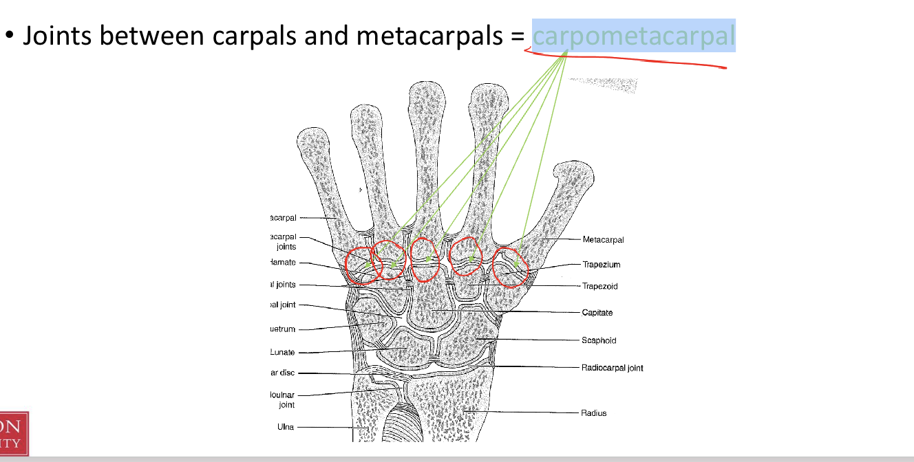
Carpometacarpal joints
Joints between carpals and metacarpals
***All carpometacarpal joints are capable of flexion/extension***
for the thumb,
has it’s own synovial joint and a greater ROM (abduction, adduction, flexion, extension, opposition)
For the fingers
share one synovial cavity
Metacarpals of the third and fourth fingers
have limited ROM,
fifth metacarpal have more ROM than 3rd + 4th.
The relatively immobile second and third joints
provide a stable axis for opposition
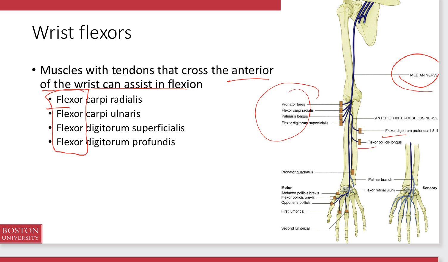
Wrist flexors
Muscles with tendons that cross the anterior of the wrist can assist in flexion
• Flexor carpi radialis
• Flexor carpi ulnaris
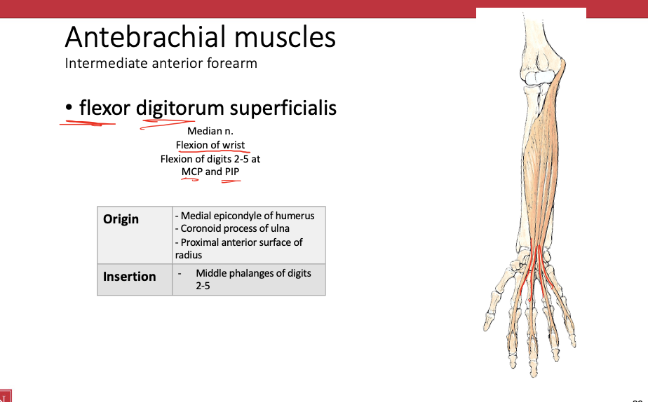
• Flexor digitorum superficialis
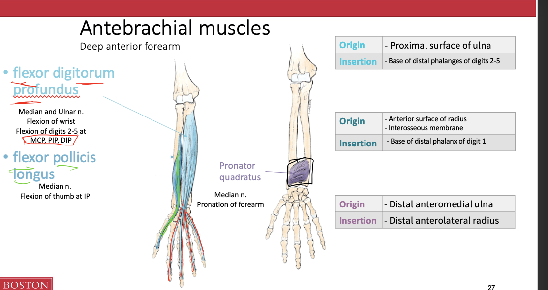
• Flexor digitorum profundis
Flexor pollicis longus
pronator teres
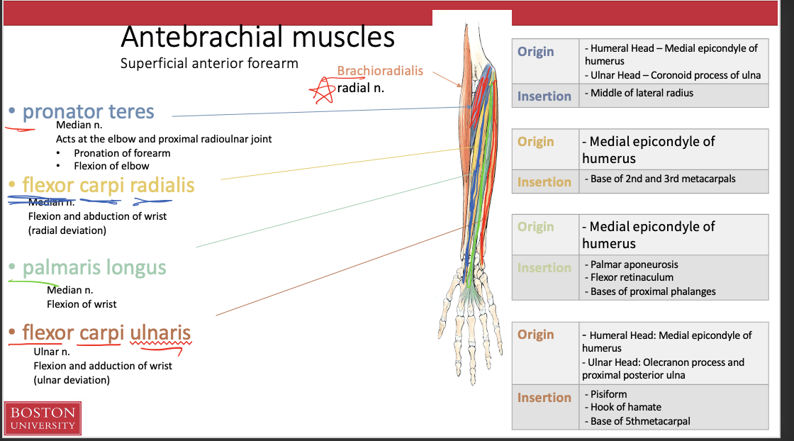
Antebrachial muscles (superficial anterior forearm)
Pronator teres
Flexor carpi radialis
palmaris longus
flexor carpi ulnaris
Antebrachial muscles (intermediate anterior forearm)
Antebrachial muscles (Deep anterior forearm)
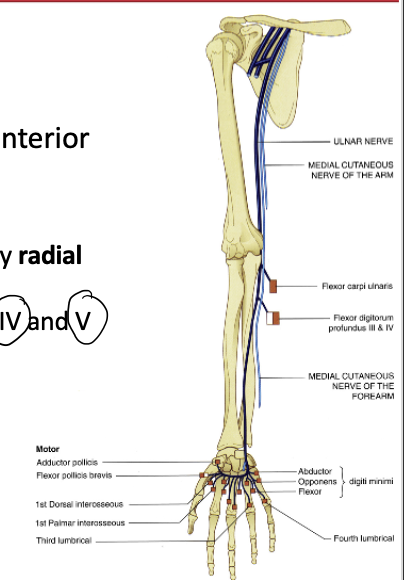
Median nerve
supplies the majority of the anterior compartment of the forearm
exceptions:
brachioradialis b/c it is lateral → radial nerve
flexor carpi ulnaris, and flexor digitorum profundi b/c = medial → ulnar nerve
Wrist extensors
Muscles with tendons that cross the posterior aspect of the wrist can assist in extension
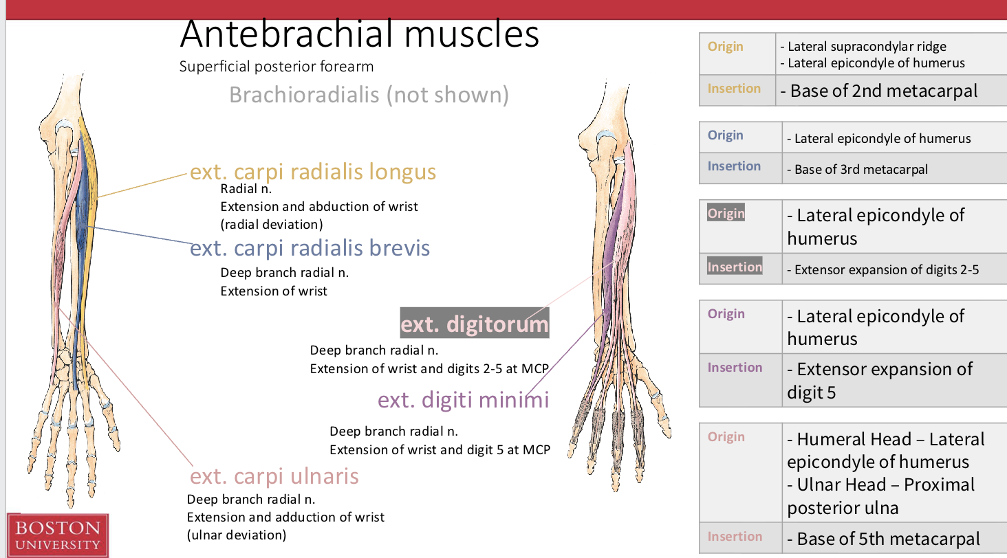
• Extensor carpi radialis longus
• Extensor carpi radialis brevis
• Extensor carpi ulnaris
• Extensor digitorum
• Extensor indicis
• Extensor digiti minimi
Antebrachial muscles (superficial posterior forearm)
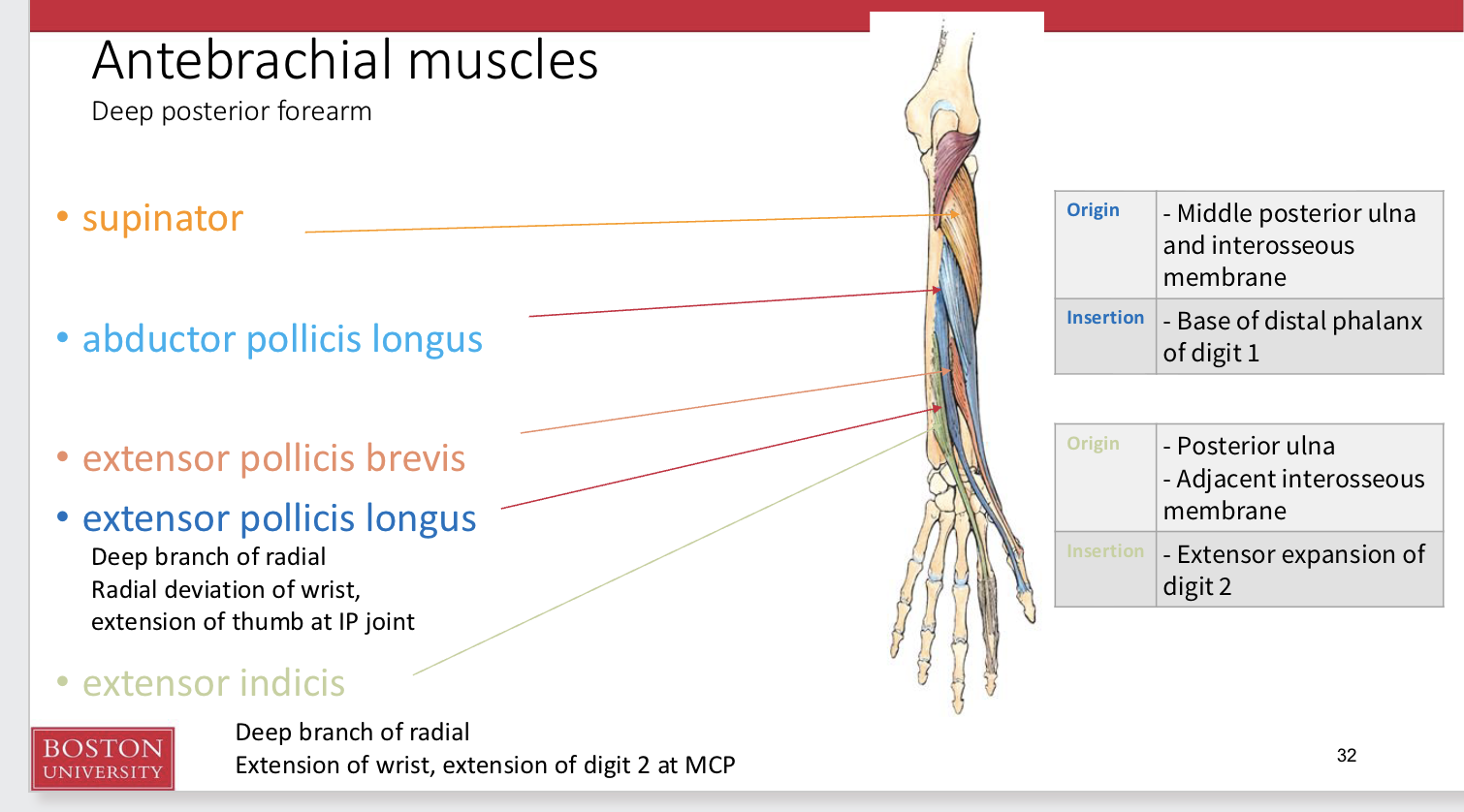
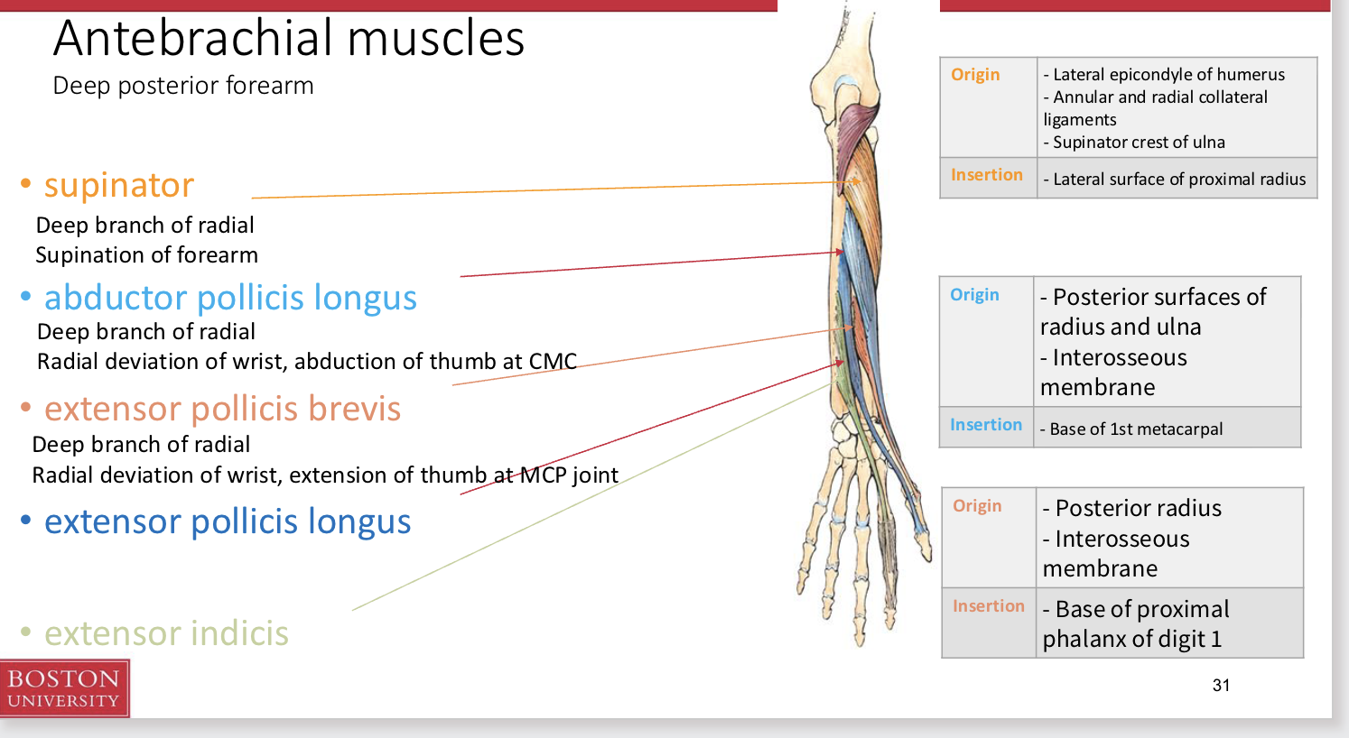
Antebrachial muscles (Deep posterior forearm)
Wrist Adductors and Abductors
ADDuctors
Muscles with tendons that cross the ULNAR side of the wrist ADDuct
Flexor carpi ulnaris
Extensor carpi ulnaris
ABDuctors
Muscles with tendons that cross the RADIAL side of the wrist ABDuct
Flexor carpi radialis
Extensor carpi radialis
Thumb (pollicis) muscles with tendons that cross the RADIAL side of the wrist assist in ABDuction
• Abductor pollicis longus
• Extensor pollicis longus
• Extensor pollicis brevis
Wrist motion in general
No muscular forces are applied directly to the proximal carpals
(scaphoid, lunate, triquetrium)
These bones essentially serve as a mechanical link between the radius
and distal carpals
They tend to collapse anteriorly when force is applied, but are
stabilized by the intercarpal ligaments
What things passes via carpal tunnel
Tendons of:
• Flexor digitorum profundus
• Flexor digitorum superficialis
• Flexor pollicis longus
Median nerve
- Pass through the carpal tunnel
Flexor carpi radialis is embedded within the tendon
Palmaris longus
Flexor carpi ulnaris are outside the tendon
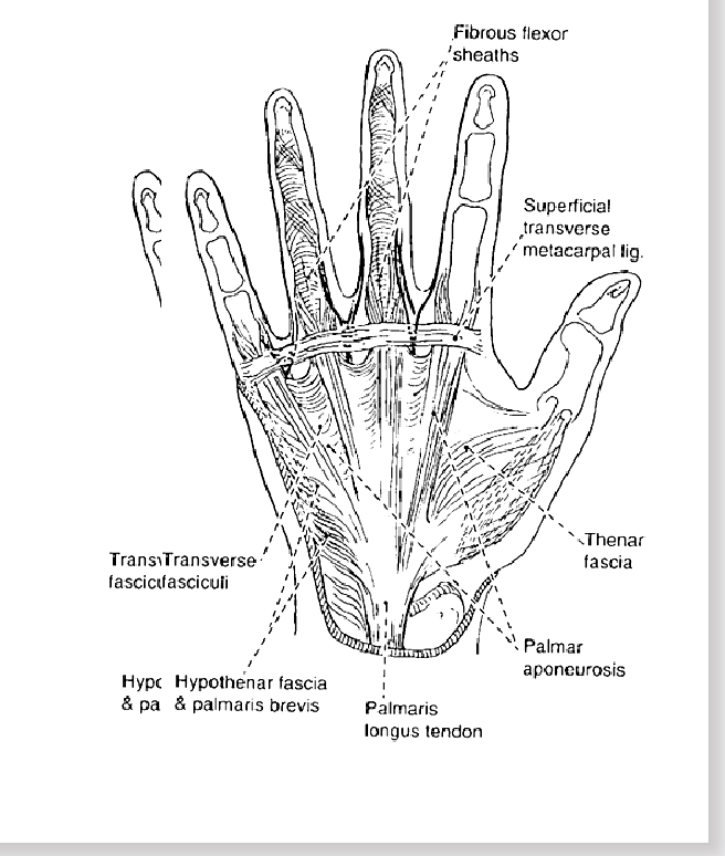
Anterior Palmar aponeurosis
Distally, the flexor retinaculum gives way to the palmar aponeurosis
This fascia covers the tendons, blood supply, nerves and muscles
It connects on it’s distal side to the superficial transverse metacarpal ligament
Posterior palmar aponeurosis
Similarly on the posterior side, the ligaments of all of the extrinsic finger extension muscles pass through the extensor retinaculum
Its function is to hold the tendons in place and not let them wander medially/laterally
Carpometacarpal joints
****All carpometacarpal joints are capable of flexion/extension*****
For the thumb
has it’s own synovial joint and a greater ROM
ex movem → (abduction, adduction, flexion, extension, opposition)
For the fingers
share one synovial cavity
Metacarpals of the third and fourth fingers have
limited ROM but fifth metacarpal have more ROM
The relatively immobile second and third joints
provide a stable axis for opposition
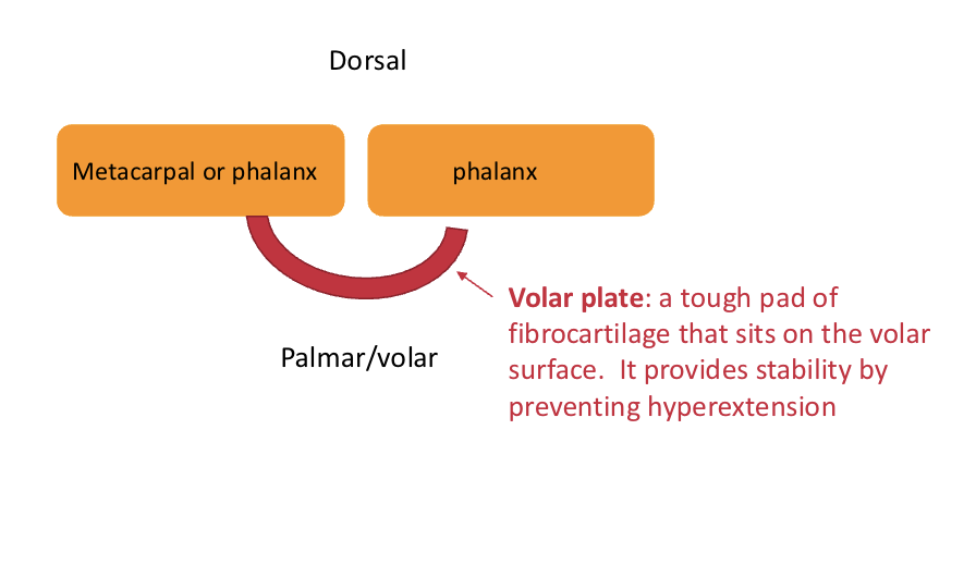
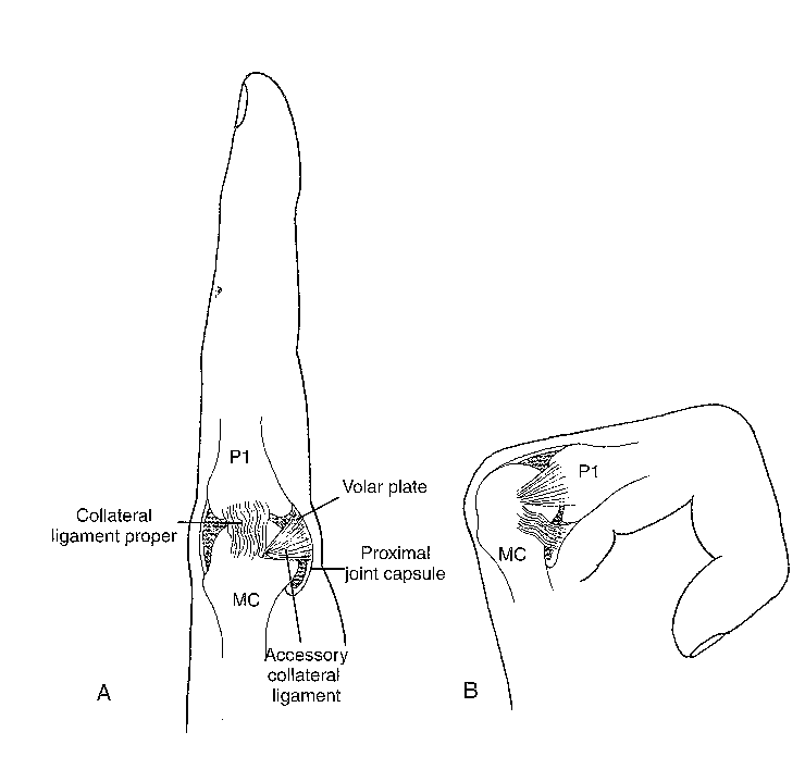
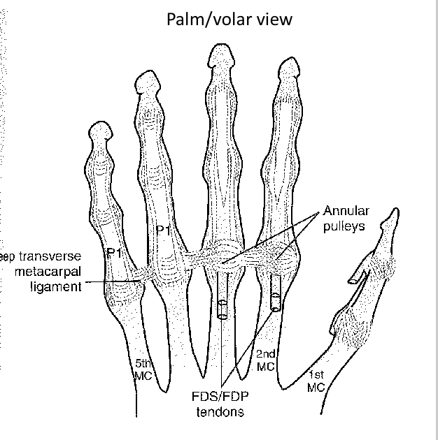
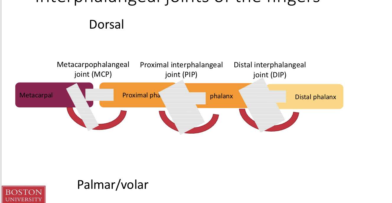
Finger joint layout basics
Metacarpophalangeal joints
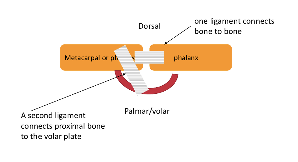
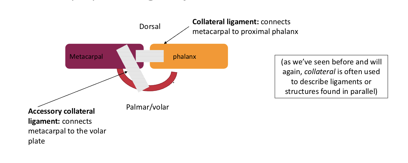
Components:
Volar plate → prevents hyperextension
Collateral ligament connects metacarpal to proximal phalanx
Accessory collateral ligament connects metacarpal to volar plate
Metacarpophalangeal joints are connected to each other by the deep transverse metacarpal ligament
Deep transverse metacarpal ligament passes connects the volar plates of the metacarpophalangeal joints
Flexor tendons pass over it
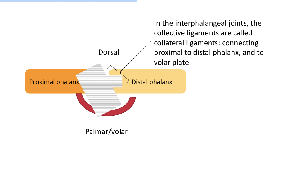
Interphalangeal joints
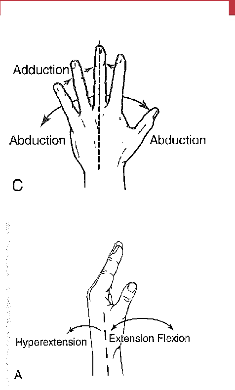
Movements of finger and thumb
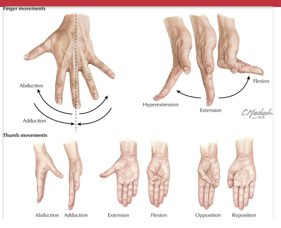
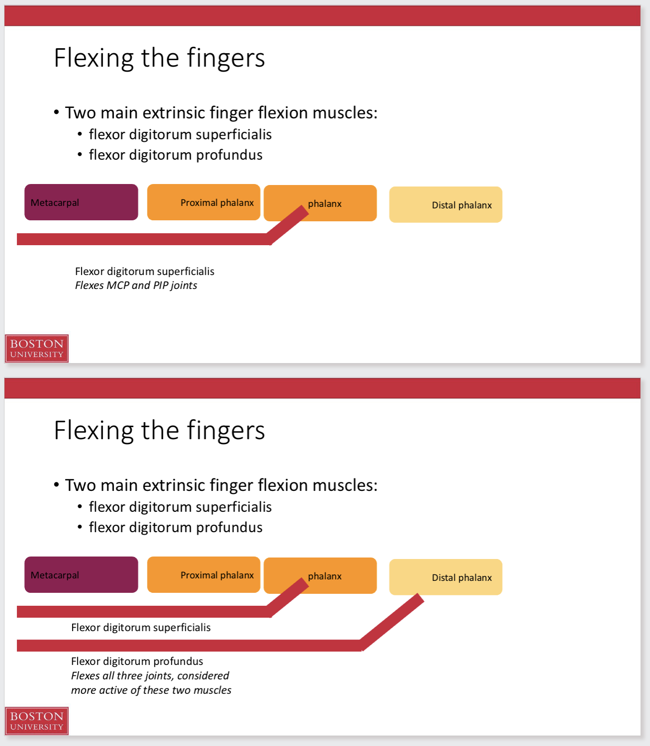
Flexing the fingers
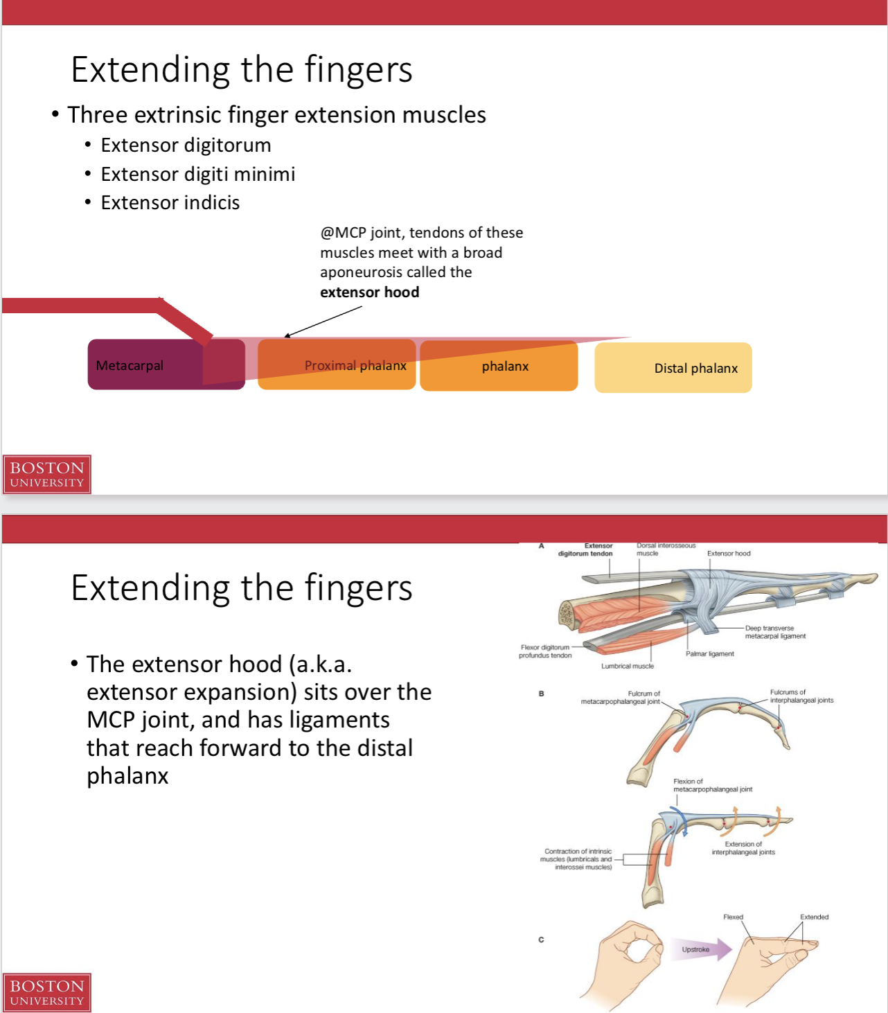
Extending the fingers
Intrinsic muscles of the hands:
• Lumbricals insert on the extensor hoods, aiding in extension
Interossei muscles:
insert onto the extensor hoods and contributing to finger extension and ab/adduction
Finger abduction and adduction
The dorsal interossei muscles contribute to abduction
The palmar interossei contribute to adduction
DAB/ PAD
Intrinsic muscles of the thumb and little finger:
Intrinsic muscles of the thumb:
Abductor pollicus brevis
Flexor pollicus brevis
Opponens pollicus
Adductor pollicus
Intrinsic muscles of the little finger:
• Abductor digiti minimi
• Flexor digiti minimi
• Opponens digiti minimi