Physiology lecture 23
1/39
Earn XP
Description and Tags
Vision and the eye - anatomy and physiology
Name | Mastery | Learn | Test | Matching | Spaced | Call with Kai |
|---|
No analytics yet
Send a link to your students to track their progress
40 Terms
LOs
• Describe the anatomy of the eye and the retina
• Explain how an image is formed on the retina
• Know what is meant by the fovea and the blind spot
• Describe two types of retinal photoreceptors: rods and cones.
• Know how the photoreceptors respond to light under different conditions
• Outline the trichromatic theory of colour vision
• Explain the process of light transduction
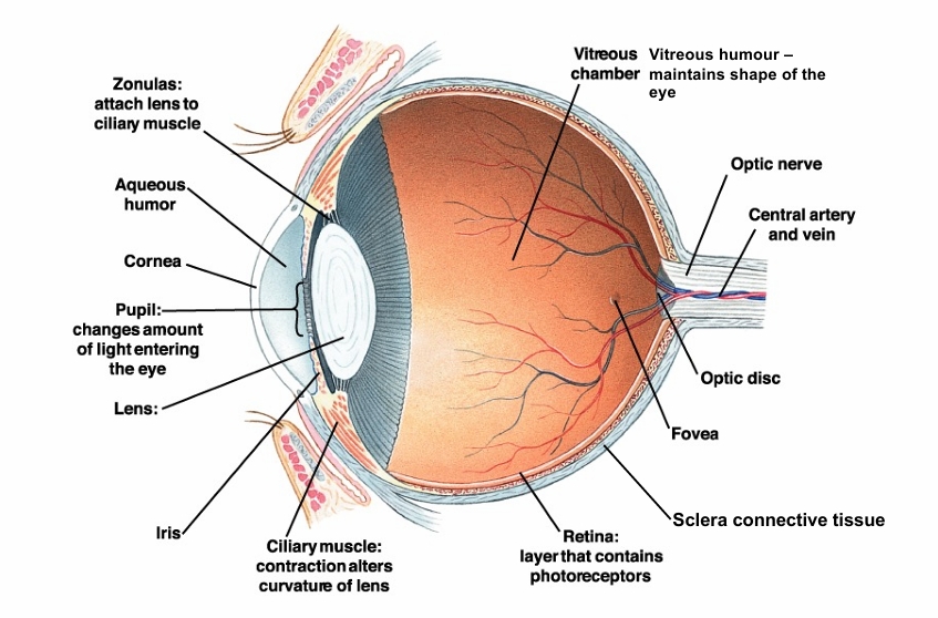
Cross section of eye
Cornea - curved surface in front of eye - role in refracting light and protective layer - fixed
Aqueous humor - fluid that provides nutrients to eye
Pupil - can open or close to let light in
Iris - can open or close to change pupil size
Lens - adjusts size to focus light and refract at diff levels
retina - the photoreceptors sense light and colour
sclera - protective tissue and maintains eye structure
The cornea
Responsible for refracting (bending) light to bring it into focus on the retina. The cornea (not the lens!) is responsible for most of refractive power of the eye. (Laser treatment acts on cornea)
Lens
Responsible for accommodation – adjusting the refractive properties of the eye to ensure objects in focus over a wide range of distance.
Iris
Pigment determines eye colour (blue eyes are actually lack of brown pigment)
Muscles (circular and radial) controlled by autonomic ns adjusts pupil diameter according to light levels, and emotional signals - attractive people
Pupil
Opening in centre of the iris allows light entry (2(light-8 dark mm range in diameter)
Retina
Layer at the back of the eye: photoreceptors, horizontal cells, bipolar cells, amacrine cells, ganglion cells and nerve fibres. (Retina part of brain).
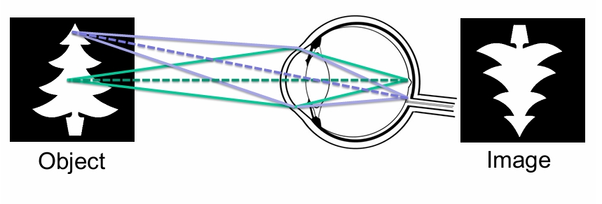
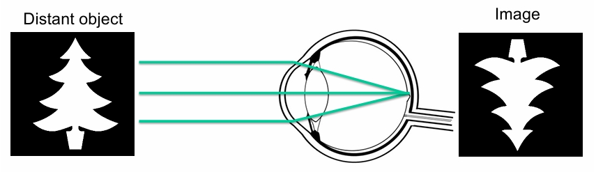
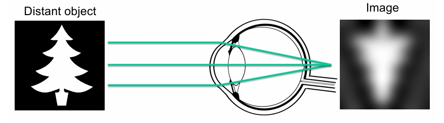
Image formation
Note that the image is inverted (& left/right reversed) on retina - brain then corrects this
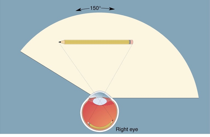
how large is eye’s visual field?
150 degrees fixed field of view
Role of the lens in accommodation
As object brought nearer to eye, light rays need to be bent more to keep object in focus
Lens flatter at longer distances, fatter/rounder to shorten focal length for close vision
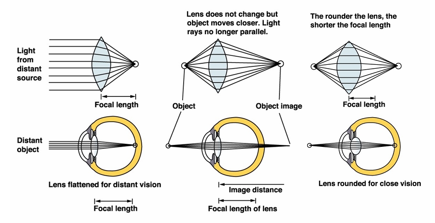
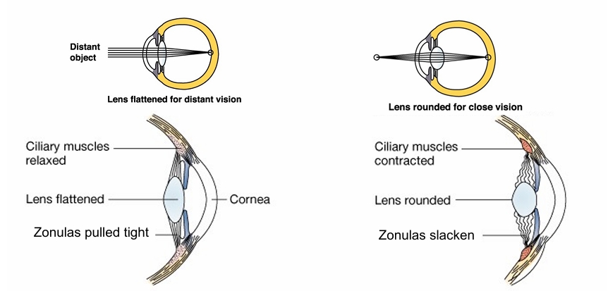
Role of the ciliary muscles in accommodation
• Lens flattened for distant vision, rounded for near vision (below),
• Contraction of ciliary muscles allows zonulas of Zinn to slacken, lens expands and becomes more rounded.
• Accommodation weakens with age. Reading glasses needed in middle age
Emmetropia
Normal focusing
The emmetropic eye can focus light from a distant object on to the retina.
• The image at the retina is sharp (in focus)
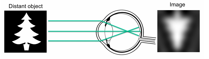
Myopia
Short sightedness
• The myopic eye has too much focal power for its length
• Light rays converge (are focused) in front of the retina
• The image at the retina is blurred (out of focus) Image . .
• Remember that distant objects are focused closer to the lens - A myopic eye will not be able to focus on distant objects, but can focus on objects at a short distance.
Hyperopia
Long sightedness
• The hyperopic eye has too little focal power for its length
• Light rays converge (are focused) behind the retina.
• The image at the retina is blurred (out of focus) Image .
• Remember that close objects are focused further to the lens - A hyperopic eye will not be able to focus on close objects, but can focus on objects at a long distance.
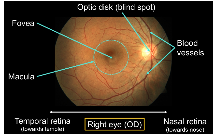
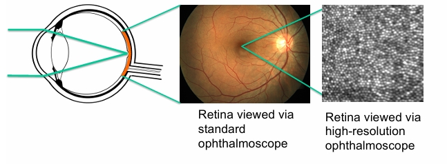
The retina – macro structure
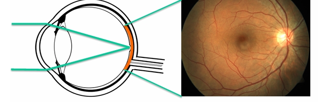
Retina viewed via an ophthalmoscope
macula - central part of retina (often affected by retinal diseases)
Middle of macula = fovea - very centre of vision
Optic disk - where optic nerve exits to brain - blind spot (lack of photoreceptors)
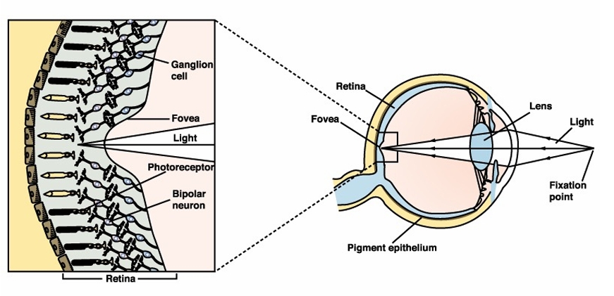
The foveal pit
In fovea cells, upper layers of retina pushed aside to allow light direct access to photoreceptors, region of highest acuity
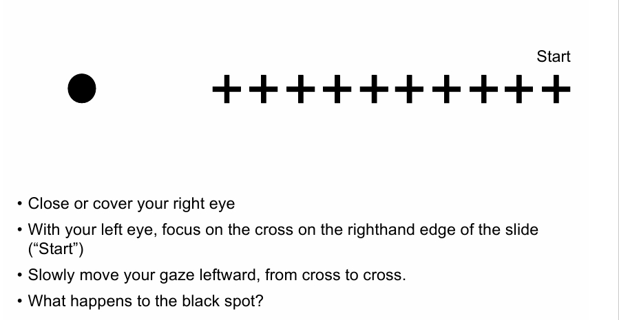
Blind spot demonstration
due to lack of photoreceptors in optic disc
The retina – macro structure (3 things)
• Fovea
– Pit in centre of macula
– Central part of visual field.
– Area of maximum acuity (highest image resolution) with highest density of cone receptors, colour vision
• Blind spot or optic disk
– Region where nerve fibres and blood vessels leave the eye
– Blind in this region
– Brain “fills in” the missing area (see demonstration of blind spot)
• Blood vessels
– Delivering oxygen and nutrients to retina
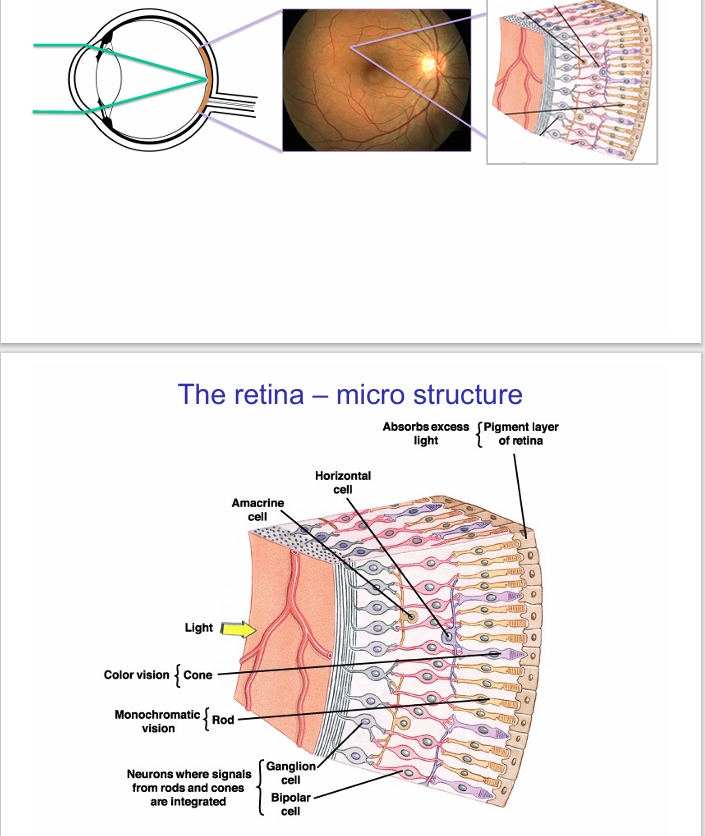
Retina microstructure
light comes in from front, interacting with photoreceptors (rods and cones) connected to bipolar cells and ganglion cells - these can integrate the rods and cones signals
Horizontal connections and amacrine cells also modulate signals
Pigment layer of retina doesn’t recognise light but absorbs the light to ensure vision can occur but also to protect the photoreceptor cells
retinal organisation - pigment epithelium
– Cells at very back of retina.
– Photoreceptors embedded in this layer.
– Cells contain melanin black pigment. Absorbs light prevents scattering stray light that would affect the image.
Retinal organisation - photoreceptors
– Rods (scotopic vision - low light level)
– Cones (photopic vision – high light levels) - colour
– More rods than cones (20:1)
retinal organisation - other cells
connecting vertically and horizontally– Bipolar cells (v), horizontal cells (h), amacrine cells (h), ganglion cells – origin of an optic nerve fibre
what does light pass to reach photoreceptors?
Light must pass through blood vessels and nerve fibres to reach photoreceptors– except at fovea for maximum acuity
The retina – micro structure PRs
Two types of photoreceptors
• Rods (scotopic vision)
• Type most sensitive to light
• Sensitive to brightness
• So only detect contrast - difference between light and dark - Function at low light levels
• Bleached at high light levels
• Cones (photopic vision)
• Three types sensitive to long (red), medium (green) and short (blue) wavelengths of light
• Enable colour vision
• Responsible for highest acuity (sharpness)
• Work only at high light levels
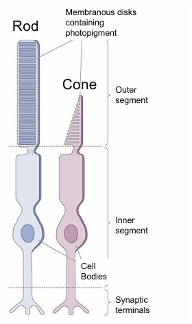
These cells connect to pigment epithelium at outer segment and bipolar cells at synaptic terminals
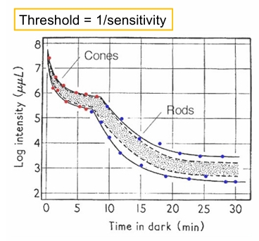
Dark Adaptation
• When moving from bright to low light level (entering darkened room) initially it is hard to see: cones are not sensitive, rods are bleached.
• Gradually visual sensitivity increases as rods recover
• This process of adjustment to low light is called dark adaptation
• Note also colour loss as switch from colour vision with cones to contrast only vision with rods
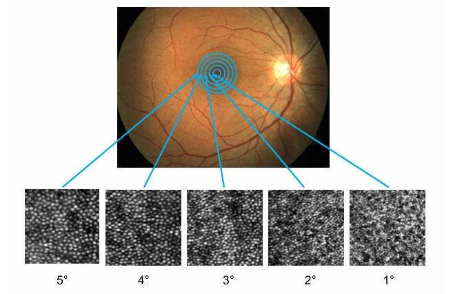
Distribution of cones
fovea - cells are really small and packed
Progressively get larger and less densely packed
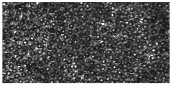
Distribution of rods
Further out there are rods - smaller cells in between cones
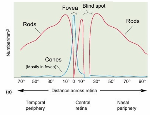
Distribution of rods and cones
Cones mostly in fovea - density decreases further away
Rods mainly absent in fovea - but peripheral mostly rods
Light transduction in photoreceptors
Light sensitive photopigments in rods and cones
– Rods - rhodopsin
– Cones - three different pigments (but wavelength (colour) sensitive)
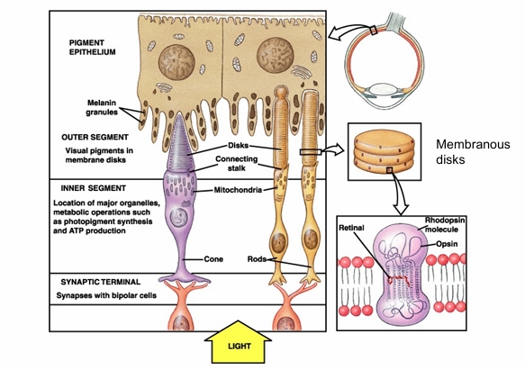
Rhodopsin and bleaching
– Opsin (Protein) + 11-CIS-retinal (derived from Vitamin A)
– In the dark, opsin and retinal are bound together
– On light exposure retinal molecule changes shape (conformational change)
– Causes it to split from opsin:
– This process is called bleaching
Structure of Photoreceptors
diagram also shows what photoreceptors are connected to
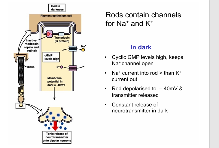
Rods channels in dark
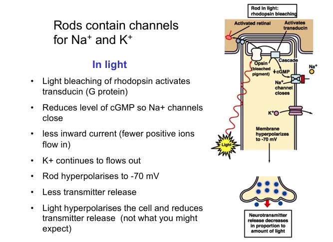
rods channels in light
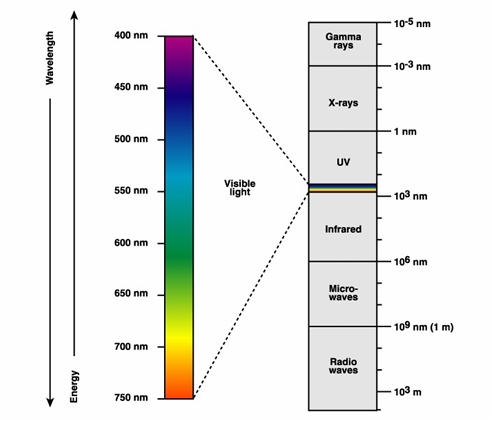
EM spec
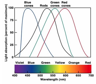
Colour vision: absorption spectra of cone photoreceptors
• Three cone types:
Light absorption (percent of maximum)
- short wavelength sensitive (’Blue’)
- medium wavelength sensitive(‘Green’)
- long wavelength sensitive (‘Red’)
• Each cone sensitive to range of wavelengths.
• The wavelength or colour is represented by unique ratio of outputs from the 3 different cone types.
• Allows many different colours to be represented using only 3 different receptors - the Trichromatic theory of colour vision.
Colour vision: Trichromatic theory
Each colour has unique ratio of absorption. Many colours are represented with only three cone types
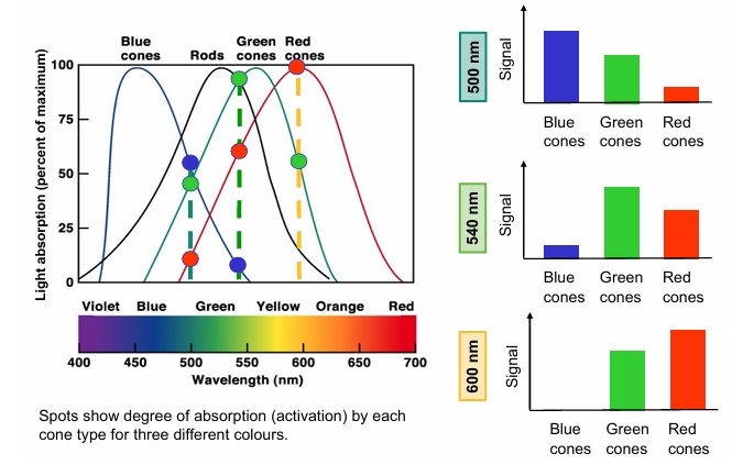
what happens in absence of a cone type or no cone types?
Absence of a cone type, or a cone with an abnormal pigment leads to colour confusion or colour blindness
Summary
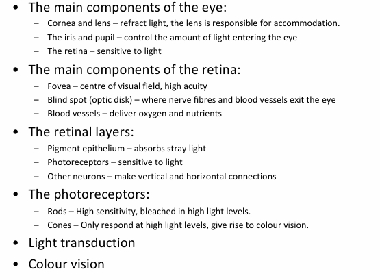
• The main components of the eye
• The main components of the retina
• The retinal layers
• The photoreceptors
• Light transduction
• Colour vision