psych 250 exam 3 master list
1/150
There's no tags or description
Looks like no tags are added yet.
Name | Mastery | Learn | Test | Matching | Spaced | Call with Kai |
|---|
No analytics yet
Send a link to your students to track their progress
151 Terms
stimulus
physical event that triggers sensory response
sensory receptor organ
organs specialized to detect certain stimuli
receptor cell
specialized cell within an organ that converts stimuli into an electrical signal
labeled lines
each sensory receptor and its pathway to the brain is “labeled” for a specific type of stimulus and location (the brain knows what kind os sensation you’re experiencing based on which neural pathway is activated, not just the signal itself)
sensory transduction
the process by which a sensory receptor converts a physical or chemical stimulus into an electrical signal (a change in membrane potential) that the NS can interpret
Receptor cells act as
transducers - convert energy from one form to another
receptor (generator) potentials
local changes in resting membrane potential
sequence of events in sensory transduction for the pacinian corpuscle
mechanical stimulus applied (pressure or vibration)
capsule deformation - the layered structure of the pacinian corpuscle compresses
membrane of the sensory neuron is stretched
mechanically gated ion channels open
Na+ ions enter the neuron —→ creates a receptor (generator) potential
if threshold is reached, an action potential is generated in the axon
action potentials travel to the CNS where the sensation is interpreted as pressure/vibration
what is the pacinian corpuscle
a specialized mechanoreceptor ion the skin that detects deep pressure and high-frequency vibration
deep ion the dermis
onion like layers of connective tissue surrounding a nerve ending
response
rapidly adapting - respond mainly to changes in pressure or vibration, not constant pressure
converts mechanical deformation into an electrical signal (sensory transduction) sent to the CNS
basically a vibration and deep pressure sensor that signals quick changes to the NS
adequate stimulus
a type of stimulus for which a given sensory organ is particularly adapted
each receptor type responds best to one kind of stimulus
photoreceptors (rods/cones) —→ light
hair cells (auditory) ——> mechanical vibration
pacinian corpuscles —→ pressure/vibration
6 aspects of sensory processing
coding - how sensory systems represent information about a stimulus, such as its type, intensity, location, or duration
stronger pressure, higher firing rate of a receptor
adaptation - the decrease in receptor response when a stimulus is constant. prevents sensory overload and allows the NS to focus on changes or new stimuli
phasic receptors - rapidly adapt (detect changes)
tonic receptors - slowly adapt (monitor continuous stimuli)
suppression - when the NS reduces or filters sensory signals, often to prevent overload or ignore irrelevant information
pathways - the neural routes sensory information takes from receptors to the brain
various pathways for different types of stimuyli
receptive fields - the specific area or set of stimuli that a sensory neuron responds to
a single touch neuron in the skin responds to a patch of skin
attention - the brains ability to selectively focus on certain sensory inputs while. ignoring others, enhancing perception of important stimuli
1/6 aspect of sensory processing - coding
how sensory systems represent information about a stimulus, such as its type, intensity, location, or duration
2/6 aspect of sensory processing - adaptation
the decrease in receptor response when a stimulus is constant - prevents sensory overload and allows the NS to focus on changes or new stimuli
phasic receptors - rapidly adapt (detect changes)
tonic receptors- slowly adapt (monitor continuous stimuli)
3/6 aspect of sensory processing - suppression
when the NS reduces or filters sensory signals, often to prevent overload or ignore irrelevant information
4/6 aspect of sensory processing - pathways
the neural routes sensory information takes from receptors to the brain
various pathways for different types of stimuli
5/6 aspect of sensory processing - receptive fields
the specific area or set of stimuli that a sensory neuron responds to
a single touch neuron in the skin responds to a patch of skin
6/6 aspect of sensory processing - attention
the brains ability to selectively focus on certain sensory inputs while ignoring others, enhancing perception of important stimuli
involves prefrontal cortex (what to focus on)
and posterior parietal cortex (where to focus)
6 sensory processing aspects summary
coding - how info is represented
adaptation - response change
suppression - filtering
pathways - routes
receptive fields - sensory “Area”
attention - focus
1/6 sensory processing aspects simple terms - coding
how info is represented
2/6 sensory processing aspects simple terms - adaptation
response change
3/6 sensory processing aspects simple terms - suppression
filtering
4/6 sensory processing aspects simple terms - pathways
routes
5/6 sensory processing aspects simple terms - receptive fields
sensory “area”
6/6 sensory processing aspects simple terms - attention
selective focus
tonic receptor
which receptor is
slow adapting
fire continuously as long as the stimulus is present
provide info about duration and intensity
pain receptors
phasic receptors
which receptor is
rapidly adapting
fires briefly at the beginning and sometimes end of a stimulus
quickly adapts and stops responding if the stimulus stays constant
pacinian corpuscle
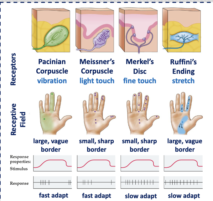
4 types of tactile receptors
messier corpuscle
merkel disc
pacinian corpuscle
Ruffini ending
what is a tactile receptor
a nerve ending that converts physical contact with your skin into electrical signals your brain can understand
4 types of tactile receptors extended version
messier corpuscle
detect light touch and low-frequency vibration
rapidly adapting (phasic)
found in areas like fingertips
merkel disc
detect pressure, texture, and shape
slowly adapting (tonic)
important for fine detail (reading braille)
pacinian corpuscle
detect deep pressure and high frequency vibration
rapidly adapting (phasic)
Ruffini ending
detect skin stretch and sustained pressure
slowly adapting (tonic)
basically
messier - light touch
merkel - detail/texture
pacinian - vibration
Ruffini - stretch
messier corpuscle
type of tactile receptor -
detect light touch and low frequency vibration
rapidly adapting (phasic)
found in areas like fingertips
merkel disc
type of tactile receptor -
detect pressure, texture, and shape
slowly adapting (tonic)
important for fine detail
pacinian corpuscle
type of tactile receptor -
detect deep pressure and high-frequency vibration
rapidly adapting (phasic)
Ruffini ending
type of tactile receptor -
detect skin stretch and sustained pressure
slowly adapting (tonic)
primary somatosensory cortex (S1)
S1 or S2?
first-stop for touch (raw data). direct input from thalamus, initial processing of tactile information
secondary somatosensory cortex (S2)
S1 or S2?
higher-level interpretation (what the touch means). input from other S and some thalamic area. higher order processing
two brain regions involved in attention
posterior parietal cortex
Cingulate cortex
synesthesia
a stimulus in one modality creates a sensation in another (seeing colors when hearing music, tasting words)
layers of skin
epidermis - outermost layer; thinnest
dermis - middle layer; nerve fibers
hypodermis - innermost layer; anchors muscles, helps shape body
epidermis
outermost and thinnest layer of skin
dermis
middle layer of skin; nerve fibers
hypodermis
innermost layer of skin anchors muscles, helps shape body
somatosensory stimuli/receptor organ/receptor cell/brain pathway
stimuli —→
receptor organ (skin, muscles, joints, tendons) —→
receptor cell ——→
brain pathway
(receptors —→
dorsal root ganglion (spinal nerve) ——→
ascend via dorsal column-medial lemniscal pathway (fine touch, vibration) or spinothalamic (ventral) tract (pain, temp) ——>
thalamus (ventral posterior nuclei) ——>
primary somatosensory cortex (s1) ——>
secondary somatosensory cortex (S2) for higher order processing
external ear
pinna, ear canal —→ collect sound
sound waves hit the tympanic membrane (eardrum) —→ vibration
middle ear
contains two protective muscles
tensor tympani and stapedius
when activated —→ stiffen ossicles —→ reduce sound transmission
protects inner ear from loud sounds
(ossicles: malleus, incus, stapes) ——> amplify vibration and concentrates vibrations onto the oval window
inner ear (receptor organ)
the true receptor organ is the cochlea
within it, the organ contains the hair cells (receptor cells). they concert mechanical vibration into neural signals
cochlea structure
scala vestiboli (vestibular canal)
scala media (middle canal) —→ contains organ of cortisol
scala tympani (tympanic canal)
two structures in external ear that collect sound
pinna, ear canal
eardrum
tympanic membrane
two protecting muscles in middle ear
tensor tympani, stapedius
what happens when tensor tympani and stapedius stiffen
reduce sound transmission, protects inner ear from loud sounds
receptor organ in inner ear
cochlea
cells that collect auditory info
hair cells
cochlea structure
scala vestiboli (vestibular canal)
scala media (middle canal) ——> contains organ of corti
scala tympani (tympanic canal)
functions of the 3 bones in the middle ear (ossicles)
ossicles - work together to transmit and amplify sound vibrations from the eardrum to the inner ear
malleus - attached to the tympanic membrane (eardrum); receives vibrations from the eardrum and passes them to the incus
incus - the middle ossicle; connects the malleus to the stapes and acts as a lever to transmit vibrations efficiently
stapes - the smallest bone; connects to the oval window of the cochlea; transits vibrations into the fluid of the inner ear, amplifying sound for detection by hair cells
ossicles
3 bones that work together to transmit and amplify sound vibrations from the eardrum to the inner ear
malleus
(bone in inner ear)
attached to the tympanic membrane; receives vibrations from the eardrum and passes them to the incus
incus
(bone in inner ear)
the middle ossicle; connects the malleus to the stapes and acts as a lever to transmit vibrations efficiently
stapes
(bone in inner ear)
the smallest bone; connects to the oval window of the cochlea; transmits vibrations into the fluid of the inner ear, amplifying sound for detection by hair cells.
scala media (part of cochlea) contains
organ of corti - receptor system that converts vibration into neural activity
basilar membrane
separates scala media and tympani
vibrates in réponse to sound
hair cells are embedded
tip links - thin fibers that connect hair cell stereocelia
vibration —→ stereo cilia sway —→ tip links move —→ opens ion channels they’re attached to ——> K+ and Ca2+ enter stereo cilia ——→ depolarization, opened Ca2+ channel at cell base —→ neurotransmitters release
organ of corti (part of scala media, which is part of cochlea)
receptor system that converts vibration into neural activity
basilar membrane
separates scala media and tympani
vibrates in response to sound
hair cells are embedded
tip links - thin fibers that connect hair cell stereocelia
tip links
thin fibers that connect hair cell stereocelia.
auditory system - pathway to brain
stimulus
receptor organ
receptor cell
brain pathway
sound —→ tympanic membrane vibrates —> ossicles (malleus, incus, stapes) amplify
stapes pushes on oval window —> fluid movement in cochlea
hair cells transduce movement into neural signals
signals travel via auditory nerve —→ brainstem (cochlear nuclei) —>
superior olivary complex (sound localization) —→
inferior colliculus —→ thalamus ——>
primary auditory cortex
parts of eye
cornea - round, transparent front of the eye
lens - flexible, transparent structure helps focus on an image on the retina
refraction - bending of light rays by the cornea and lens to form the image on the retina
fovea - part of the retina where vision is the sharpest
visual receptor organ
eye
cornea
round transparent front of the eye
lens
flexible, transparent structure helps focus on an image on the retina
refraction
bending of light rays by the cornea and lens to form the image on the retina
fovea
part of the retina where vision is the sharpest
photoreceptors
light-detecting cells
rod cells
cells that respond to light of any wavelength, detect lower light level
cone cells
type of cell that responds to different wavelength light, detect color
ganglion cell axons
the fibers of retinal ganglion cells that bundle together to form the optic nerve
retinal circuit
photoreceptors (rods and cones) —→ detect light and transduce it into electrical signals
bipolar cells —→ relay signals from photoreceptors
ganglion cells —→ generate action potentials
output
axons of ganglion cells form the optic nerve, which carries visual info to the brain
key idea -
photoreceptor —→ bipolar —→ ganglion = flow of visual information out of the retina
photopigments
light-sensitive molecules in photoreceptors (rods and cones)
made of opsin and retinal
absorbs light and starts visual transduction
photopigment = opsin (protein) + retinal (light-reactive part) ——> absorbs light ——> starts vision
retinal
light-sensitive molecule derived from vitamin A
changed shape when it absorbs light
shape change Is what triggers the electrical signal in the photoreceptor
opsin
a protein that surrounds retinal
determines which wavelengths of light are absorbed (color sensisitivty in cones)
visual system neural pathway
stimuli (light (electromagnetic energy)) ——>
receptor organ (eye (specifically retina)) ——>
receptor cell (photoreceptors: rods (low light) and cones (color, detail))
brain pathway
light —→ cornea —→ pupil —→ lens —→ focused on retina
photoreceptors transduce light ——> signal to bipolar cells —→ ganglion cells
ganglion cell axons form the optic nerve
partial crossing at the optic chasm
—> lateral geniculate nucleus (thalamus)
→ primary visual cortex (occipital lobe)
phototransduction (what happens in photoreceptors)
light enters the eye and hits photoreceptors (rods/cones)
activates rhodopsin (a photopigment)
rhodopsin = retinal and opsin
retinal changes shape when it absorbs light
triggers a cascade —→ sensory transduction
leads to hyper polarization of the photoreceptor
what is rhodopsin made of
retinal and opsin
ganglion cells to primary visual cortex
Pathway from ganglion cells to the primary visual cortex
main route for conscious vision
ganglion cell axons form the optic nerve
partial crossing at the optic chasm
continue as optic tracts to the lateral geniculate nucleus (thalamus)
project via optic radiations to the primary visual cortex (occipital lobe)
the two major visual processing pathways after the primary visual cortex
dorsal stream (where/how pathway)
ventral stream (what pathway)
dorsal stream (where/how pathway)
path - from visual cortex to parietal lobe
function - processes motion, location, and spatial relationships
helps guide actions
key idea - where is it? how to interact?
ventral stream (what pathway)
path - from visual cortex to temporal lobe
function - processes object recognition, shape and color
helps identify what something is.
key idea - what is it?
multimodal perception
the brain combines information from multiple senses to form a single, unified, perception.
cross modal phenomenon
one sense influences or alters how another else is perceived.
electrical brain potentials
used to classify levels of arousal, sleep states
3 ways to record sleep
electroencephalography (EEG) - records electrical activity in brain
electrooculography (EOG) - records eye movements
electromyography (EMG) - records muscle activity
electroencephalography (EEG)
records electrical activity in brain
electrooculography (EOG)
records eye movement
electromyography (EMG)
records muscle activity
phases of sleep cycle
wake - EEG pattern of activity in an awake person contains many frequencies - beta activity of desynchronized EEG
non-REM - 3 stages, characteristic activity patterns
stage 1 - small-amplitude EEG, irregular frequency, lowering heart rate, muscle tension
stage 2 - defined by bursts of 14-18 EEG waves - sleep spindles
stage 3 slow wave sleep (SWS) - defined by large-amplitude, very slow delta waves
REM (rapid-eye-movement) sleep
EEG activity is like an awake person
muscles are relaxed and limp
brainstem area inhibits motor neurons
characterized by rapid eye movements under closed lids, irregular breathing and pulse rates, vivid dreams.
stage 1 sleep
stage of sleep with small-amplitude EEG, irregular frequency, lowering heart rate, and muscle tension
stage 2 sleep
stage of sleep defined by bursts of 14-18 EEG waves-sleep spindles
state 3 slow wave sleep (SWS)
stage of sleep defined by large-amplitude, very slow delta waves
REM (rapid-eye-movement) sleep
EEG activity is like an awake person
muscles are relaxed and limp
brainstem area inhibits motor neurons
characterized by rapid eye movements under closed lids, irregular breathing and pulse rates, vivid dreams
Sleep cycles
4-5 cycles of sleep stages (90-110 minutes)
cycles early in the night have more stage 3 SWS
later cycles have more REM sleep
the last REM is just before waking up
how do sleep patterns change across the lifespan?
at puberty - shift in circadian rhythm of sleep —→ get up later in the day
with age, total time asleep decreases, and number of awakenings increases
largest decline is the loss of time spent in stage 3
at age 60, only half as much time is spent in SWS as age 20
at age 90 its gone
vivid dreams in REM
vivid dreams - REM sleep, visual imagery, sense that dreamer is there
predicted if there is rising high-frequency EEG activity in posterior cortex region
nightmares - long, frightening dreams awaken the sleeper from REM sleep
night terrors - sudden arousals from NREM sleep, marked by fear and autonomic activity
brain regions most involved
amygdala - intense emotions
hippocampus - involved in memory and dream content
brainstem (especially pons) - generates REM sleep and dreaming activity.
vivid dreams
REM sleep, visual imagery, sense that dreamer is there
predicted if there is rising high-frequency EEG activity in posterior cortex region