Thoracic Cage Deformites, Obesity
1/34
There's no tags or description
Looks like no tags are added yet.
Name | Mastery | Learn | Test | Matching | Spaced | Call with Kai |
|---|
No analytics yet
Send a link to your students to track their progress
35 Terms
Thoracic cage deformities
Respiratory impairment within thoracic cage deformities is due to a combination of factors
1. Elastic WOB is increased due to stiff thoracic cage
2. Diaphragm and other muscles of ventilation are at a
mechanical disadvantage from abnormal geometry
3. Lung volumes are reduced, and lung growth can be limited
in children
Manifests as: sleep disordered breathing (sleep hypoventilation) and progresses to daytime hypercapnia and pulmonary hypertension if untreated
Scoliosis
spine is curved to one side, typically appearing as an S or C
shape (lateral curvature)
Kyphosis
posterior curvature (convex) of spine occurring in the thoracic
and sacral regions (“humpback/hunchback”)
Kyphoscoliosis
Combination of both these thoracic deformities
• In severe cases, the deformity can compress the lungs &
restrict alveolar expansion → hypoventilation & atelectasis
• Impaired ability to cough & mobilize secretions
Kyphoscoliosis alteration
• Lung restriction/
compression
• Mucous accumulation
• Atelectasis
• Cor Pulmonale
CXR
Mediastinal shift in the same direction as the lateral curvature
increase density of compressed areas
kyphoscoliosis etiology
Affects 1 -2% of population - mostly young children
may present from birth due to congenital or
neuromuscular abnormalities including
Spina Bifida & Muscular
Dystrophy
• However, the majority (80 – 85%) of cases have no known cause
Risk factors Kyphoscoliosis
Gender – female more than male
Age – the younger the child is when first diagnosed, the greater the chance of curve progression
Angle of the curve – the greater the curvature of the spine, the greater the chance that the curve progression will worsen
Location – curves in the middle to lower spine are less likely to progress than those in the upper spine
Height – taller people have a greater chance of curve progression
Spinal problems at birth – children with congenital scoliosis have a greater risk for worsening of the curve
Kyphoscoliosis management
Braces
Surgery
Other Approaches
braces
during growth years to limit curve progression in
idiopathic deformities
Surgery
performed to correct severe deformity (> 40 – 50
degrees) and prevent further curvature
• Spinal fusion
• Rod instrumentation
Other approaches
• Electrical stimulation of muscles for strength
• Chest physio for secretions
• Broncho-dilators, antibiotics, anti-inflammatories as needed
• Adequate oxygenation as always
Ankylosing Spondylitis (AS)
A systemic rheumatic disease that affects the spine and thoracic cage
Chronic joint inflammation ultimately leads to fusion of the vertebral bodies and ribs
Typically leading to severe kyphosis and a dramatic decrease in thoracic cage compliance
in severe cases of AS, complete fusion and ridgity of the spine can occur .. what is the percentage of patients develop parenchymal lung disease
10% form PLD at apical fibrocystic changes that can decrease gas exchange and
provide location for chronic infection (especially fungal)
Clinical findings of AS
Extraparenchymal Restriction → decrease chest expansion & VC
Treatment AS
• Ant-inflammatory
• Analgesics
• Exercise programs in early stages to promote range of motion
Pectus Excavatum
Congenital chest wall deformity, producing a concave or caved-in appearance in the anterior chest wall
Most common type of congenital chest wall abnormality
occurs in estimated 1 in 300-400 births (M:F 3:1)
Pectus Carinatum
spectrum of protrusion abnormalities of the anterior chest wall (Pigeon Chest)
moderate to severe cases to the chest wall is rigid held outward position
respirations are inefficient and need to use accessory muscles for respirations rather than normal chest muscles during strenuous exercise Respirations
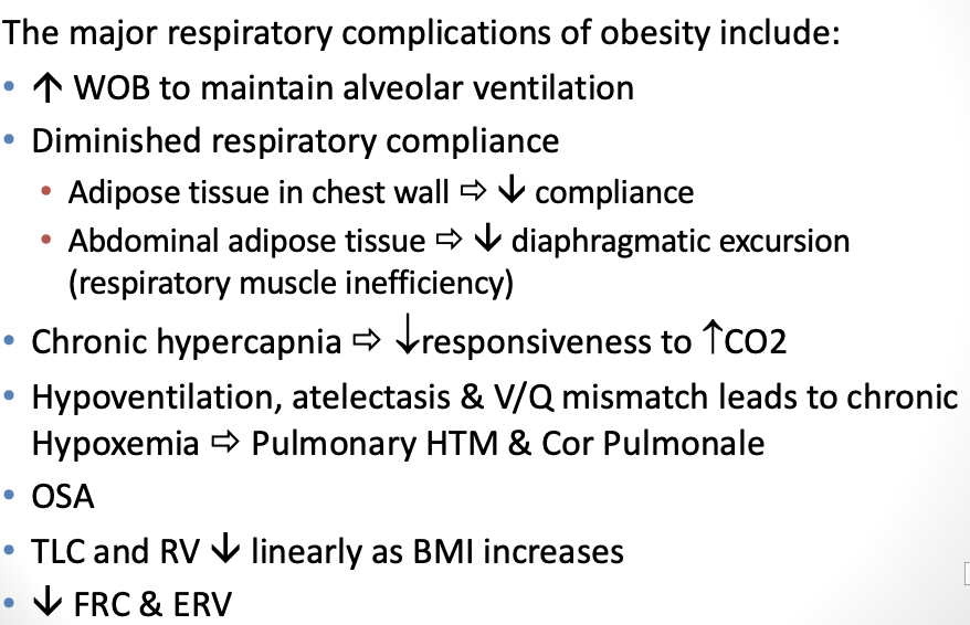
Obesity
progressive chronic disease that is characterized by abnormal or excessive fat accumulation that may impair health
Overweight is classified as BMI over___ and over __ is obese
25, 30
BMI
ratio of weight to height (kg/m2)
• normal weight (BMIs 18.5 to 24.9)
• overweight (BMIs 25 to 29.9)
• obese (BMI 30 and over)
• Class I (30.0 – 34.9)
• Class II (35.0 – 39.9)
• Class III (≥ 40.0)
waist circumference
provides an indicator of abdominal fat.
• WC at or above 102 cm (40 in.) for men, and 88 cm (35 in.) for women, is
associated with an increased risk of developing health problems
Subcutaneous fat
Located in the fatty tissue just beneath the skin
• Behaves like the fat elsewhere in the body
Visceral fat
Located around the internal organs
• Cells release free fatty acids into the liver, heart, and other organs
• local immune responses in visceral adipose tissue →
chronic systemic inflammation
health-related conditions
Type 2 diabetes
HTN
Cardiovascular disease
Undernutrition
Dyslipidemia
Osteoporosis
Hypertension
Infertility
OSA
Gallbladder disease
Certain cancers ( prostate.breast, colon, endometrial)
Determinants of Obesity
Social determinants: socioeconomic status/poverty.
For adults, education is a strong determinant; for children, household income plays a major role.
Age of Onset
increase size and number of adipose cells in obese children
Genetics
2 – 8 x higher for a person with a family history as opposed to a person with no family history of obesity
Endocrine Disorders – (e.g., hypothyroidism, polycystic ovarian syndrome, Cushing's syndrome )
Respiratory patho
Asthma
Obesity has been identified as a major risk factor for the
development of asthma
tends to not response well for treatment particularly controller medication
One possible explanation is that obesity causes chronic inflammation throughout the body, including the lungs.
• Adipose tissue secretes a large number of proinflammatory cytokines and factors modulating immune function
Obesity hypoventilation Syndrome (OHS)
a combination of obesity, a body mass index greater than
or equal to 30kg/m2 with awake chronic hypercapnia (PaCO2 >45mm Hg) & sleep-disordered breathing
Chronic daytime alveolar hypoventilation
arises from a complex interaction between sleep-disordered breathing, decrease respiratory drive, and obesity-related respiratory impairment (decrease ERV & compliance)
Central chemosensitivity is lost, impairing their ventilatory response to hypoxemia and hypercapnia
is associated with significant morbidity and mortality
~ 90% of patients with OHS also have OSA
Clinical manifestation
Dyspnea
• Cyanosis
• increase HR
• HTN (Pulmonary & Systemic)
• Pitting pedal edema (Cor Pulmonale)
• Peripheral vascular disease (emboli/stroke)
• increase surgical risk and postoperative complications
Lab findings with Class 3 obesity
• PFT: Extra parenchymal Restriction
• ABG: Chronic hypoventilation and hypoxia
• Lipid panel: Hyperlipidemia, high triglycerides
• Blood Work: ^Hct, ^ Hgb, (secondary polycythemia---
chronic Decrease O2)
• increase lipoproteins
• CXR: Wide chest wall, elevated diaphragm, enlarged heart
Treatment - Behavioural and psychological interventions
• Cognitive Behavioural Therapy (CBT)
• Medical Nutrition Therapy
• Physical Activity
pharmacological treatments
regulating appetite, metabolism, cravings
surgical interventions
• Gastric Bypass – reduces stomach size and reroutes digestion
• Sleeve Gastrectomy – removes a portion of the stomach, limiting food intake and
affecting hunger hormones
• Duodenal switch – alters digestion to limit calorie absorption (more extensive
procedure)
Respiratory management
• Bronchodilators and steroids as needed
• Oxygen as needed
• CPAP or BiPAP as needed for OSA