BIOM1001 MOD4 LEC1 Functions & Anantomy of Respiratory System
1/19
There's no tags or description
Looks like no tags are added yet.
Name | Mastery | Learn | Test | Matching | Spaced | Call with Kai |
|---|
No analytics yet
Send a link to your students to track their progress
20 Terms
What are the Functions of Respiratory System
Respiration
External Respiration
Internal Respiration
Cellular Respiration
Ventilation
Perfusion
What is Respiration
Intake of Oxygen and release of Carbon dioxide
Cells need oxygen to make energy (ATP), occurring through cellular respiration in mitochondria
C6H12O6 (glucose) + 6O2 —> 6CO2 + 6H2O
What is External Respiration
Ventilation : exchange of air between atmosphere and lungs
Gas Exchange : Exchange of gases between lungs and bloodstream
Gas Transport : transport of O2 and CO2 by blood
What is Internal Respiration
Gas exchange : Exchange of O2 and CO3 between blood and cells in body
What is Cellular Respiration
Metabolism within cells
What is Ventilation
Mechanical movement of muscles and thoracic cavity causing air flow into and out of the lungs
What is Perfusion
Passage of blood through blood vessels in organ or tissue
What is included in the Conducting Zone
Upper respiratory system: mouth, nasal cavity, pharynx, larynx
Lower respiratory system: Trachea, bronchi, bronchioles
What is in the Respiratory Zone
Alveoli and respiratory bronchioles - involved in gas exchange
What does Upper respiratory system do?
Route for incoming and outgoing air
conducts air to alveoli, deep in lungs
Removes debris and pathogens from incoming air
Warms and humidifies incoming air
What is in the Respiratory Epithelium
Found in conchae, meat uses, paranasal sinuses of nose and trachea
GOBLET CELLS
produce mucous to trap debris
CILIA
removes mucous and debris from nasal cavity
Constant beating motion ; sweep material to throat to swallow
CAPILLARIES
warms air by convention
The Larynx - what are the major cartilage pieces
Cartilaginous structure connecting pharynx to trachea
EPIGLOTTIS
elastic cartilage that covers trachea during swallowing
THYROID CARTILAGE
largest ; forms the Adam’s apple
CRICOID CARTILAGE
ring shaped and supports airways
The Larynx - muscles?
GLOTTIS
Includes vestibular folds, vocal chords and space between
VESTIBULAR FOLDS
false vocal chords (protect airways)
Mucous membrane
TRUE VOCAL CHORDS
Vibrates to produce sound
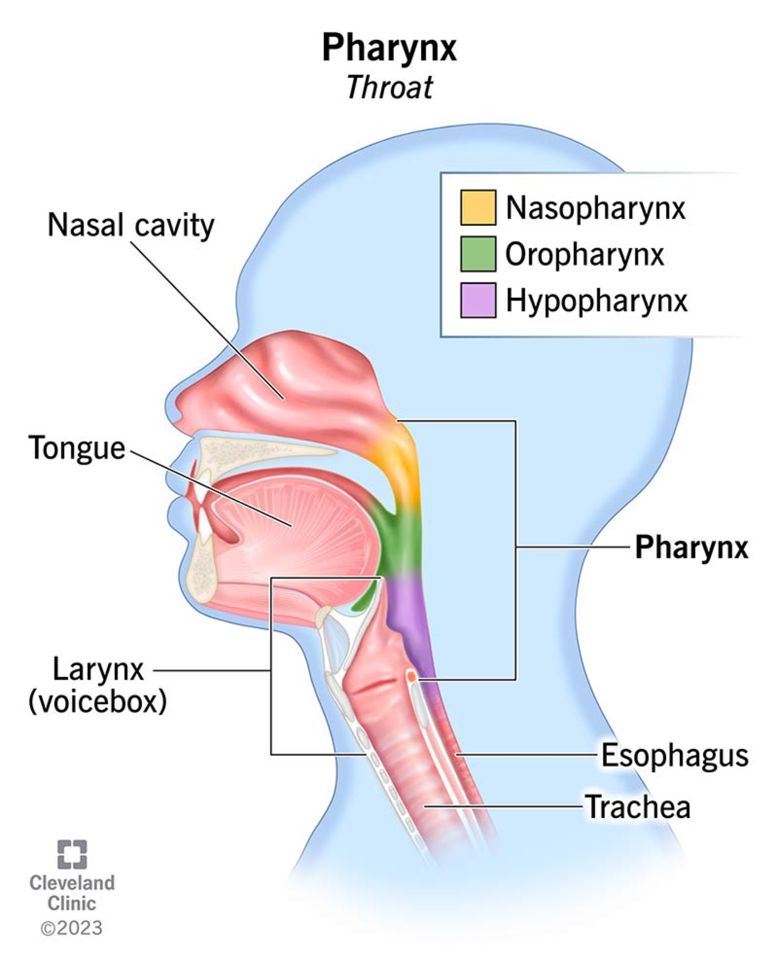
The Pharynx - what are the 3 regions?
NASOPHARYNX - behind nasal cavity
Air passage only
contains pharyngeal tonsils to trap pathogens
Connects Eustachian tubes - ear pressure
Uvula and soft palate close when swallowing
OROPHARYNX - Behind Mouth
Air + food passage
Palatine and lingual tonsils for immune defence
Lined with stratified squamous epithelium for friction protection
LARYNGOPHARYNX - behind larynx
Air and food pathway
Opens anteriorly into larynx, posteriorly to esophagus
What is included in the anatomy of the thorax? (6)
Bones
Muscles
Pleural membranes
Visceral pleural
Parietal pleural
Pleural cavity
Bones and Muscles in the Thorax (2)(6)
BONES
spine + ribcage (sternum & ribs)
MUSCLES
Diaphram
Externalcoastals and Internalcoastals
Sternocleidomastoids
Scalenes
Abdominal muscles
What are the 2 Pleural Membrane and what do they do?
1) Visceral Pleura
attached to the lungs surface
2) Parietal Pleura
Attached to the ribs
Small amount of pleural fluid between the membranes to reduce friction
Helps lungs stay inflated
The Visceral Pleura
covers the surface of lungs
Helps maintain shape, prevents lungs collapse
The Parietal Pleura
Lines thoracic cavity
Covers lungs, heart + major blood vessels
What is the pleural cavity and what does it do?
The pleural cavity is the space between the visceral and parietal pleural, containing pleural fluid.
Reduces friction by creating a slippery surface through which the membranes slide across one another.
Surface tension sticks the lungs to thoracic wall, holding the lungs open.