lab quiz 4
1/42
There's no tags or description
Looks like no tags are added yet.
Name | Mastery | Learn | Test | Matching | Spaced | Call with Kai |
|---|
No analytics yet
Send a link to your students to track their progress
43 Terms
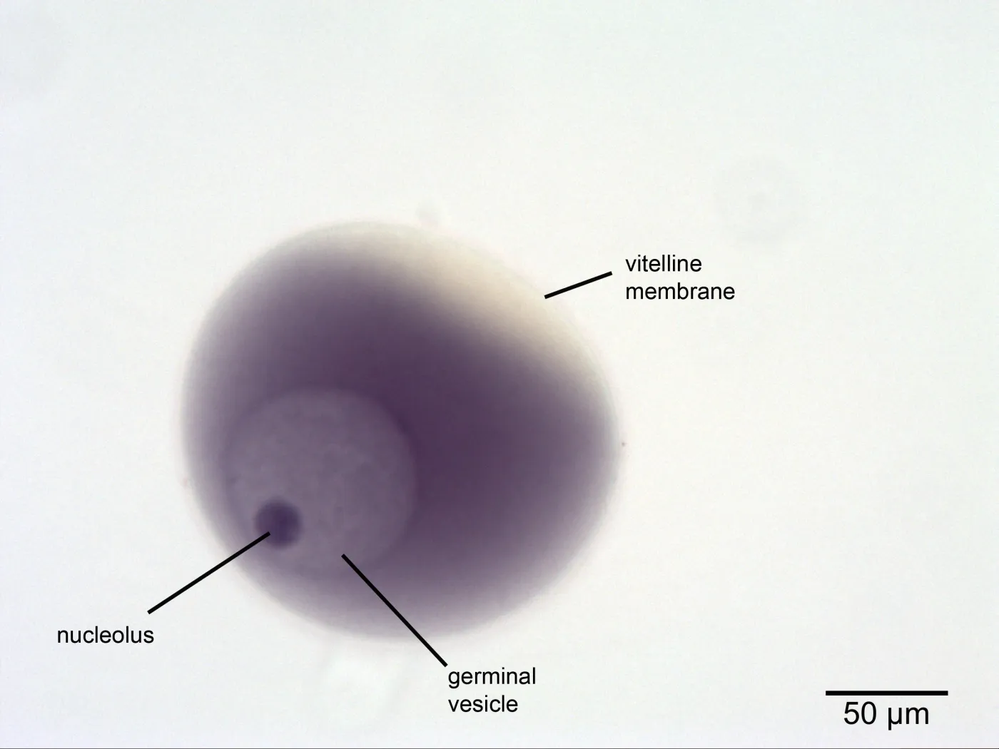
unfertilized egg
has nucleus and nucleolus(germinal vesicle)
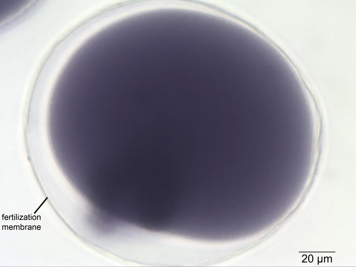
fertilized egg
no nucleus, has fertilization membrane
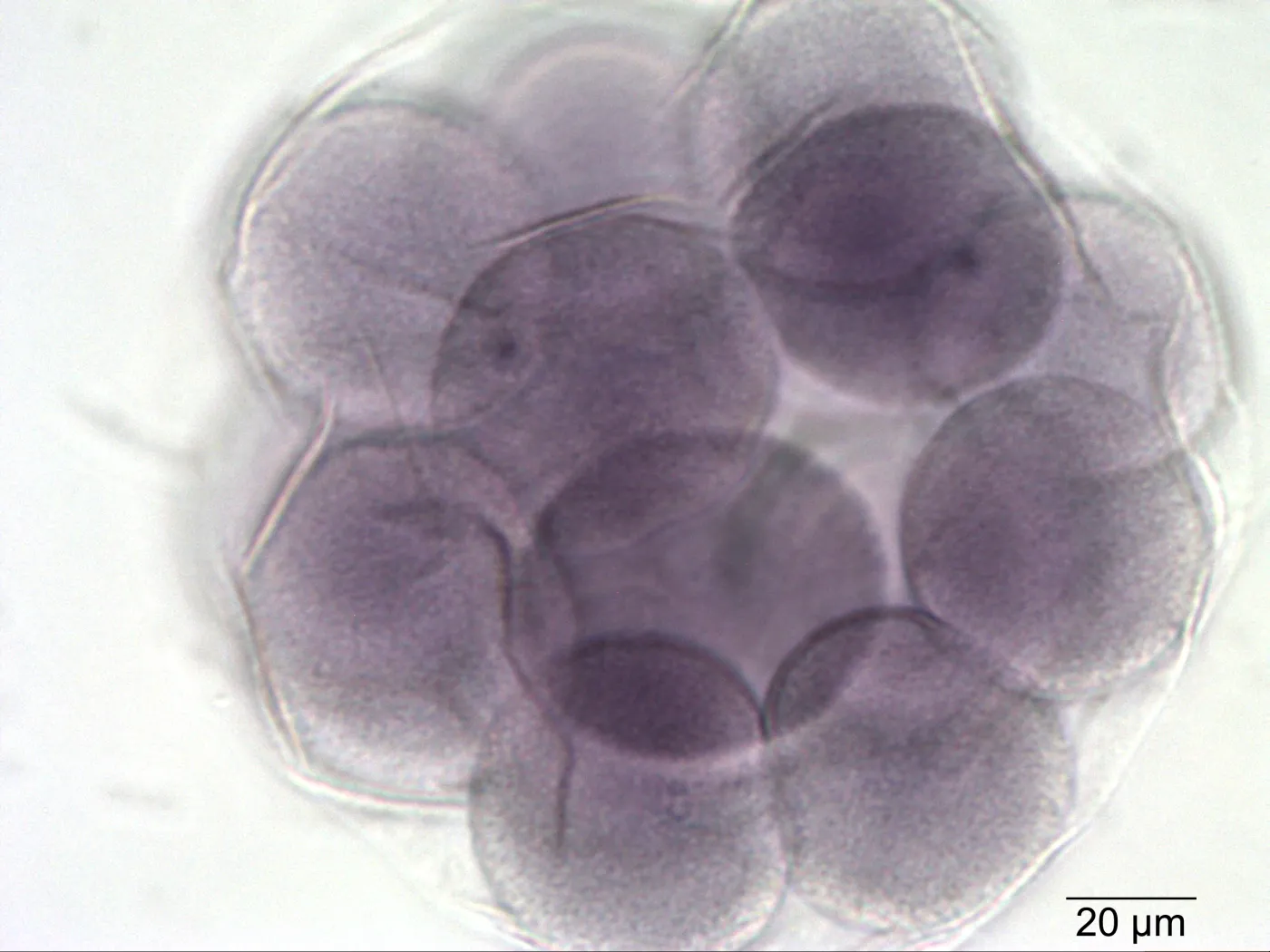
cleavage
rapid mitotic cell divisions after fertilization
zygote
diploid cell formed by the fusion of a haploid sperm and egg during fertilization
unicellular; right after they meet
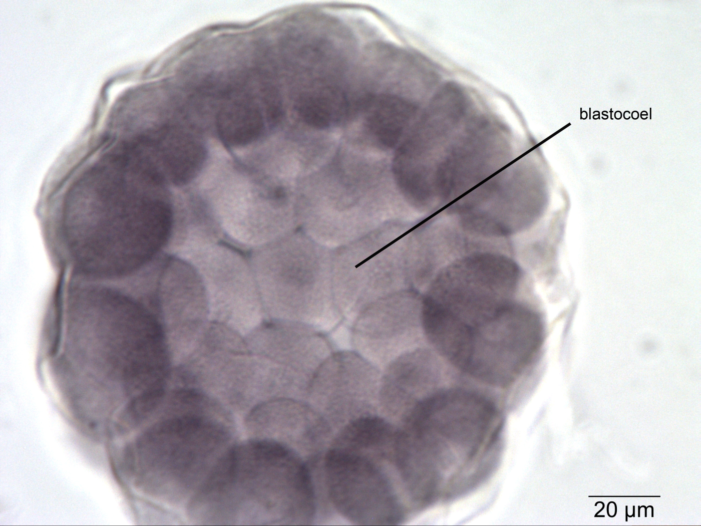
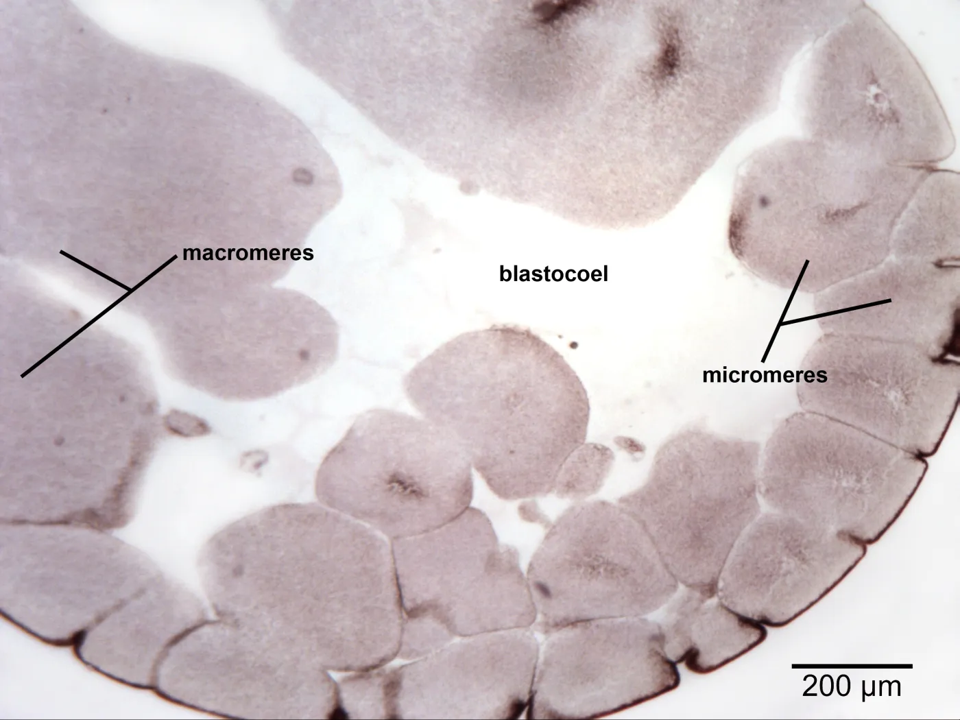
blastula
When cleavage process transforms the large single-celled zygote into a multicellular structure
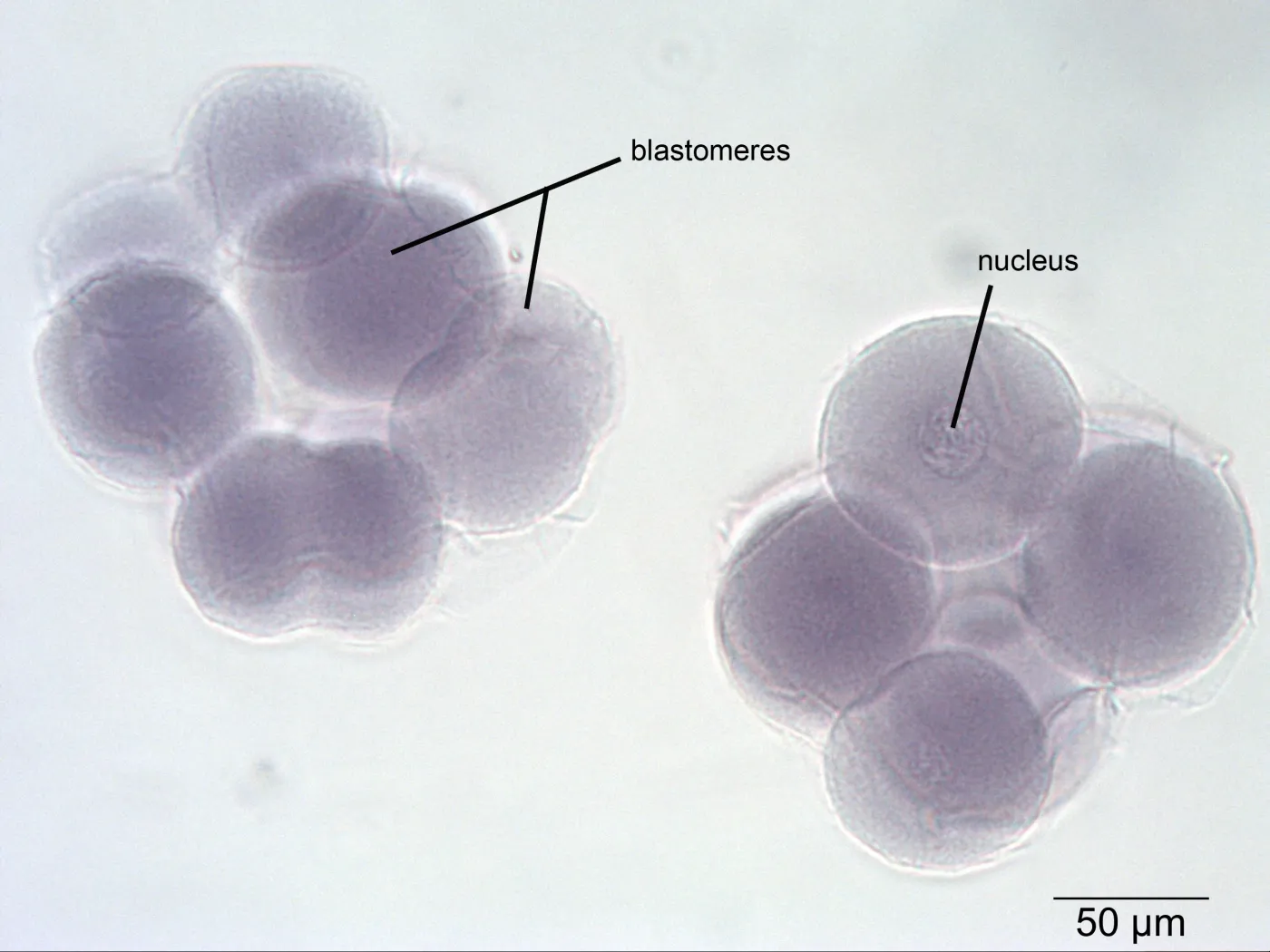
blastomeres
smaller cells that makes blastula
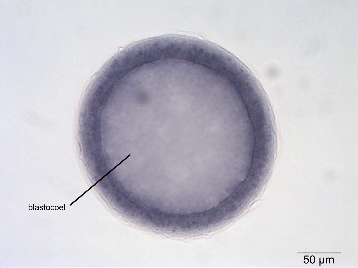
blastocoel
fluid-filled cavity that develops within the mass of blastomeres in the blastula.
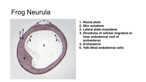
gastrula
after blastula
embryo starts taking shape and laying down the foundation for all future tissues and organs. In triploblasts, it’s characterized by extensive cell movement and the formation of the three primary germ layers:

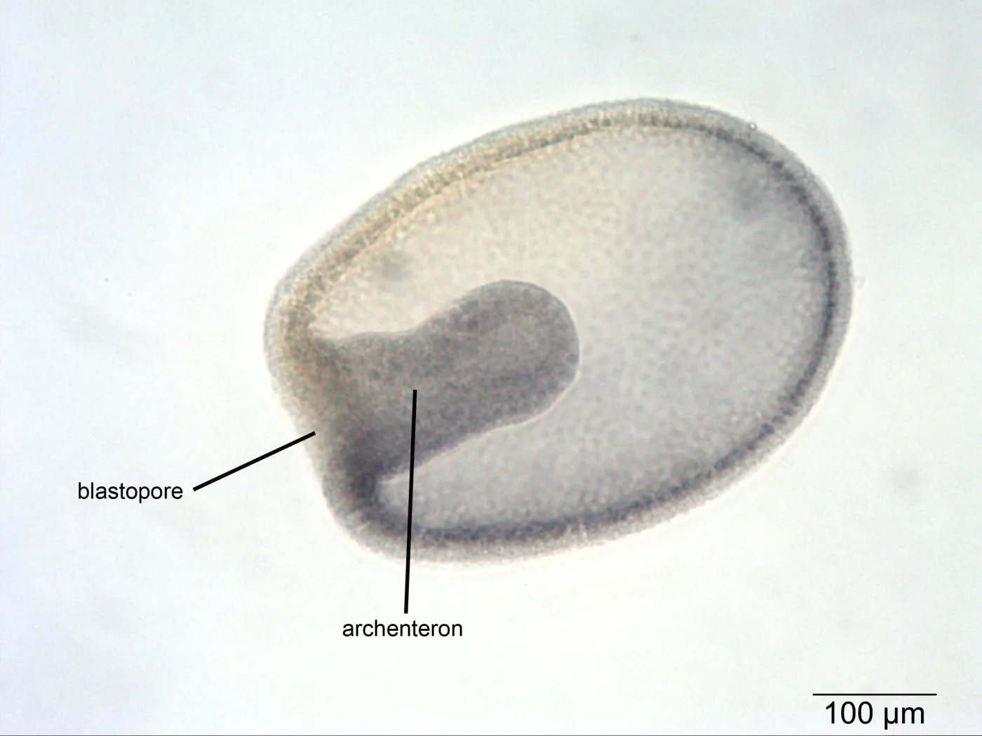
blastopore
first opening that forms in an early animal embryo during the gastrulation stage
It is crucial for defining the embryo's body axis and forming germ layers (endoderm, mesoderm).
The blastopore determines the animal's classification. In protostomes (e.g., mollusks, worms), the blastopore becomes the mouth, while in deuterostomes (e.g., humans, starfish), it becomes the anus.

archenteron
primitive gut, is the internal cavity formed during the gastrulation stage of embryonic development. It forms via the inward folding (invagination) of cells, developing into the animal's digestive tract (lumen)
late cleavage
The unequal cleavage sets up the animal-vegetal polarity, which influences where the body axes and germ layers will form.
gastrulation
It creates the primary germ layers—ectoderm, mesoderm, and endoderm—which are essential for organogenesis
The process of gastrulation begins at the blastula stage, which has:
• A blastocoel (fluid-filled cavity).
• An animal pole (pigmented, less yolk, smaller cells).
• A vegetal pole (larger cells, more yolk).
blastula stage
stage in early animal embryonic development that produces the blastula
blastoderm
the cells that surround a fluid-filled cavity known as the blastocoel
blastocyst
Specific to placental mammals. It represents a more advanced stage than a simple blastula, having differentiated into the trophoblast (outer layer) and the inner cell mass (embryoblast).
animal-vegetal polarity,
a fundamental asymmetry in animal eggs and blastula-stage embryos, dividing them into two distinct hemispheres: the animal pole and vegetal pole.
This polarity determines early developmental axes, with the animal pole forming the embryo proper (ectoderm) and the vegetal pole forming inner structures (endoderm/mesoderm), often defined by uneven yolk distribution
the nervous system
neural plate
thickened layer of ectodermal cells
the precursor to/becomes brain and spinal cord

neural folds
ridges of ectoderm that arise from the side edges of the neural plate
They elevate, converge, and fuse at the midline to form the neural tube, Help shape the tube

neural tube.
It forms from the folding of the neural plate into a cylinder, which seals itself shut to create a hollow structure
becomes CNS (brain + spinal cord)
neurulation
the process right after gastrulation that forms the nervous system!
makes neural places, tube and folds.
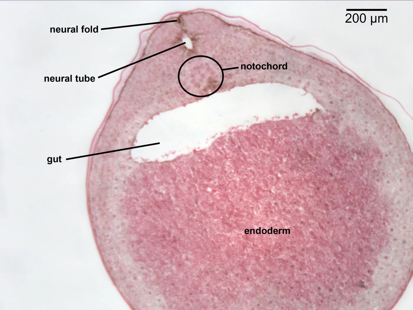
notochord
a cartilaginous skeletal rod supporting the body in all embryonic and some adult chordate animals.

gut
formed from endoderm lining the yolk sac which is enveloped by the developing coelom

somites
acting as essential segmentation units for developing vertebrae, ribs, skeletal muscles, and the dermis

hensons node
It acts as a specialized signaling center that initiates, organizes, and patterns the embryonic axis, equivalent to Spemann’s organizer in amphibians.
As it regresses caudally, it leaves behind the notochord and neural tissue.
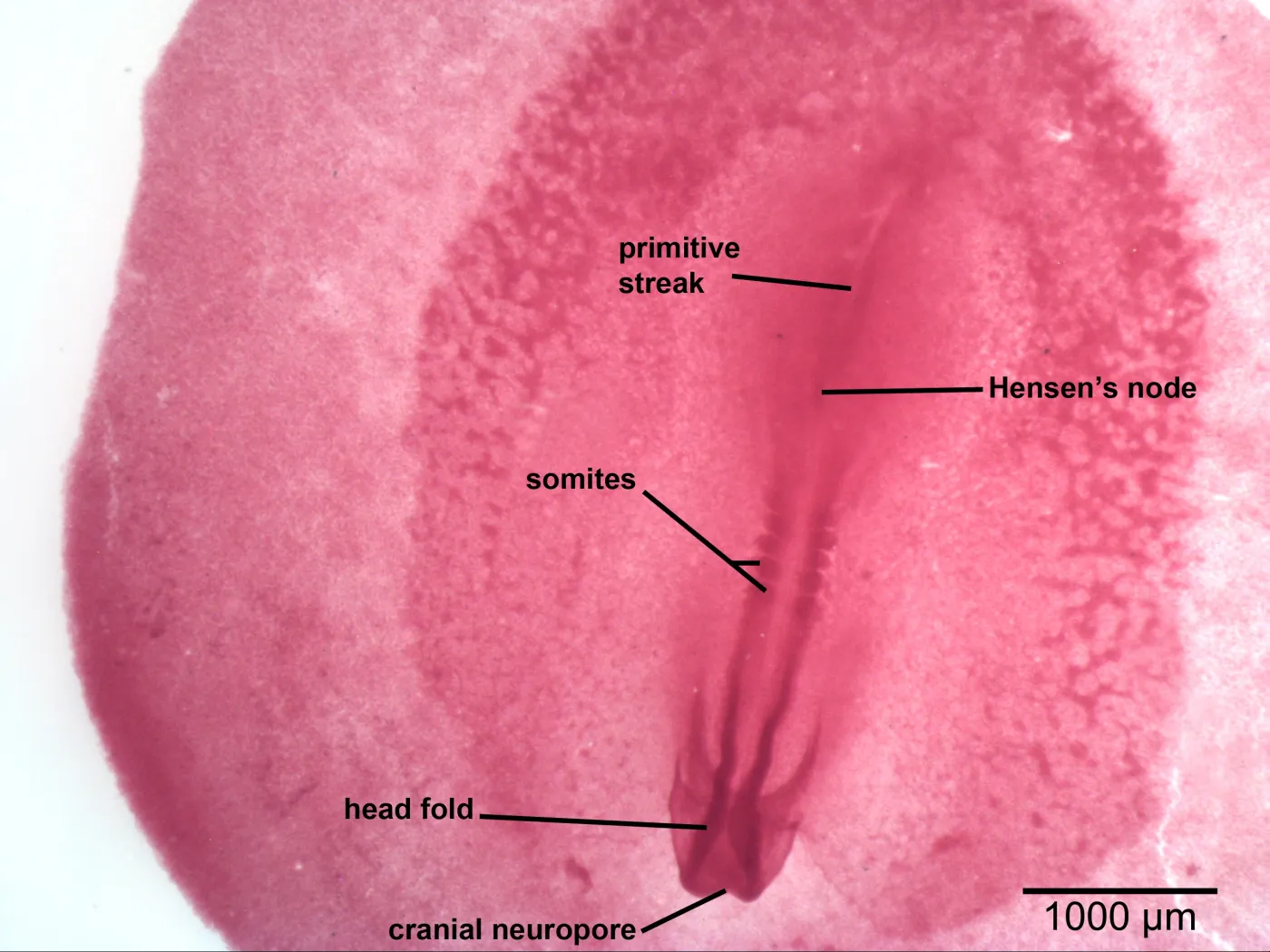
head fold
It involves a crescent-shaped anterior folding of the blastoderm
forming the foregut, and establishing the heart region.

cranial neuropore
the final rostral opening of the neural tube,
crucial for proper brain development.
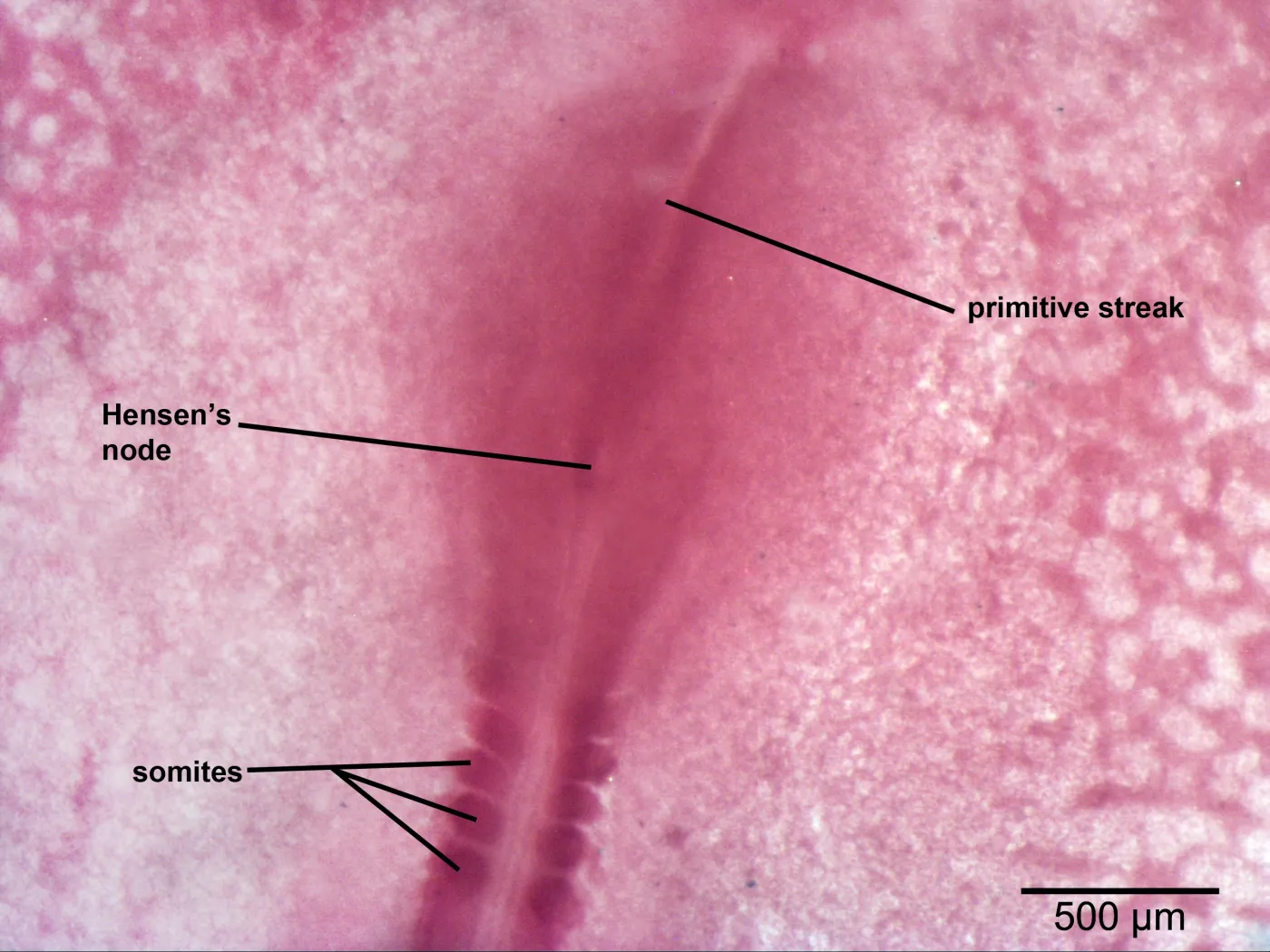
primative streak
marks the start of gastrulation, establishing the embryo’s cranial-caudal (head-tail) and bilateral symmetry
conduit for cells to migrate inward and form the three primary germ layers (ectoderm, mesoderm, endoderm)
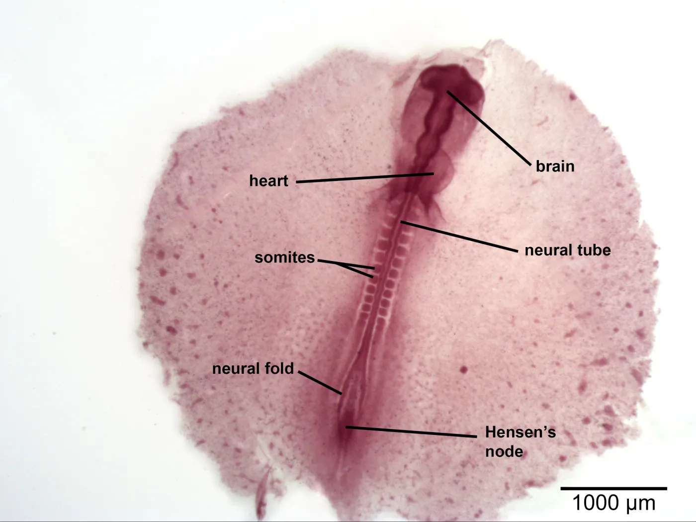
neural fold
transient, paired ridges of ectoderm that arise from the lateral edges of the neural plate during embryonic development.
They elevate, converge, and fuse at the midline to form the neural tube, which serves as the precursor to the brain and spinal cord

neural tube
from the folding of the ectoderm to form a cylinder

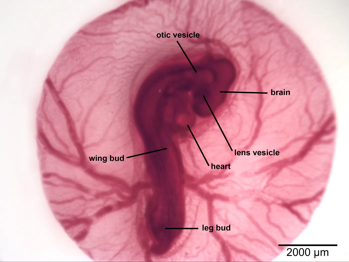
heart

brain

lens vesicle
a transient, hollow sphere of surface ectodermal cells formed during embryonic eye development

otic vesicles
a hollow, pear-shaped structure formed during embryonic development from the invagination of the otic placode, serving as the precursor for the entire membranous labyrinth of the inner ear
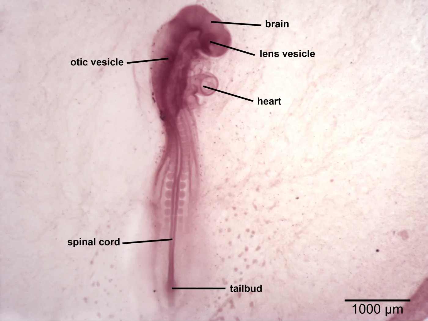
tailbud
mesenchymal cells at the posterior end of vertebrate embryos that drives the development of the caudal body, including the tail, posterior neural tube, and somites

spinal cord

wing bud
small protrusions on the flank, developing into wings through signaling centers

leg bud
appear around 72 hours (Day 3) of incubation, emerging as mesenchymal swellings from the lateral plate mesoderm
guide the development from limb bud to functional leg