Hepatic Circulatory Disorders and Infiltrates
 Call Kai
 Learn
Learn Practice Test
 Spaced Repetition
 Match
Match Flashcards
 Knowt Play
1/66

There's no tags or description
Looks like no tags are added yet.
Name | Mastery | Learn | Test | Matching | Spaced | Call with Kai |
|---|
No analytics yet
Send a link to your students to track their progress
67 Terms
What is the primary cause of passive hepatic congestion?
Right-sided heart failure, which leads to elevated pressure in the caudal vena cava and hepatic vein.
Where does congestion occur within the hepatic lobule during passive congestion?
Centrilobular congestion of the sinusoids.
What are two common causes of right-sided heart failure in dogs leading to hepatic congestion?
Valvular endocardiosis of the tricuspid valve and canine heartworm (Dirofilaria immitis).
What is the sequence of events from congestion to liver damage?
Congestion leads to hypoxia, which causes centrilobular degeneration, atrophy, and loss of hepatocytes.
What is the term for the 'enhanced lobular pattern' seen in the liver due to chronic hypoxia?
Nutmeg liver.
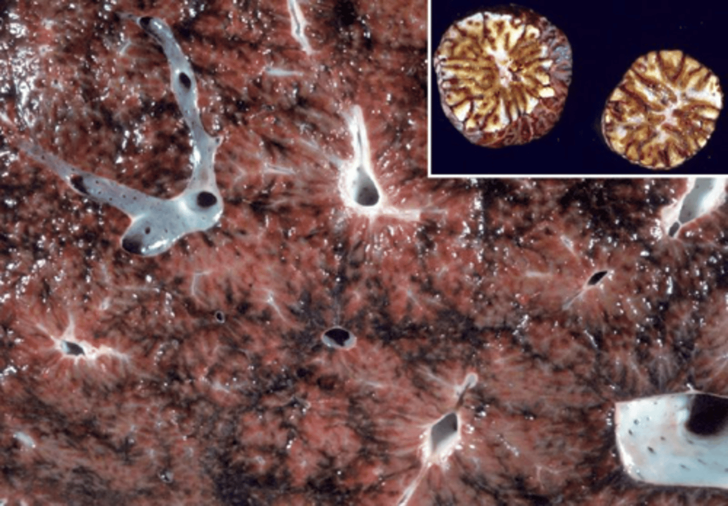
Besides passive congestion, what other process can cause an enhanced lobular pattern in the liver?
Zonal hepatic necrosis.
What gross morphological changes are observed in a liver with passive congestion?
The liver lobes are enlarged with rounded edges.
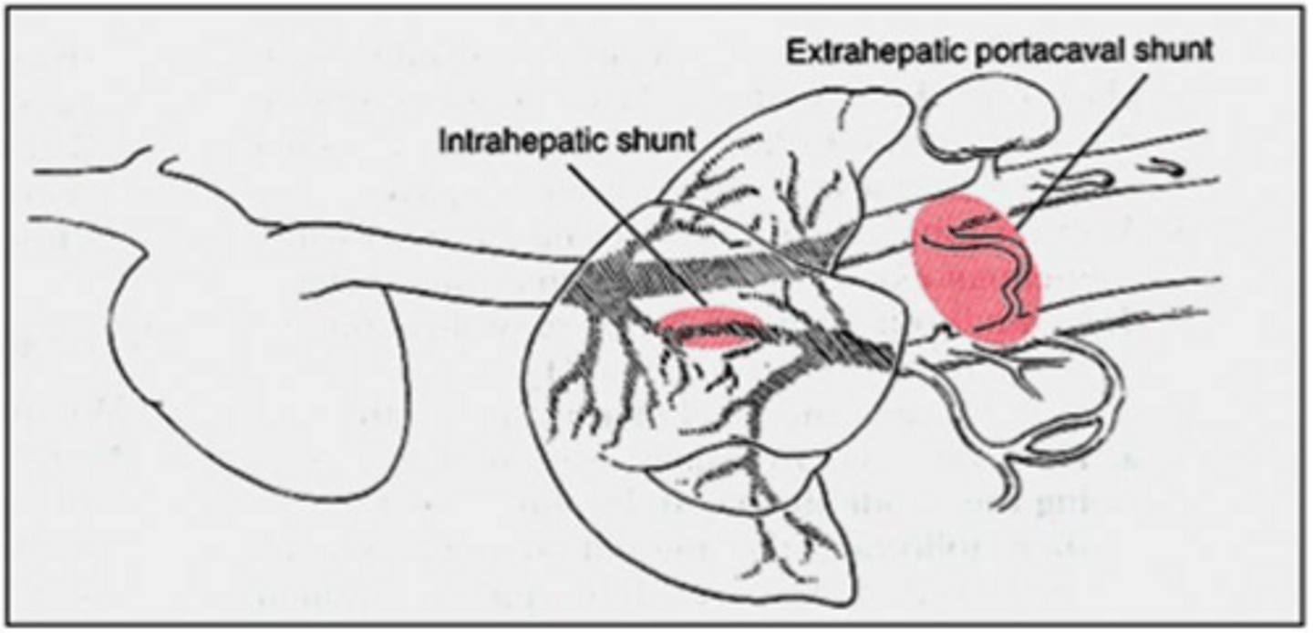
What is a congenital portosystemic shunt (PSS)?
An abnormal vascular structure allowing portal blood to bypass the liver and drain directly into the systemic circulation.
What are the common clinical signs of hepatic encephalopathy in animals with PSS?
Ataxia, seizures, blindness, and head pressing.
Why does a portosystemic shunt cause hepatic encephalopathy?
It leads to hyperammonemia because blood bypasses the liver's detoxification processes.
What is the gross appearance of the liver in an animal with a congenital PSS?
The liver is small, a condition known as microhepatica.
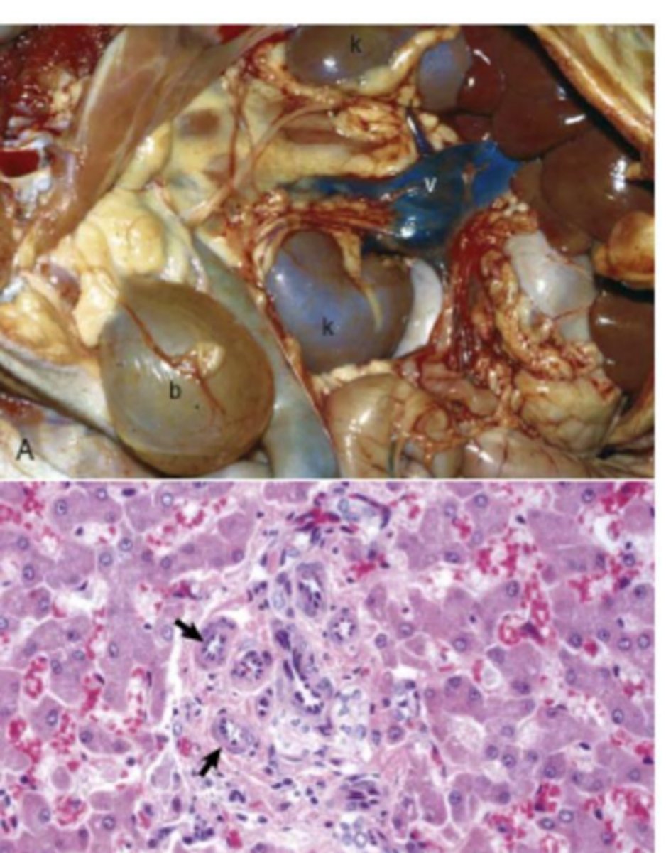
What are the histological findings in a liver with a congenital PSS?
Lobular atrophy, portal miniaturization with small or absent portal veins, and reduplication of arterioles.
What is the most common cause of intrahepatic shunts?
Failure of the ductus venosus (a fetal vessel) to close.
In which type of dogs are intrahepatic shunts most common?
Large breed dogs.
What substances can accumulate inside or outside the hepatocyte in infiltrative liver disorders?
Lipids, glycogen, amyloid, iron, copper, bile pigment, and products of lysosomal dysfunction.
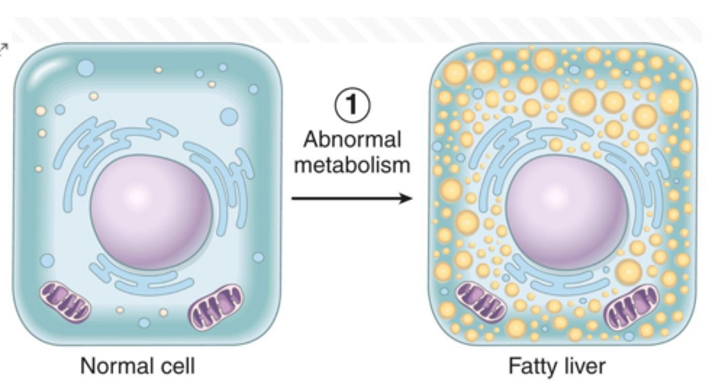
What is the effect of chronic hypoxic injury on hepatocytes regarding lipid metabolism?
It leads to steatosis (fatty degeneration).
What is the definition of portal vein hypoplasia?
A congenital condition characterized by the underdevelopment of the portal vein.
What is the clinical significance of acquired vascular shunts?
They are typically secondary to portal hypertension.
What is the definition of telangiectasis in the liver?
A circulatory disorder characterized by the dilation of capillaries or terminal arteries.
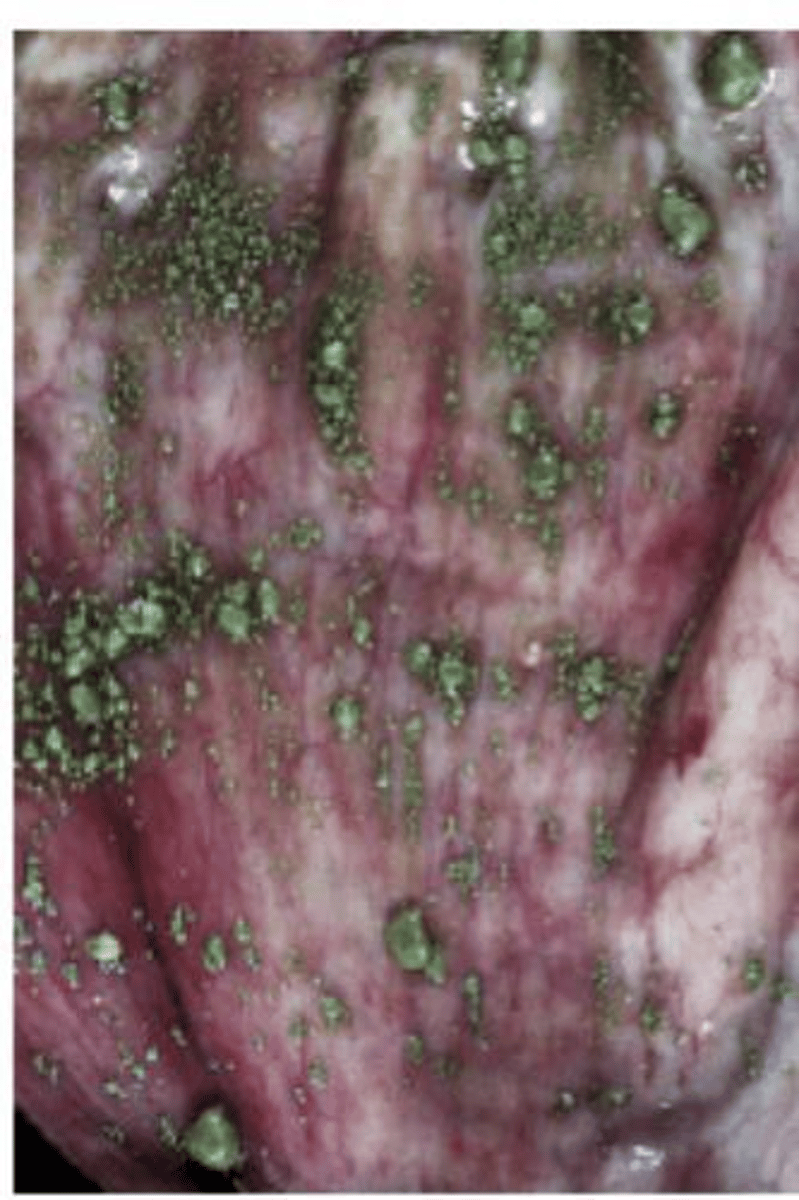
What is the relationship between portal hypertension and acquired shunts?
Portal hypertension increases pressure in the portal system, forcing blood to find alternative pathways (acquired shunts) to reach systemic circulation.
What is the primary physiological consequence of stunting in animals with PSS?
The liver fails to receive adequate portal blood flow, which is necessary for normal growth and metabolic function.
What are the two common anatomical sites for extrahepatic portosystemic shunts (PSS)?
Portal vein to caudal vena cava anastomosis and portal vein to azygos vein anastomosis.
In which animal population are congenital portosystemic shunts most commonly found?
Small breed dogs and cats.
What metabolic abnormality is commonly associated with dogs suffering from PSS?
Abnormal ammonia metabolism.
What type of crystalluria is a clinical hallmark of PSS in dogs?
Ammonium biurate crystalluria.
What is another name for congenital portal vein hypoplasia?
Hepatic Microvascular Dysplasia.
List three dog breeds predisposed to congenital portal vein hypoplasia.
Yorkshire terriers, Maltese dogs, and Cairn terriers (also Tibetan spaniels, shih-tzus, and Havanese).
What are the primary physiological consequences of congenital portal vein hypoplasia?
Diminished hepatic perfusion and portal hypertension.
What are the typical physical findings in an animal with portal vein hypoplasia?
Microhepatica (small liver) and ascites.
Why is radiology recommended when diagnosing portal vein hypoplasia?
Because its histologic features are indistinguishable from those of a portosystemic shunt (PSS).
What are two major categories of causes for portal hypertension?
Thrombosis/occlusion within the portal vein/hepatic outflow and intrahepatic causes (e.g., fibrosis, nodular regeneration).
What clinical conditions can develop as a result of persistent portal hypertension?
Ascites and the development of acquired portosystemic shunts.
What are the four main mechanisms of abnormal intracellular accumulation in hepatocytes?
Inadequate removal/degradation, excessive production of an endogenous substance, deposition of abnormal exogenous material, and genetic enzyme deficiencies.
What are the two forms of protein involved in hepatic amyloidosis?
Amyloid light chain protein (AL) and amyloid associated protein (AA).
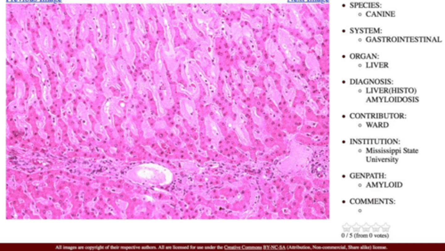
What is the primary cause of secondary (reactive) amyloidosis?
Prolonged systemic inflammation.
Which dog and cat breeds are specifically noted for inherited or familial amyloidosis?
Shar-Pei dogs, Abyssinian cats, and Siamese cats.

What are the gross pathological features of a liver affected by amyloidosis?
Enlarged, rounded edges, friable, and pale.
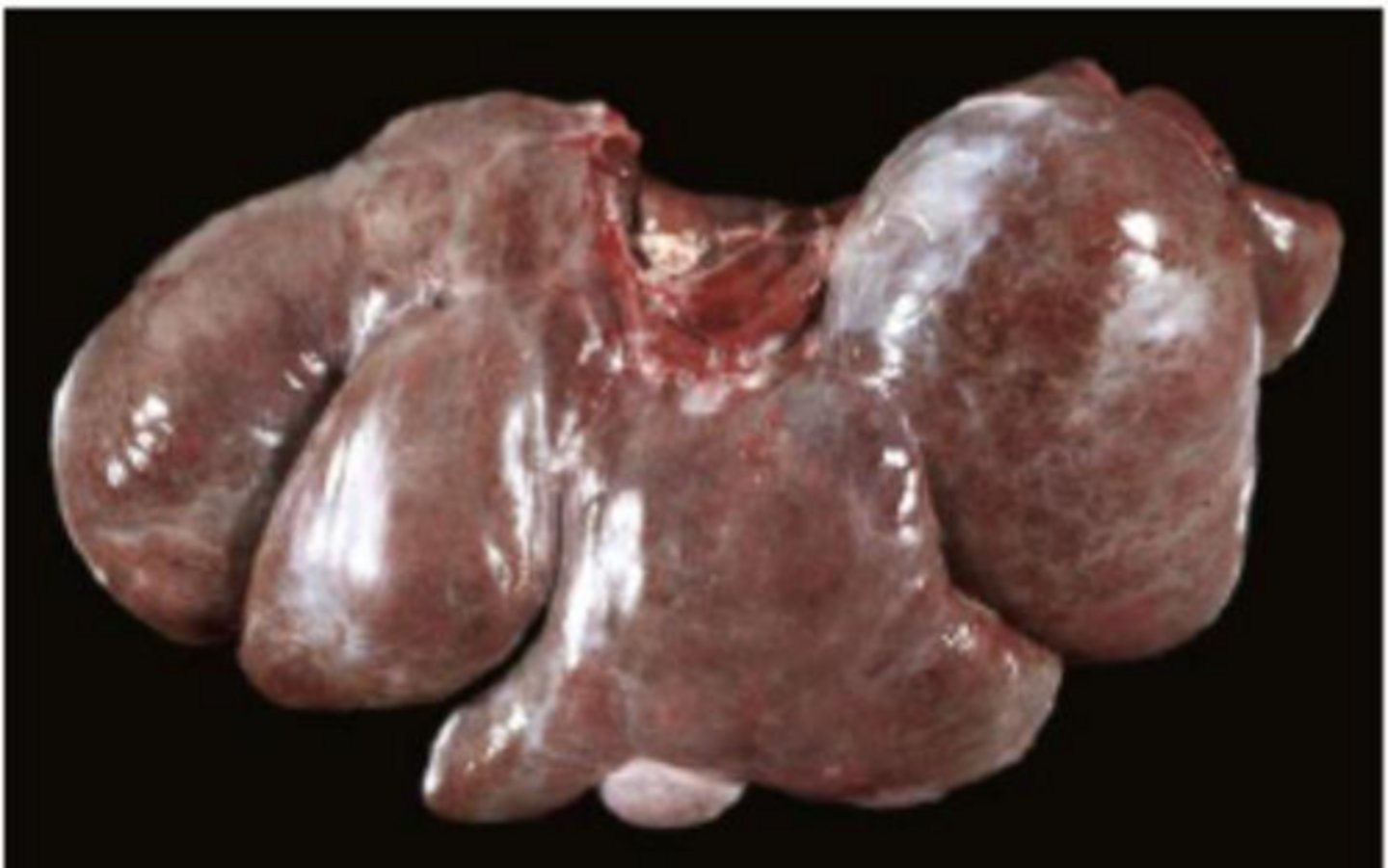
Where does amyloid deposition typically begin within the liver tissue?
In the space of Disse, eventually extending into the sinusoids.
What special stain is used to identify amyloid deposits?
Congo red.
What is the primary mechanism of injury in hepatocellular copper accumulation?
Copper accumulation leads to reactive oxygen species, causing oxidative injury to mitochondria and cellular membranes, resulting in centrilobular necrosis.
Which dog breeds are commonly associated with copper-associated hepatopathy?
Bedlington terriers and Labrador retrievers.
What is the clinical significance of acquired portosystemic shunts?
They are typically a consequence of persistent portal hypertension and appear as multiple tortuous blood vessels.
What is the result of severe amyloid deposition in the liver?
Pressure atrophy and necrosis of hepatocytes, potentially leading to hepatic failure.
What is the difference between congenital PSS and acquired PSS regarding their etiology?
Congenital PSS is a developmental vascular anomaly, whereas acquired PSS develops secondary to persistent portal hypertension.
Which gene mutation is associated with copper accumulation in Bedlington terriers?
Mutation in the COMMD1 gene.
What are the histological consequences of copper accumulation in the liver?
Oxidative injury, hepatocellular necrosis, chronic inflammation, fibrosis, nodular remodeling, and eventual cirrhosis.
Why are sheep particularly susceptible to copper toxicosis?
Copper storage is poorly regulated in sheep, and they are often sensitive to low dietary levels of molybdenum and sulfur.
What clinical event can be triggered by the rapid release of copper into the blood in sheep?
Hemolysis.
What triggers the release of copper from the liver in sheep?
Stress or illness.
Which special stain is used to identify and quantify copper granules in a liver biopsy?
Rhodanine special stain.
What is another name for Iron Storage Disease?
Hemochromatosis.
Which stain is used to identify iron accumulation in the liver?
Prussian blue reaction.
What substance accumulates in the liver during cholestatic disease?
Bile pigment.
What is the primary cause of Lysosomal Storage Diseases?
Defects in lysosomal function due to missing enzymes, typically inherited as autosomal recessive disorders.
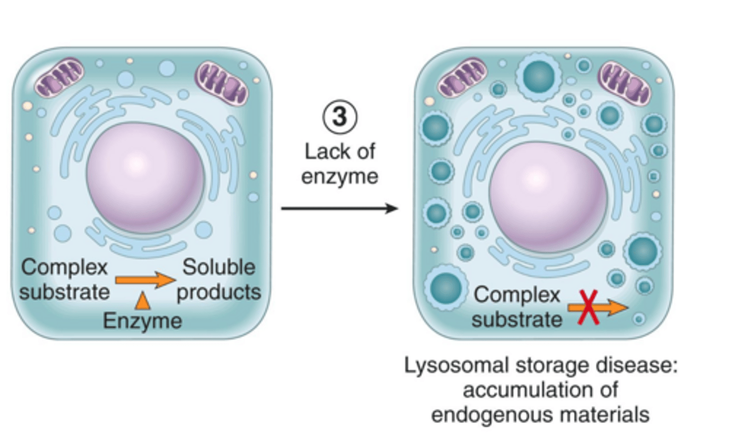
What are the cellular effects of Lysosomal Storage Diseases?
Accumulation of substances within lysosomes, leading to cytoplasmic swelling and vacuolization of hepatocytes, macrophages, and neurons.
What are the two main types of canine degenerative vacuolar hepatopathy (VH)?
Glycogen-type VH and Lipid-type VH.
What conditions are associated with Glycogen-type vacuolar hepatopathy in dogs?
Stress, Cushing's disease, genetic storage disease, and glucocorticoid administration.
What conditions are associated with Lipid-type vacuolar hepatopathy in dogs?
Hypoxia, certain toxins, and metabolic/endocrine diseases like hypothyroidism and diabetes mellitus.
Why is serum ALP often elevated in canine degenerative vacuolar hepatopathy?
Severe hepatocyte swelling can block bile canaliculi, leading to intrahepatic cholestasis.
What are the gross liver characteristics of canine steroid-induced hepatopathy?
The liver is enlarged, pale, and displays an enhanced lobular pattern.
What is the primary clinical context for feline hepatic lipidosis?
Occurs in obese cats following a period of anorexia.
In what species and physiological state is bovine fatty liver disease most common?
Dairy cows in late gestation or peak lactation, especially after inappetence or anorexia.
What gross physical property is characteristic of a liver affected by severe hepatic lipidosis?
The liver is diffusely swollen, yellowish, greasy, and small pieces may float in water or formalin.
What does histology reveal in cases of hepatic lipidosis?
Well-demarcated intracytoplasmic vacuoles in hepatocytes.
Which type of hepatocellular accumulation is characterized by discrete vacuoles that push the nucleus to the periphery?
Hepatic lipidosis.
Which type of hepatocellular accumulation is associated with prolonged steroid treatment in dogs?
Glycogen accumulation (steroid-induced hepatopathy).
What are the four main categories of hepatocellular accumulations discussed in the summary?
Amyloid, Copper, Iron, and Glycogen/Lipid.