Spinal cord tracts
1/24
There's no tags or description
Looks like no tags are added yet.
Name | Mastery | Learn | Test | Matching | Spaced | Call with Kai |
|---|
No analytics yet
Send a link to your students to track their progress
25 Terms
peripheral nervous system
2 types of info
Afferent (sensory)
Somatic
-info taken from skin, retina or ear(membranous labyrinth)
Visceral
-info taken from thoracic and abdominal organs, olfactory epithelium or from taste buds
Efferent (motor)
Somatic
- to skeletal muscle
Visceral
-to cardiac muscle- to smooth muscle- to exocrine glands
spinal pathways
All spinal pathways involve a sequence of neurons
Excitability is transmitted from one neuron to the next in the sequence
Pathways are either ascending (carrying information from receptors to the brain) or descending (conveying information from the brain to spinal cord neurons)
spinal pathways
Tract is a bundle of functionally related axons in the CNS
Tracts are found in the white matter (white cos myelinated neurons for insulation)
Tracts carrying sensory information are ascending, and motor commands are descending
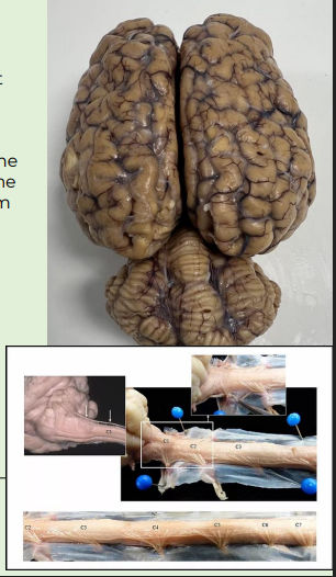
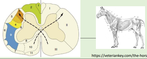
And divides into dorsal, lateral, and ventral funiculus (or columns)
Nerve tracts are named according to their origin spinothalamic tract etc.
These funiculi have a common origin, destination, and function
pic shows I = dorsal funiculus, II is lateral funiculus, III is ventral funiculus = white matter
in the grey matter we have dorsal horn lateral horn and ventral horn
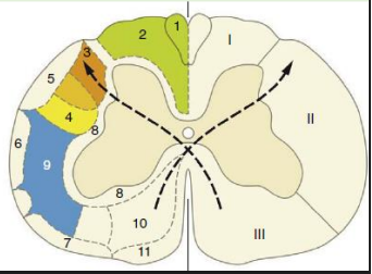
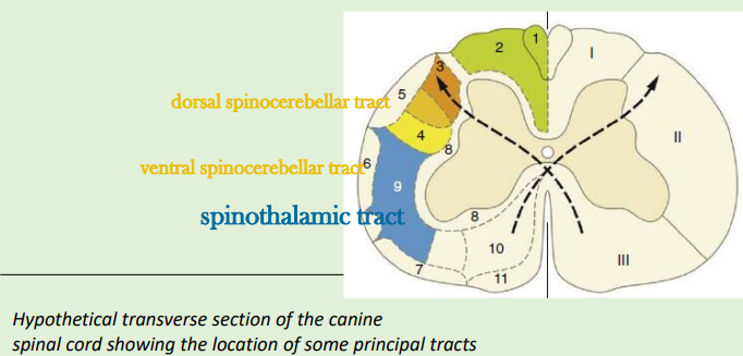
ascending tracts - just need to know spinothalamic tract not the others
each tract is named from origin
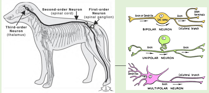
Begin with the primary afferent neuron- terminate in the brain
Information about pain is carried in – spinothalamic tract
Spinocerebellar tracts in which funiculus? lateral and entral part of the column
spinothalamic pathway (pain and temperature)
Information about pain is carried in – spinothalamic tract
Begin with the primary afferent neuron (first-order neuron) - info arriving from environment, taken by first order neuron, synapsing in spinal cord, then second info transfered to the brain
Terminate in the brain
most correct term is pseudounipolar - but one axon
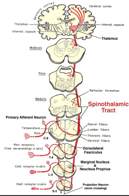
spinothalamic pathway cont
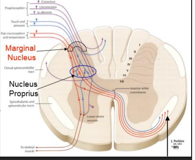
Pain is transmitted from primary afferent axons to the spinal
cord dorsal horn (marginal nucleus or nucleus proprius)
Throughout the length of the spinal cord, projection neurons are concentrated in the marginal nucleus and nucleus proprius of the dorsal horn
Primary order neurons synapse with secondary neurons
Axons of second-order neurons cross the midline and join other axons that also carry pain sensation
These axons form the Spinothalamic tract
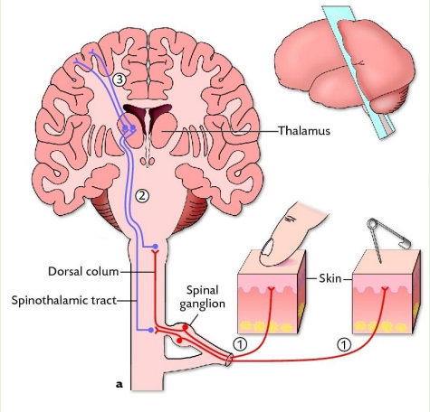
spinothalamic patway cont
The spinothalamic tract to reach the contralateral thalamus (third- order neuron)
Thalamic projection neurons send axons to the somesthetic area of the cerebral cortex
Axons terminating in the lateral thalamus mediate discriminative aspects of pain
Axons terminating in the medial thalamus mediate the motivational-affective aspects of pain (emotional aspects and attention to and memory of pain
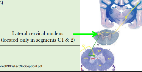
spinocervicothalamic pathway
Spinocervicothalamic tract (spinocervical) - stops in cervical region
Important (most) in cats and dogs - carnivores
Concerned with the transmission of superficial pain and tactile sensations, considered the primary conscious pathway in carnivores
(Spinoreticular tract is primarily concerned with deep pain and visceral sensations)
synpases at lateral cervical nucleus then it will cross
First order neurons: Spinal Ganglion
Second-order neurons: Marginal Nucleus or Nucleus Proprius
Axons of these second-order neurons ascend ipsilaterally to the upper cervical spinal cord to synapse on third-order neurons located in the lateral cervical nucleus
Axons from third-order neurons in the lateral cervical nucleus cross the midline and ascend to the contralateral thalamus to terminate on fourth- order neurons
Axons of the fourth- order neurons project to the cerebral cortex
motor systems
Motor system function
Maintain posture and gait
Provide a stable platform for movement
Voluntary movement and locomotion
Visceral motor function
Somatic motor activity is regulated by separate groups of nerve cells, designated the lower and upper motor neurons
descending tracts
Nerve tracts are named according to their origin
Caudally projecting neurons generally terminate on interneurons
Rubrospinal tracts
Vestibulospinal tracts
Reticulospinal tracts
Tectospinal tracts
Corticospinal tracts
Corticobulbar tracts
Motor systems often functionally grouped into two categories:
Ventromedial motor system- axial and proximal limb muscles, especially extensors
Dorsolateral motor system- Distal limb muscles, especially flexors
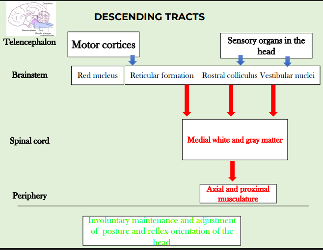
descending motor tracts
Rubrospinal , Vestibulospinal, Reticulospinal, Tectospinal tracts
Caudal projection tracts that originate from brainstem nuclei
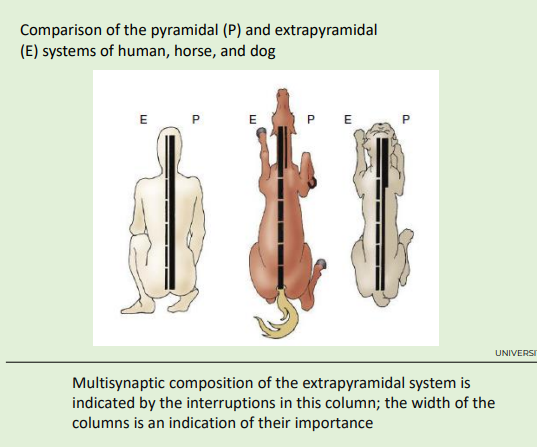
These four tracts are often referred to as the extrapyramidal motor system (pass through the medulla oblongata outside the ventral pyramids- called pyramids cos more tracts passing that region)
It is the most important system in domestic species
Rubrospinal tracts- voluntary and skilled movements in non-primate mammals, very important in quadrupeds
Vestibulospinal tracts- subconscious posture control- keeping the body subconsciously upright against gravity
Reticulospinal tracts- subconscious posture control- muscle tone
Tectospinal tracts- essential for automatic orientation of the head and eyes to auditory and visual stimuli
descending tract rubrospinal tract example
dorsal lateral spinal chord of white matter
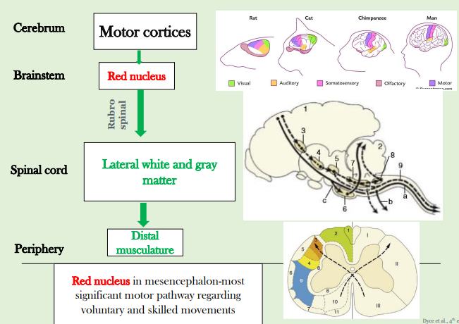
all of them originate brain stem
corticospinal tract - descending motor tract
Originates in the cerebral cortex
Fine motor skills requiring concentration and conscious thoughtprimates and carnivores
Voluntary skilled movements are mainly dependent on this tract
Most elaborate and dexterous voluntary movement
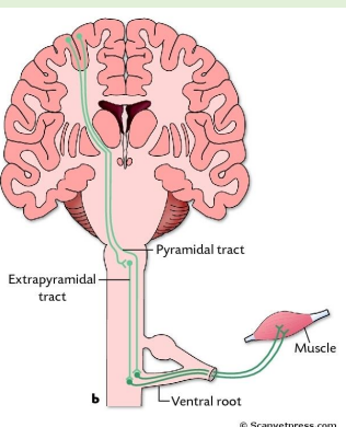
Axons pass through the ventral portion of the medulla oblongata in a region characterised by pyramid section, hence named the pyramidal system
pic shows extrapyrimadal is more developed in domestic specieis like horse e.g. rubrospinal tract more important
spinal cord tract functions
Connects with spinal nerves, through afferent & efferent axons in spinal roots
Communicates with the brain, by means of ascending and descending pathways that form tracts in spinal white matter – serving as a link
Gives rise to spinal reflexes, pre-determined by interneuronal circuits- integrating reflex activity
lower motor neurons
Located within the ventral column of the gray substance of the spinal cord and within the somatic motor nuclei of cranial nerves that contain somatic efferent components
Their axons are conveyed within the spinal and relevant cranial nerves to the skeletal muscles, where each terminates on a group of muscle fibers
LMNs provide the efferent limbs of simple reflexes, but are mostly directed by upper motor neurons
Their axons project into the peripheral nervous system via cranial or spinal nerves to connect with muscle
When stimulated LMN induces muscle contraction
‘Workers ‘
upper motor neurons
UMNs are completely contained within the central nervous system
The UMN cell body is located in the motor nucleus of the brain stem or the motor cortex of the forebrain
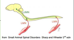
Their axons connect to the lower motor neurones either by synapsing on them directly or indirectly, via interneurons
Upper motor neurons do not project onto muscle fibers
UMNs initiate, regulate, modify and terminate the activity of the LMN
UMN may inhibit or facilitate LMN
‘managers’
LMNs make connection to the muscle
UMNs connect from the brain
they synapse
upper and lower motor neurons
the alpha motor neuron is the principal or most common type of lower motor neuron (LMN)
Disease of lower motor neurons causes stereotypical clinical signs
Upper motor neurons lie completely in the central nervous system and control lower motor neurons
Signs of upper motor disease differ from signs of lower motor neuron disease
reflexes
A reflex is an inherent, subconscious, relatively consistent response to a particular stimulus
Reflex arc involves sensory input, connection in the CNS to the UMN, the LMN, neuromuscular junction and muscle
There are number of different types of reflexes; somatic; autonomic etc.
Reflexes utilise inputs from exteroceptors, interoceptors or proprioceptors
They use cranial or spinal nerves and may affect striated or smooth muscle
Understanding reflexes and their modifiability is important in clinical settings
what five basic components do all reflexes contain?
Sensory receptor
Sensory neuron
Synapse in CNS
Motor neuron
Target organ (Effector organ)
spinal cord-medulla spinalis-reflexes
Examples of spinal reflexes, involving spinal nerves and the spinal cord, include:
Myotatic reflex (muscle stretch): muscle stretch is resisted by reflex contraction of the muscle
Withdrawal (flexor) reflex: limb flexes to withdraw from a noxious stimulus
Panniculus (cutaneous trunci) reflex: pricking skin triggers contraction of cutaneous trunci (panniculus) m.
Perineal (anal constriction) reflex: mild compression of the skin of the perineum or anus with forceps causes contraction of anal sphincter and flexion of the tail
Myotatic reflex (muscle stretch): muscle stretch is resisted by reflex contraction of the muscle
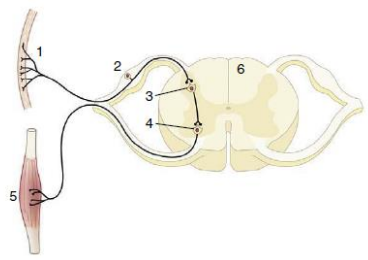
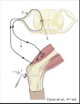
Patellar reflex- the most reliable pelvic limb reflex
Stretch stimulus on the tendon (1) travels via the afferent neuron (2) to the spinal cord
Impulse is then transmitted to the efferent neuron (3), which stimulates the quadriceps muscle (4)
Myotatic reflex (muscle stretch)- Patellar reflex
Motor component of this reflex arises from L4-L6 in the spinal cord, which is the origin of the femoral nerve
The femoral nerve innervates the quadriceps muscle- effector muscle of this reflex
Simultaneously, it also causes reciprocal inhibition
Therefore, this reflex evaluates the integrity of the spinal cord segments (L4–L6 and sensory/afferent and motor/efferent) as well as the femoral nerve
spinal cord reflexes
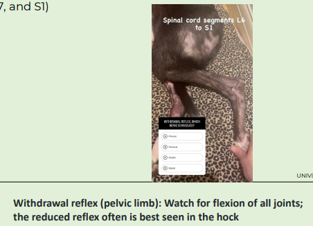
Withdrawal or flexor reflex: limb flexes to withdraw from a noxious stimulus
Test for primarily for the sciatic nerve and its spinal cord segments (L6, L7, and S1)
Elicited by applying a noxious stimulus to the distal part of the limb
Observe withdrawal ( flexion) of the entire limb
Initiated by free nerve endings- axons enter dorsolateral fasciculusbifurcate
Collateral branches enter gray matter to synapse on interneurons and projection neurons (other limbs will extend to compensate if standing
Panniculus (cutaneous trunci) reflex
pricking skin triggers contraction of cutaneous trunci (panniculus) m.
The sensory pathway from the skin enters the spinal cord and ascends bilaterally to the C8 to T1 spinal cord segment, where it synapses with the lateral thoracic nerve, resulting in a contraction of the cutaneous trunci muscles bilaterally
Perineal (anal constriction) reflex
mild compression of the skin of the perineum or anus with forceps causes contraction of anal sphincter and flexion of the tail
Evaluates S1 to S3 spinal nerves and, peripherally, the pudendal nerve
brain and spinal reflexes
Most spinal reflexes can be overridden at least temporarily by higher brain centers
Impulses may be sent down descending pathways to the efferent neurons supplying the involved muscles to override the input from the receptors, actually preventing the muscle from contracting despite a harmful stimulus