The Nervous System
1/73
There's no tags or description
Looks like no tags are added yet.
Name | Mastery | Learn | Test | Matching | Spaced | Call with Kai |
|---|
No analytics yet
Send a link to your students to track their progress
74 Terms
4 functions of the nervous system
Controls physiological parameters in the body
Works with the endocrine system to maintain homeostasis
Initiates voluntary movements
Is the origin of thought, memories, & emotions in the brain
The nervous system is organized into what 2 division? What do these divisions consist of?
Central Nervous System (CNS) - brain & spinal cord
Peripheral Nervous System (PNS) - the rest of the nerves; responsible for afferent & efferent communication
4 components of the peripheral nervous system (PNS) & what they do
Receptors - bring info in
Afferent nerves - bring info to the CNS
Efferent nerves - bring info out of the CNS
Effectors - place that talks to brain & spinal cord and back
Afferent
Info coming INTO the CNS; arriving
Efferent
Info coming OUT of CNS; exiting
Visceral
Referring to organs, smooth muscle, cardiac muscle
Somatic
Referring to skin, skeletal muscle, or joints
Nerve
Structure of PNS that you can see with the naked eye that carries info in and out of the CNS
(The CNS does not contain any nerves!)
Autonomic nervous system
Involuntary nervous system pertaining to organs
2 divisions of the autonomic nervous system
Sympathetic division
Parasympathetic division
Sympathetic nervous system
Fight or flight response
Parasympathetic division
Conserves energy; rest & digest because it promotes rest and increases activity of digestive organs
Neuroglia (glial cells)
Supporting cells of the nervous system; they do NOT send signals, but instead, they take care of neurons
4 glial cells of the CNS
Astrocytes
Microglia
Ependymal cells
Oligodendrocytes
2 glial cells of the PNS
Satellite cells
Schwann cells
Astrocytes
Star-shaped cells in the CNS that are meant for guidance in early development; involved in maintaining ionic gradients & blood brain barriers (aka helps prevent toxins & germs from coming out of the blood & into the neuron environment)
Microglia
CNS cells that eat up any junk or germs they might find in between the cells
Ependymal cells
CNS cells that line the cerebral ventricles; have cilia to help circulate cerebrospinal fluid
Oligodendrocytes
CNS cells that have wrap around the axons of neurons; acts as the myelin sheath for neurons in the CNS
Satellite cells
PNS cells that surround the cell bodies to ensure a proper environment
Schwann cells
Cells that produce the myelin sheath for axons in the PNS
Neurons
Cells that live a very long time that don’t go through mitosis (amitotic); Have a high metabolic rate, meaning they are constantly making ATP, so they require lots of glucose, oxygen, and blood flow
5 structures of a neuron
Cell body (aka soma)
Dendrite
Axon
Axon hillock
Axon collateral
Telodendria
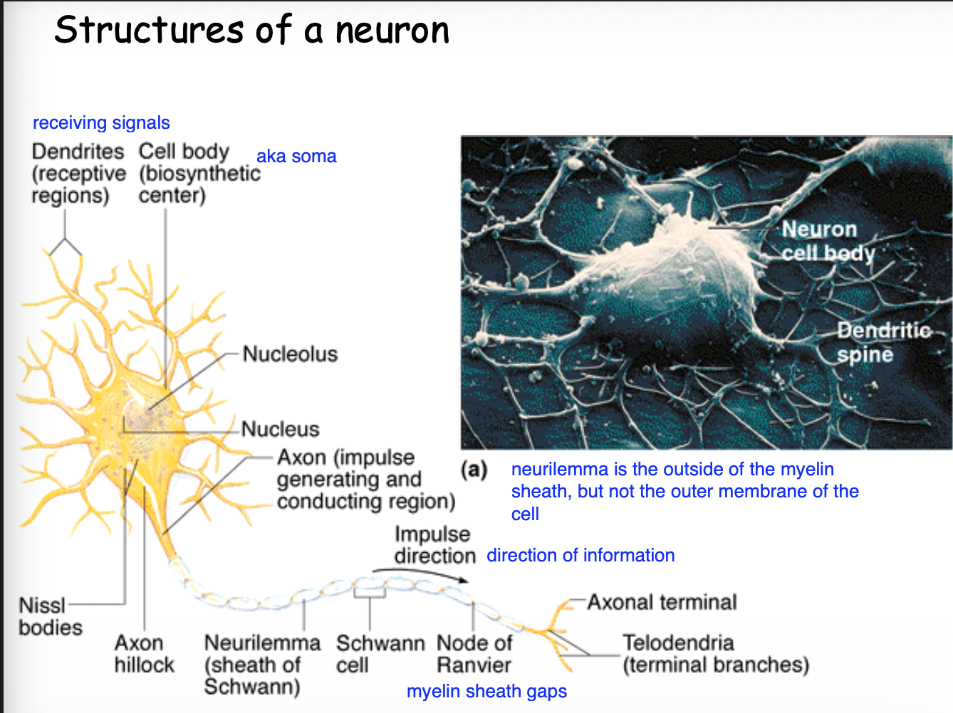
Cell body (soma)
Contains the nucleus and nucleolus; responsible for integration of signals
Dendrite
Branch-like extensions of the neuron that receiving electrical signals, carrying them to the cell body
Axon
Fiber-like extension that carries electrical impulses away from the cell body
Axon hillock
Cone-like attachment of the axon to the cell body
Axon collateral
Branches of the axon itself
Telodendria
Axon terminals that have synaptic knobs at their ends
3 components on a nerve fiber
Myelin sheath
Neurilemma
Nodes of Ranvier (myelin sheath gaps)
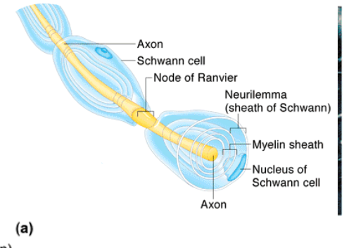
Myelin sheath
Made up of Schwann cells that insulates axons & speeds up the transmission of an action potential
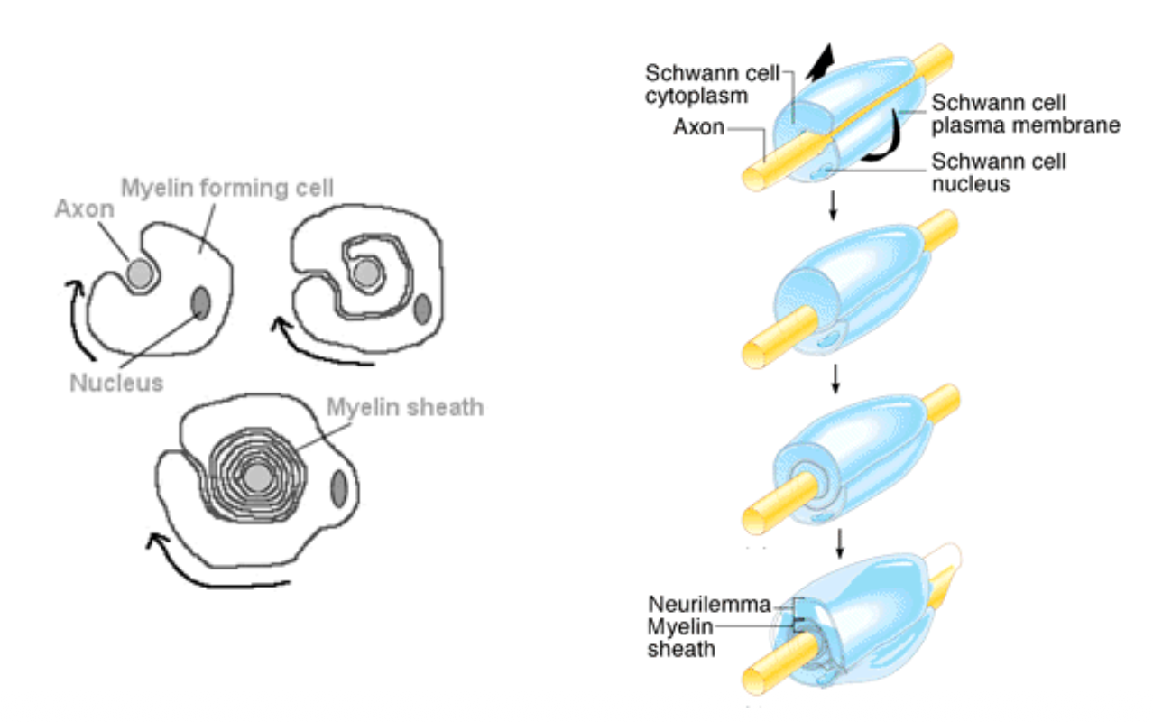
Neurilemma
Plasma membrane of a Schwann cell
Nodes of Ranvier
Gaps in between each of the myelin sheaths
What is axonal transport?
What are the 2 kinds & what do they do?
A way to transport intracellular items along a neuron’s axon
Anterograde (amazon prime) - moving proteins from soma to axon terminal, similar to how regular impulses would move
Retrograde (amazon returns) - removing viruses from the axon terminal to the soma
3 types of neurons according to structural classification
Pseudounipolar
Bipolar
Multipolar
Pseudounipolar neurons (3)
Cell body with a stunted process, then a really long process coming off of it
Most sensory neurons are like this
Cell bodies of these cells can be found in the dorsal root ganglion
Bipolar (4)
2 processes coming off of each side of the cell body
1 process serves a dendrite purpose
1 process serves as an axon
Can be found in sensory systems (ex. retina of the eye)
Multipolar (2)
What the standard neuron we think of looks like
Neuron with soma, lots of dendrites, 1 axon
3 functional classes of neurons + what structural neurons encompass them & their functions
Afferent (sensory; pseudounipolar & bipolar) - info toward CNS
Efferent (motor; multipolar) - info away from CNS
Interneuron (multipolar) - info within CNS
Cluster of cell bodies in the CNS
Nucleus
Cluster of cell bodies in the PNS
Ganglion
Bundle of nerve fibers (axons or dendrites) in the CNS
Tract
Bundle of nerve fibers (axons or dendrites) in the PNS
Fascicle
4 structures of nerves & what they are
Endoneurium - connective tissue covering each nerve fiber
Fascicle - a bundle of axons/dendrites
Perineurium - connective tissue surrounding each fascicle
Epineurium - connective tissue surrounding the whole nerve
3 functional classes of nerves & what they do
Afferent nerves - carry impulses towards the CNS
Efferent nerves - carry impulses away from the CNS
Mixed nerve - contains both afferent and efferent fibers
What 2 things determine electrical flow?
Ions inside & outside the cell
Permeability of the cel membrane
Excitable Cells (5)
Neurons & muscle cells
Overall, high concentration of K+, but low concentration of Na+
Outside - high concentration of Na+, low concentration of K+
Inside - high concentration of K+, low concentration of Na+
This unequal distribution is a resting membrane potential
Resting membrane potential of most neurons
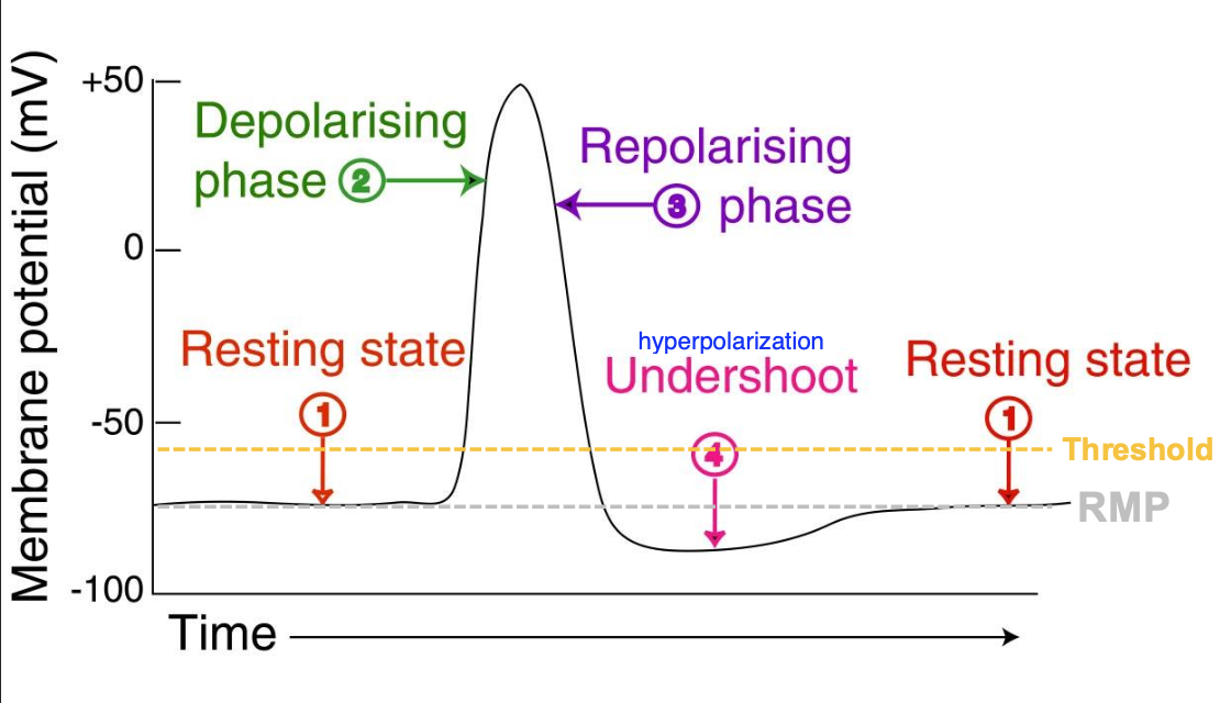
-70 mV (polarized)
What is the net charge on the inside of the membrane vs. the outside?
Inside - net negative charge
Outside - net positive charge
Polarized
Inside of the cell is negative compared to the outside of the cell
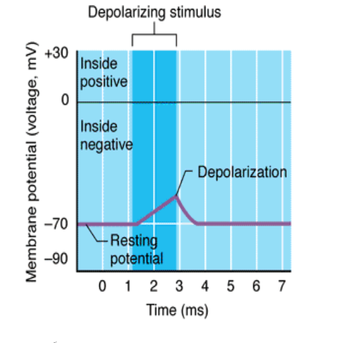
Depolarized
The charge inside the cell moves closer to zero (more positive); excitatory event
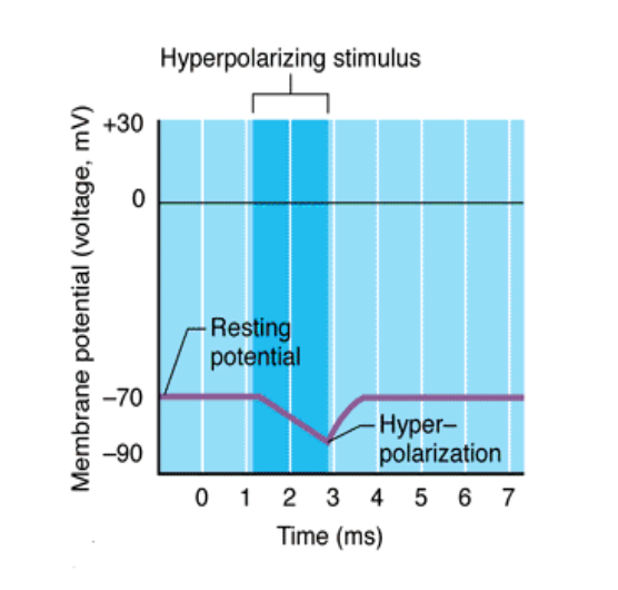
Hyperpolarized
Inside of the cell becomes more negative than its normal resting membrane potential; inhibitory event
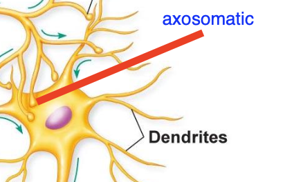
Axosomatic synapse
Synapse onto a cell body
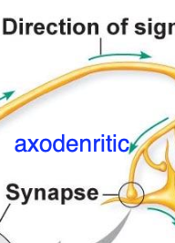
Axodendritic synapse
Synapse onto a dendrite
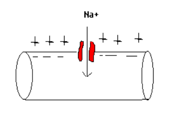
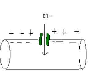
Ligand-gated (chemically gated) ion channels
Stimulus (ligand) binds to receptor, causing a channel to open, allowing electrically-charged ions to move across the membrane
(ex. neuromuscular junction)
Depolarization in a ligand-gated (chemically-gated) ion channel (3)
Inside of the cell is negative, outside is positive
Na+ comes into the cell, bringing its positive charge
Inside of the cell becomes more positive, meaning membrane potential moves closer to 0
Hyperpolarization in a ligand-gated (chemically-gated) ion channel (3)
Inside of the cell is negative, outside is positive
Cl- comes into the cell, bringing its negative charge
Inside of the cell becomes more negative, meaning membrane potential becomes moves farther from 0
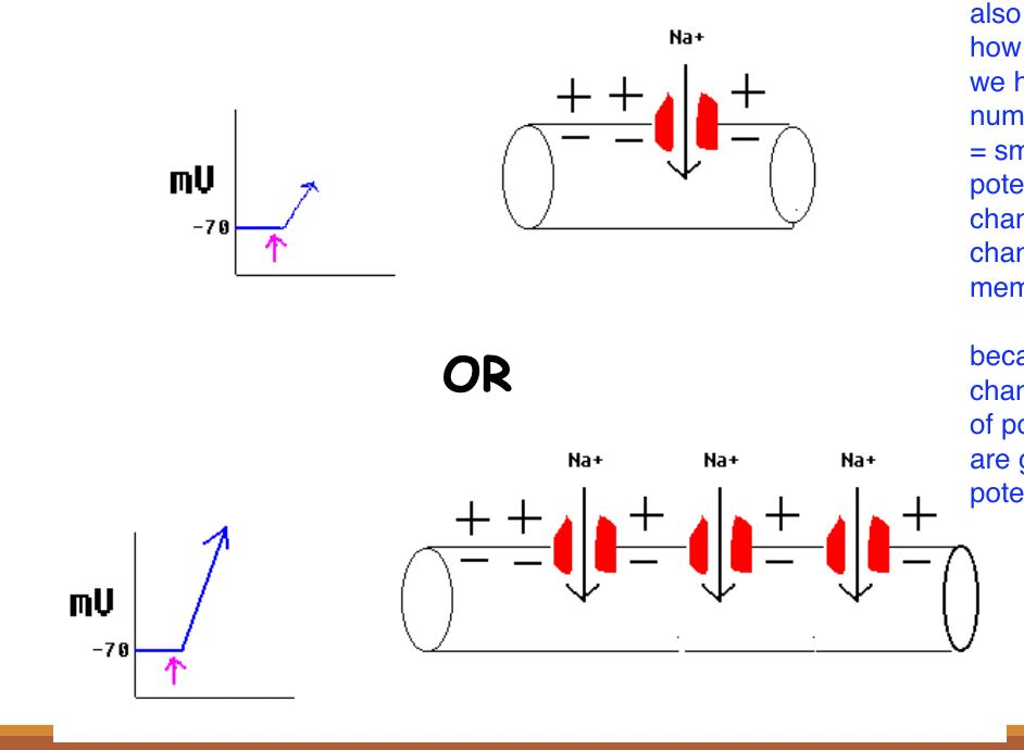
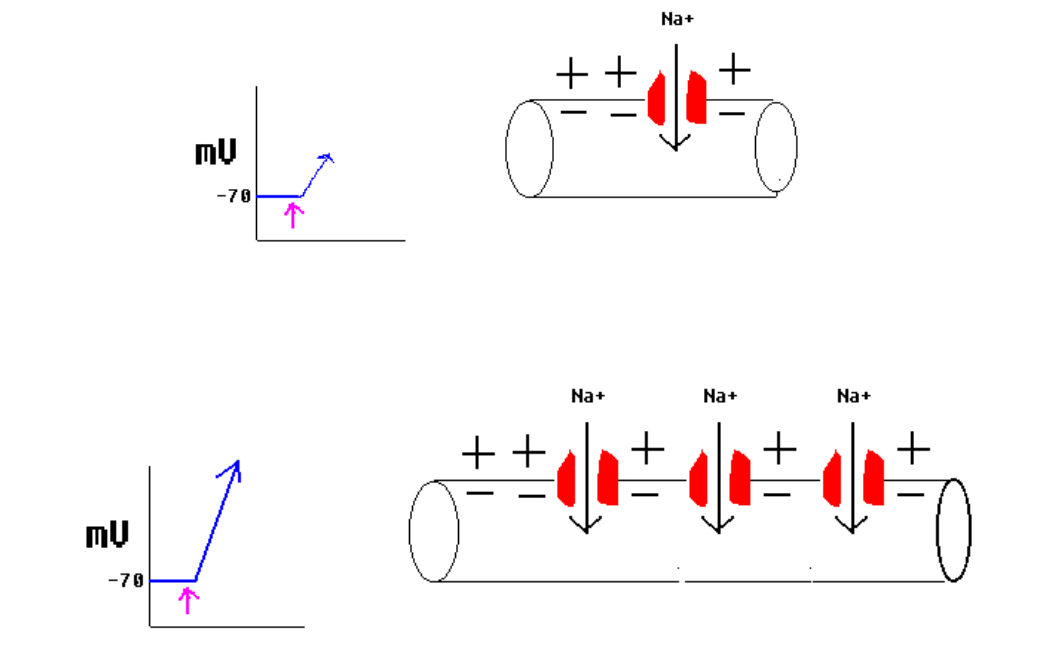
Graded Potentials
Membrane potentials we can change depending on how many channels we have
Small number of open channels = small change in membrane potential
Many open channels = big change in membrane potential
3 types of graded potentials
Postsynaptic potential
Receptor potential
Pacemaker potential
Postsynaptic potential
Graded potential produced by responding to binding of neurotransmitter & opening of a chemically-gated ion channel that could cause either excitatory (EPSP) or inhibitory postsynaptic potential (IPSP)
Receptor potential
Graded potential produced at the ends of afferent neurons when they are stimulated by light, heat, or mechanical energy (mechanically-gated ion channels)
Pacemaker potential
Graded potential involving leaky ion channels that are naturally open in certain specialized cells
Mechanism of chemical synapse (ligand-gated ion channels) (5)
Neurotransmitter binds to receptor (chemically gated ion channel)
Channel gate opens
Ions move in, causing depolarization/hyperpolarization (if Na+ is coming into the cell/if K+ is coming into the cell)
If a certain amount of positive charge reaches the axon hillock, and action potential will be initiated there
4 characteristics of graded potentials
They fade out (decremental)
They vary in amplitude (the more that open, the greater the change in membrane potential)
They can be bidirectional
They can be a depolarization or a hyperpolarization
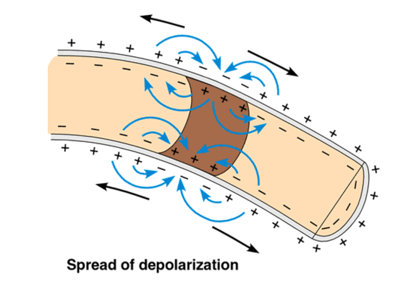
Decremental property of graded potentials
They lose strength & die out as it moves away from initial sight of stimulus
Amplitude property of graded potentials
More channels = larger change in voltage
Bidirectional property of graded potentials
Current spreads in both directions from the site of stimulation
What type of polarization does EPSP inputs cause?
Depolarization
What type of polarization does IPSP inputs cause?
Hyperpolarization
What happens when the membrane potential reaches threshold?
Action Potential
Action Potential (AP)
All of northing depolarizations that occur when excitatory graded potentials reach the axon hillock; Depend on voltage-gated ion channels to continue the AP down the axon
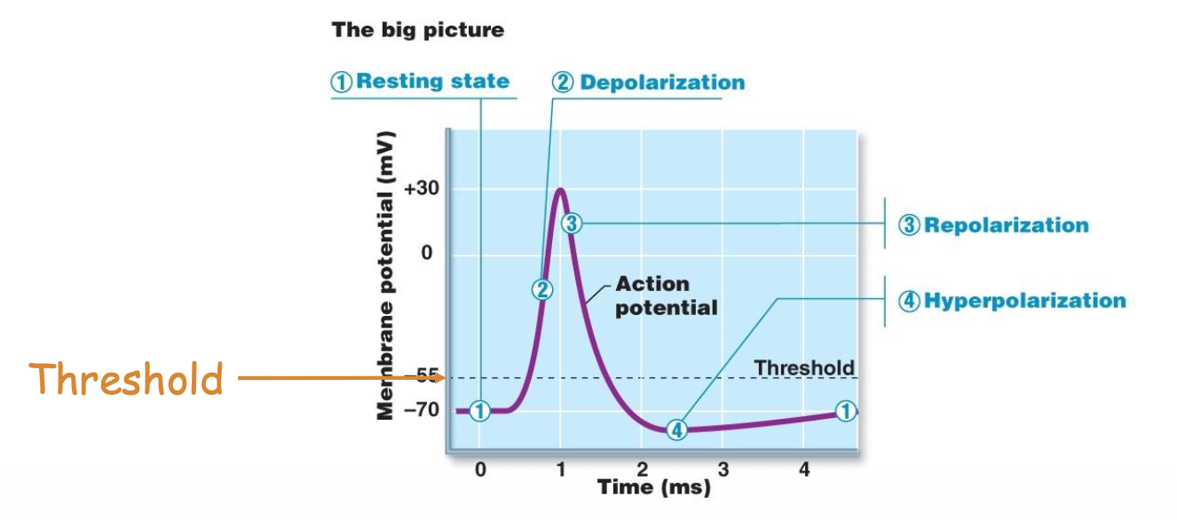
4 characteristics of action potentials
Maintains strength for the entire length of the axon
Always have the same amplitude
Unidirectional; always flows along the axon, starting at the axon hillock & going to the axon terminal
Characterized by a sharp depolarization followed by a brief period of hyperpolarization
5 Steps of Action Potential
Resting state
Depolarizing phase
Repolarizing phase
Undershoot
Resting state