Practicals
1/47
There's no tags or description
Looks like no tags are added yet.
Name | Mastery | Learn | Test | Matching | Spaced | Call with Kai |
|---|
No analytics yet
Send a link to your students to track their progress
48 Terms
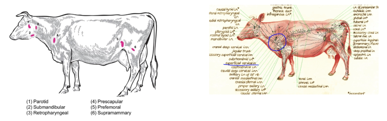
List the steps of a cow clinical exam
Place cow in head bail and lock
DISTANCE
Signalment (breed, sex, ID tags)
BCS /10
Describe mentation, posture and demeanour
Symmetry of limbs, abdomen and udder (stage of production)
Evaluate skin and hair coat
Take respiratory rate
Remove back chain
TAIL PT1
Take temperature
Check tail tone, anal tone and coccygeal pulse
Evaluate vulval mucus membranes
Palpate mammary gland and supramammary lymph nodes
Put chain back up and put left side rail down
LEFT SIDE
Visually assess HL and claw
Visually assess lateral udder and ventral abdomen
Palpate pre-femoral lymph node
Auscultate rumen contractions over 2 minutes and palpate layers
Ping paralumbar fossa and ballot layers of rumen
Ping ribs 9 - 13
Auscultate lung field
Auscultate heart
Perform Wither’s pinch test x2
Palpate pre-scapular lymph node
Visually assess FL and claw
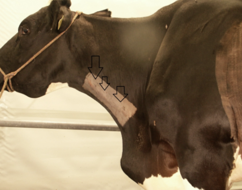
Examine brisket area
Evaluate jugular fill/pulses
Evaluate hydration with skin tent
Put left side rail up again
HEAD
Check symmetry of head
Visually check ears
Evaluate eyes, including 3rd eyelid
Check nostrils and nasal planum
Check muzzle and open mouth, palpate tongue
Palpate submandibular tissue and lymph nodes
Put right side rail down
RIGHT SIDE
Evaluate jugular region
Examine brisket area
Visually assess FL and claw
Palpate pre-scapular lymph node
Auscultate heart
Auscultate lung field
Check for palpable liver
Ping ribs 9 - 13
Auscultate and ping right paralumbar fossa
Palpate pre-scapular lymph node
Visually assess lateral udder and ventral abdomen
Visually assess HL and claw
Put right side rail up again and take back chain down
TAIL PT2
Describe collection of urine sample
Describe collection of milk sample
Describe collection of faecal sample
Discuss vaginal and rectal exam procedure
Put back chain up again and release cow from head bail
5 Features to assess when examining the farm
Clinical examination begins at the gate to observe:
General state of farm and geography
Condition of herd
Amount and types of feed present
Track/race condition
Facilities
Negative welfare indicators (eg. animals in deep mud, lack of shade or water)
→ Gives idea of what to expect of the client or animal
D1: Signalment
Importance
Example:
Bloat in cow vs. calf
Photosensitisation timing
Importance: Confirm correct patient and guide DDx list → Focus history questions and examination of relevant body systems
Examples:
Bloat
Cow = Ruminal bloat
Calf = Abomasal bloat
Photosensitisation
Late summer - autumn = Facial eczema
Spring = St John’s wort or brassicas
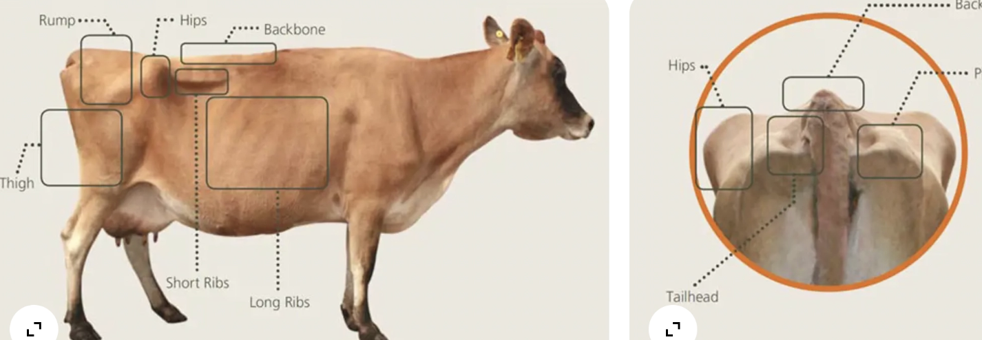
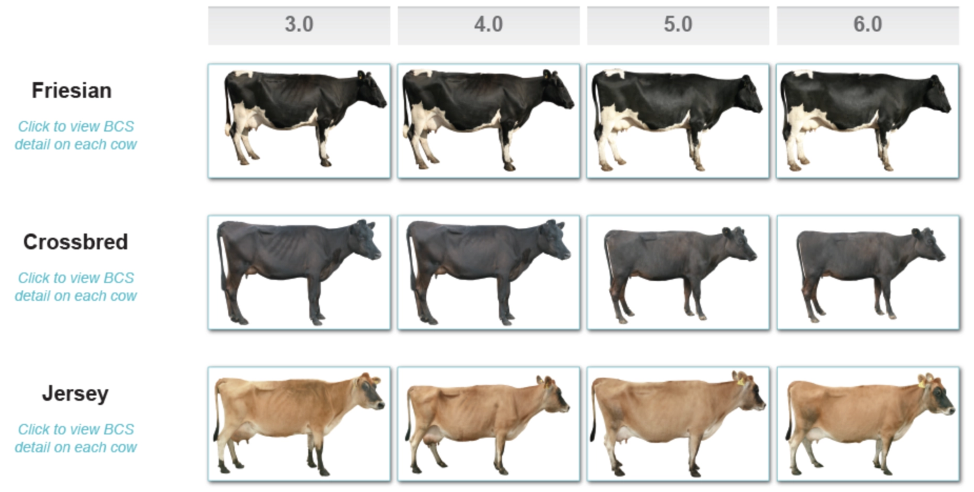
D2: BCS
10 features to assess
Ideal BCS
Features:
Spine
Short ribs
Long ribs
Hips
Pins
Rump
Tail height
Thigh
Ideal: 5 (5.5 for heifer) at calving
D3: Mentation, Posture and Demeanour
Examples to describe mentation
Examples to describe posture
Examples to describe demeanour
6 Signs of aggression
Mentation:
BAR/QAR
Obtunded = Reduced response to environmental stimuli
Comatosed = Unconscious
Posture:
Standing squarely with neutral head carriage and bearing weight x4
Head down/tilted/deviated/elevated/opisthotonic
Wide-based stance
Demeanour:
Relaxed, agitated, compulsive behaviours, aggression
Eating and chewing cud = Relaxed
Aggression:
High head
Pricked ears
Direct staring
Holding ground
Pawing
Bellowing or snorting
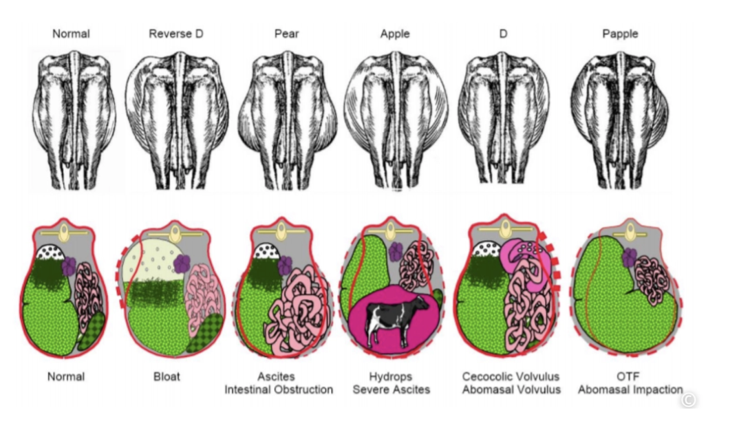
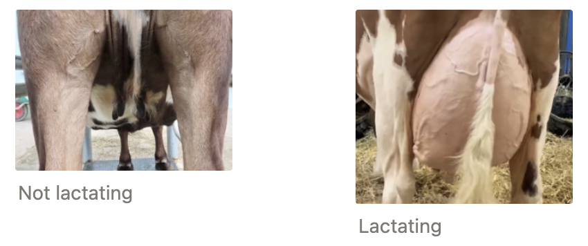
D4: Symmetry of Limbs, Abdomen and Udder
Types of abdominal distension (+ DDx)
Example description of udder
Abdominal Distension:
Udder: This cow is not lactating, the udder is small, shrunken and appears empty
D5: Skin and Hair Coat
4 Features to assess
Normal description
Describe the following lesions
Features:
Lesions
Colour changes
Faecal staining
Ectoparasites
Normal: The hair and coat is appropriate for the season and there is a little mud present on the limbs. Colour is normal and there are no lesions, faecal staining or ectoparasites observed.
Lesions:
A: There is a large amount of perineal staining, faeces appears yellow and pasty to liquid. There are patches of alopecia on the caudal stifle, the skin appears erythematous
B: The Jersey cow has a large number of proliferative, keratinised lesions that range from 0.5 - 10cm along the ventral aspect of the neck, jaw and side of face including the ears, periorbital skin, but not the node. The masses are grey with a rough, dry and irregular surface
C: The back of a Friesian cow shows significant areas of photosensitivity lesions including alopecia and erythema of the non-pigmented skin and large areas of dry skin sloughing off, with some reddened ulcerative lesions beneath
D6: Respiratory Rate
Where to observe
Normal reference range
3 Abnormal features to observe
Example normal description
Where: Thorax, nostrils or flank
Normal: 12 - 30brpm
Abnormal:
Dyspnoea (eg. open-mouth breathing with neck extended, nostrils flared and back arched with elbows abducted)
Noises (eg. coughing, sneezing, snuffling, wheezing)
Nasal discharge
Example: The RR is 12breaths/minute which is within the reference range of 10 - 30 brpm. She appears eupneic (no increased effort while breathing)
The RR is 48 breaths per minute, which is above the reference range. Loud snuffling noises can be heard from the nose, loudest on inspiration,
T1: Temperature
Normal
Method
Normal: 38 - 39.3˚C (~38.5˚C)
Method: Insert thermometer all the way until the window and against the rectal mucosa to obtain an accurate reading
T2: Tail Tone, Anal Tone and Coccygeal Pulse
Define tail tone
Coccygeal pulse location
Normal pulse rate
Example normal description
Tail Tone: Level of resistance when grasping and elevating the tail (present/absent/weak/strong)
Coccygeal Pulse: Coccygeal artery in the ventral groove of the tail at the level of the vulva (present/absent/strong/weak/bounding)
Pulse: 48 - 84bpm
Example: The tail tone is strong and the pulse is palpable and as a regular rhythm
T3: Vulval Mucus Membranes
Location
4 Features to assess
Example normal description
Location: Beyond the mucocutaneous junction
Features:
Colour
Hydration
CRT
Discharge
Example: The vulval MM are shiny/slippery and a salmon pink colour with a CRT of 1 - 2 seconds
Abnormal = Tacky, white, red, brown, yellow, blue, petechiae
T4: Supramammary Lymph Nodes and Udder Palpation
Method
5 Features to assess
Example normal description
Method: Keep head high and stoop slightly (never crouch or kneel)
Can perform at end as some animals resent
Palpate ventral surface and up lateral sides of ALL 4 quarters
Features:
Hardness or swelling
Lumps
Temperature (hot/cold)
Pain
Symmetry
Example: The cow is dry, all four quarters are soft, non-painful and relatively symmetrical. There are four teats, and the supramammary lymph nodes are not palpable
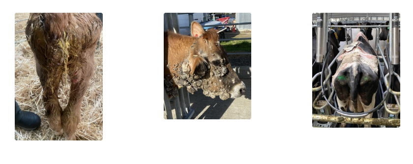
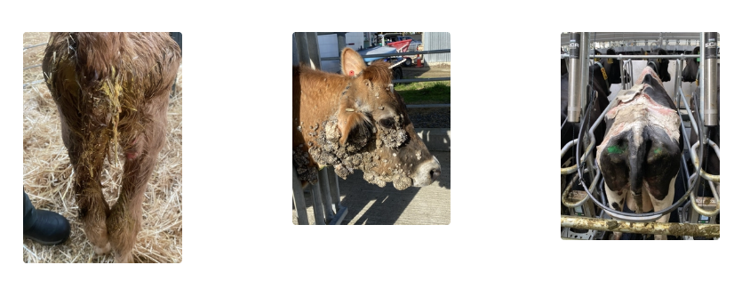
LHS1+10/RHS12+3: Visually Assess Limb and Claw
Method
4 Features to assess for
Example normal description
Method: Do NOT palpate
If further exam required → Restrain leg with ropes AFTER full clinical exam
Features:
Swelling of joints, bones and tendons
Redness or lesions on skin
Muscle atrophy
Medial and lateral claws
Example: There is mud and debris along the lateral side of the left hindlimb, she is standing with the left foot slightly in front of the right
vs. There is moderate swelling over the lateral aspect of the hock, the skin is missing over this swelling, the claws appear even
LHS2/RHS11: Visually Assess Lateral Udder and Ventral Abdomen
Method
6 Features to assess
Example normal description
Method: Do NOT get too close or crouch
Features:
Skin
Contour of abdomen
Milk vein for damage
Umbilical region for hernia
Prepuce in males
Udder: Symmetry and teat condition
Example: The skin appears smooth with an even ventral abdominal contour. The milk vein is not prominent in this dry cow. The udder is fairly symmetrical and empty. Teats are mildly dry.
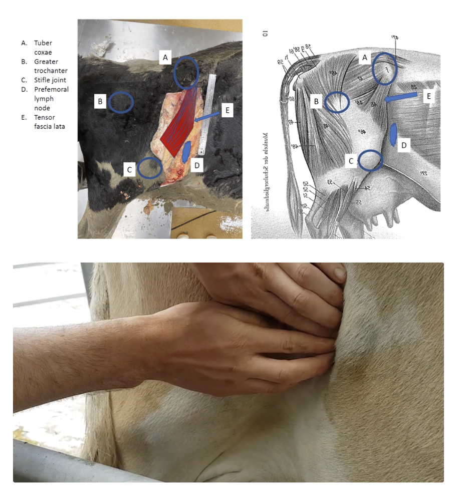
LHS3/RHS10: Palpate Pre-Femoral Lymph Node
Method/location
4 Features to assess for
Example normal description
Method: Run hands caudal to cranial across flank in front of the quadriceps muscle
Location: Between the tuber coxae and stifle (slightly cranial and dorsal to stifle)
Features:
Texture
Mobility
Size
Pain
Example: The pre-femoral lymph node is smooth, freely moveable and 10 × 5cm (large in LATU cows due to fat coverage)
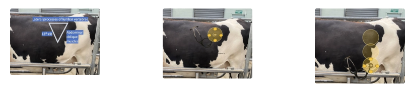
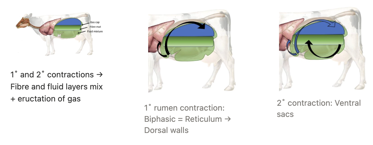
LHS4: Auscultate Rumen Contractions
Method
Normal rate of contraction
Example normal description
Method: Listen over left paralumbar fossa for ≥1.5 - 2 minutes (hear length of time between two contraction)
Left paralumbar fossa = Triangle created by the 13th rib cranially, tuber coxae caudally, transverse process of the lumbar vertebrae dorsally and the abdominal oblique muscles ventrally
Normal: ONCE every 30 - 90 seconds
Example: There were 3 strong contractions over 2 minutes
vs. Only 1 rumen contraction was auscultated over 2 minutes and there is poor rumen fill
LHS5/RHS9: Percuss/Ballot/Succuss Rumen
Method/definitions
3 Layers of the rumen
Example normal description
Method:
Percuss = Ping down left paralumbar fossa to assess layers of rumen and detect LDA
Ballot = Punch down left paralumbar fossa to feel layers of rumen
Succuss = Ballot with auscultation of layers (listen for splashes)
Layers:
Gas cap
Fibre mat
Fluid
Example: The fibre mat is doughy and extends up to here, and there is no splashing on succussion
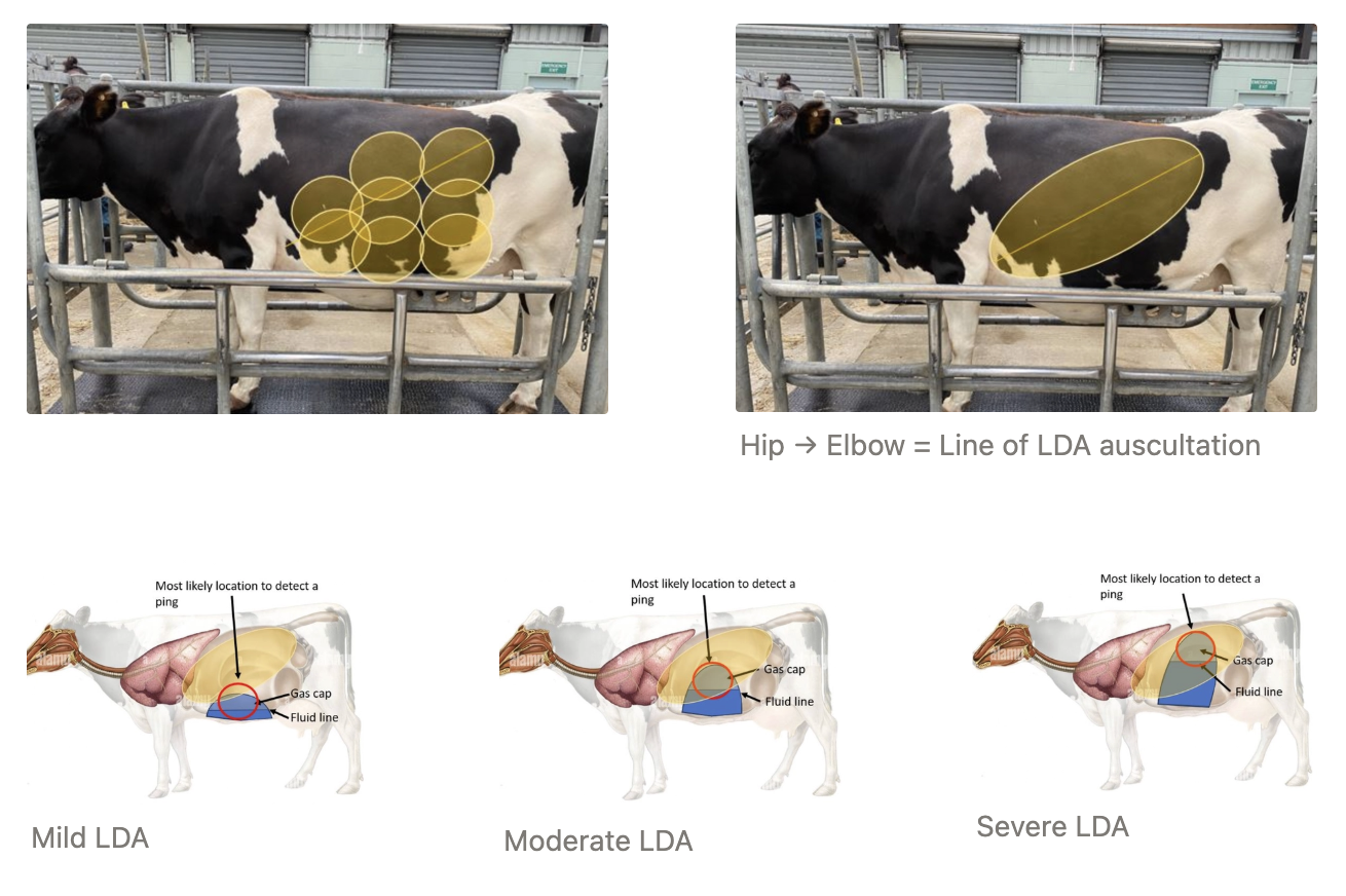
LHS6/RHS8: Ping ribs 9 - 13
Function
Location
Example normal description
Function: Detect LDA/RDA = High-pitched sound
Location: #1 location for LDA = Line from olecranon to tuber coxae
Normal abomasum ventral, to the right of midline
Example: No high-pitched pings were auscultated and there is no evidence of an LDA
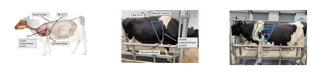
LHS7/RHS6: Auscultate Lung Fields
Difficultly with cows
Location
Example normal description
Difficulty: Reduced auscultation field due to interference of noise from GI caudally, depth of thoracic process muscles dorsally, shoulder muscles cranially and heart ventrally
Location: Bordered by the caudal scapula/triceps cranially, line from rib 10 - 11 → olecranon caudoventrally and epaxial muscles dorsally
Listen to dorsal, mid and ventral aspects
Place stethoscope between triceps and chest wall to access more cranial regions = Important for right cranial lung lobe (1st lobe off tracheal bronchus)
Example: Soft bronchovesicular sounds are auscultated, no crackles or wheezes can be heard.
LHS8/RHS5: Auscultate Heart
Location
Normal heart rate
2 Features to assess for
4 Valves (+ location)
Example normal description
Location: Under rib 3 - 6 (under elbow → move limb forwards)
Normal: 40 - 60bpm in paddock (48 - 84bpm during examination)
Features:
Arrhythmia
Murmur
Valves:
Pulmonic (3rd intercostal space LHS)
Aortic (4th intercostal space LHS)
Mitral (5th intercostal space LHS)
Tricuspid (5th intercostal space RHS)
Example: The heart rate is 68bpm, within the reference range of 48 - 84bpm. The beats are strong, regular, with no murmurs detected.
OR The heart is difficult to hear, but the sounds are regular and the heart rate is 48bpm, within the reference range of 48 - 84 bpm.
LHS9: Wither’s Pinch Test
Method
Normal (negative response)
Positive response
2 Alternative tests
Method
Result
Method: Lift skin behind wither’s off spine with both hands → Perform twice for interpretation
Normal: Dipping = Cow drops down
Positive Result: Cow does NOT dip after x2 tests → Indicates cranial abdominal or caudal thoracic pain
Alternatives:
William’s Test = Place right hand over left paralumbar fossa and stethoscope close to thoracic inlet → When rumen contraction occurs, vet can feel movement in paralumbar fossa and should listen for grunt in thoracic inlet = Positive for pain
Bar Test = Bar under abdomen at xyphoid process and lifted at each end → Grunt = Positive for pain
LHS11/RHS4: Palpate Pre-Scapular Lymph Node
Method
Normal size
Method: Place hands in middle of scapula with fingers towards head → Run hands cranially until you feel the end of the shoulder muscles → Even more cranial advancement to feel lymph node
Normal: 2 - 3cm x 6 - 8cm
LHS12/RHS2: Examine Brisket
3 Features to assess for
How to ID oedema
Features:
Oedema
Masses
Trauma
Oedema: Pitting
LHS13/RHS1: Evaluate Jugular Fill and Pulse
Normal pulse
Abnormal pulse
How to assess jugular fill
How to differentiate pathological from physiological jugular distension
3 DDx
Normal: Jugular pulse seen from level of thoracic inlet to <1/2 neck height
Abnormal: Jugular pulse >1/2 up neck
NORMAL to extend further when head down
Jugular cording
Fill: Hold off jugular vein at thoracic inlet to observe jugular fill (assess central venous pressure and volaemic status)
Differentiation: Hold off jugular vein at head and wipe blood into heart → Assess if jugular pulse returns
DDx:
RCHF (eg. endocarditis)
Traumatic reticuopericarditis
Johne’s disease
LHS14: Evaluate Hydration
4 Ways to assess hydration
Normal
Dehydrated
Skin tent (neck or upper eyelid)
Normal: Skin elastic and will return to normal position after 1 second
Dehydrated: Tent remains as skin elasticity reduces
Eye position
Normal: Eyes are not sunken and no gap between eyeball and eyelids
Dehydrated: Enophthalmos and skin surrounding eye shows tension on underlying bones
Viscosity of saliva
Normal: Saliva with viscosity of canola oil = Well-formed bubbles observed
Dehydrated: Saliva sticky and of thicker viscosity, fewer bubbles form
Jugular fill
H1: Symmetry
Additional feature to assess
Example normal description
4 DDx for head tilt
Feature: Length of horns, shape and direction of growth
Example: The cow’s face is symmetrical. The ears are moving freely, the eyes are both open and comfortable. There is no heat tilt present.
vs. The left ear and left eyelids appear to be drooping on one side, the nose appears to be deviated to the right of midline slightly
Head Tilt:
Otitis media/interna (eg. Mycoplasma bovis)
Listeriosis
Thromboembolic meningoencephalitis (TEME, Histophilus somni)
Trauma
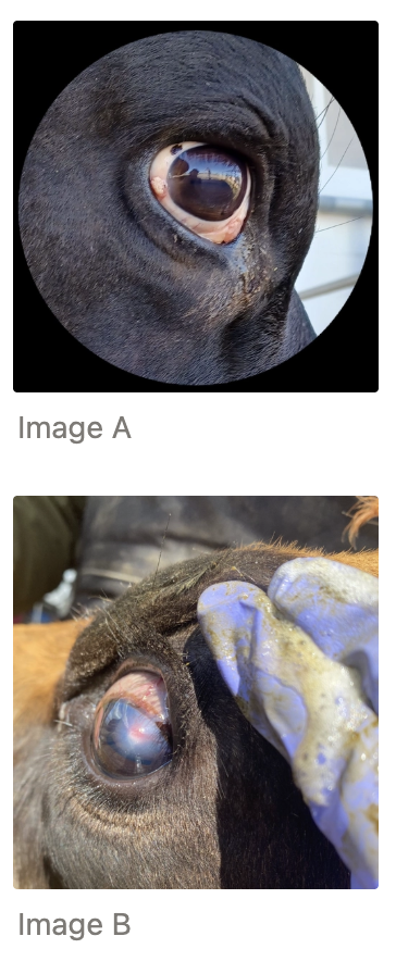
H3: Examine Eyes
Importance
Example normal description
8 Abnormalities
How to assess the 3rd eyelid
Importance: Detect SCC early to improve welfare and longevity
Example: Open, comfortable with clear, bright and visual corneas
Abnormalities:
Discharge or epiphora (excessive tear production)
Swelling
Blepharospasm
Corneal opacity, neovascularisation, hyperaemia
Foreign body
Growths
Corneal discolouration
Entropion, ectopic cilia
3rd Eyelid: Place thumb on upper eyelid and gently press through skin onto globe to regress the eyeball into the eye socket slightly
Use other forefinger to gently pull lower eyelid down so the 3rd eyelid is exposed/visible
Provide descriptions for the following images
A: 3 x 5mm raised, irregular yellow to pink mass on the lower lateral limbus of the right eye. A second area of thickened, pink to yellow mass in the region of the 3rd eyelid involving all visible tissue
B: Focal 2 x 2mm slightly raised, pink to red lesion on the dorsal aspect of the cornea around 1 - 2 o’clock, surrounded by corneal opacity extending about 1.5 - 2cm in diameter on the left eye. Enlarged scleral vessels on the dorsal limbus and scleral region on the bulbar conjunctiva
H4: Examine Nostrils and Nasal Planum
Method
7 Abnormalities
Example normal description
Method: Use finger to gently palpate inside each nostril for foreign bodies
Abnormal:
Foreign body (pruritis = nasal catarrh)
Unilateral airflow
Ozena
Abnormal discharge (eg. mucoid, purulent, haemorrhagic)
Photosensitisation
Nodular mucosa
Snuffling
Example: Beads of sweat, bilateral airflow and symmetrical with no foul odours
vs. Loud snuffling is heard on examination, the nares appear oedematous, roughened and hyperaemic bilaterally. There is mild mucopurulent nasal discharge from the left side, which also has reduced airflow
H5: Examine Muzzle, Open Mouth and Palpate Tongue
Method
5 Features to assess for
Method:
Place one hand gently into the corner of the cow's mouth with the palm of the hand facing upwards
Gently apply pressure to the roof of the cow's mouth with fingers
Grab tongue with free hand and pull away from body
Rest hand grasping tongue gently against corner of mouth
Features:
Oral ulceration
Odour
Food
MM colour
Missing/worn/erupting incisors
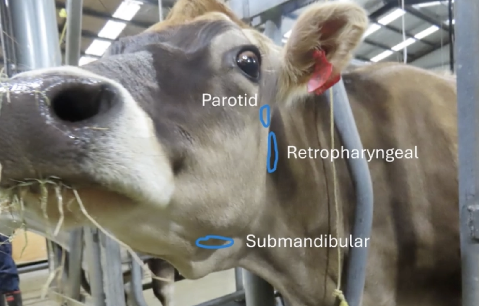
H6: Palpate Submandibular Tissue and Lymph Nodes
Method
Example normal description
4 DDx for enlarged submandibular lymph nodes
Method:
Place hand on corner of ramus
Run hands down towards the chin to feel for masses/swelling
Repeat on other side
Example: Submandibular lymph node is the size of a mandarin, soft and freely movable with no pain
Parotid and retropharyngeal lymph nodes not palpable if healthy
Enlarged Submandibular Lymph Nodes:
Woody tongue
Lumpy jaw
TB
Enzootic bovine leukosis
5 DDx for submandibular swellings (+ description on palpation)
Extra fat | Soft, non-painful, body temperature, does not pit when squeezed |
SC oedema | Soft, non-painful, pits when squeezed |
Actinomycosis (lumpy jaw) | Hard, immobile swelling on the jaw bone |
Abscess | Firm, painful, hot |
Enlarged submandibular lymph nodes | Moveable lumps which may be tender and does not pit when squeezed |
Describe the significance of the following ear tags
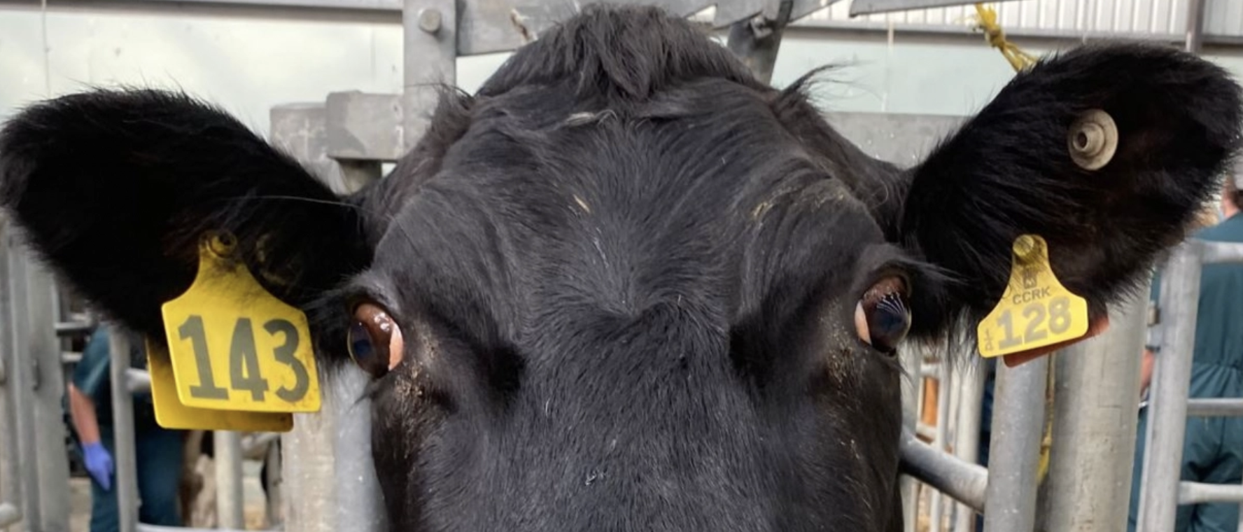
Right Ear = Herd number (#143)
Left Ear = Calf tag (123rd calf born in 2014 in CCRK herd of origin)
Vaginal Examination
Method
Indication
Normal discharge
Abnormal discharge
Method:
Perform BEFORE rectal exam to reduce risk of faecal contamination introduced into vagina
Clean and dry vulva of gross contamination
Lube hand and pass through vulva (MM should be smooth)
Palpate cervix and assess tone and size of os
Scoop along vaginal floor to collect discharge to visual and olfactory assessment
Indication: Calving and post-calving
Normal: Clear and odourless
Abnormal: White, cloudy, yellow, mucoid, haemorrhagic, foetid
Urine Sample
Method of collection
3 Features to assess
Example description
Method: Rub perineum region directly under vulva to stimulate urination females
Do NOT hold tail → Clamping
Not possible in male
Features:
Colour
Clarity
Ketones (ketostix)
Example: The urine is dark yellow, clear, voided in a strong stream and has a strong odour
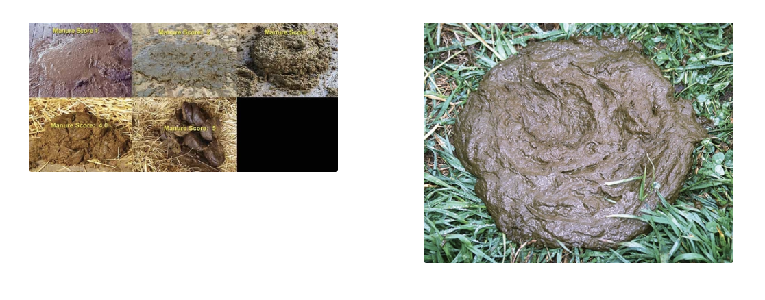
Faecal Sample
3 Pieces of information it provides
5 Features to assess
Information:
Diet
Hydration
GI function
Features:
Amount/Volume = Depends on nutrition, hydration, rate of passage of ingesta, obstructions, infectious agents
Absent OR present (+ to ++++)
Consistency = Depends on nutrition (long stem fibre vs. soluble CHO), hydration and GI function
Numerical scale OR description (eg. liquid, firm, dry, formed)
Normal: Some fibre present after squeezing out liquid component = Remnants of cereal grain husks
Colour = Depends on nutrition, hydration, GI function and hepatic function
Green, olive green, brown, yellow, orange, grey, black
Additional Components = Depends on disease
eg. Ulcers, Salmonella, rumen acidosis, rumenitis
Melaena, haematochezia, mucus.fibrin, gravel/sand, palm kernel husks, undigested feed, bubbles/foam
Odour
Normal: Fermented grass
Abnormal: Sour or dead tissue
List 6 additional diagnostic tests to perform on-farm
More specific examination → Sedated oral exam, ophthalmic exam, otic exam
Blood tests → CBC/biochemistry or BVD Ag ELISA, Johne’s ELISA
FNA → Contents, cytology and culture
Swab → Culture
Biopsy → Histology or staining
Imaging → U/S, radiographs, CT/MRI
5 DDx for jaundice
Theileriosis
Leptospirosis
Post-parturient haemoglobinuria
Onion/brassica toxicity
Facial eczema
What 3 other organs can be pinged in the right paralumbar fossa?
Abomasum (RDA)
Small intestine
Caecum
What types of disease may cause enlargement of the popliteal lymph node?
Foot infections / lesions
Foot rot (Fusobacterium necrophorum)
Digital dermatitis (Treponema spp.)
Sole abscesses or penetrating wounds
Trauma/cellulitis of distal hindlimb
Abscess
Large regions of non-pigmented skin with alopecia, erythema and crusting/flaking with ulcerative to erosive scabbed lesions with large area of necrotic epidermis
4 DDx?
Facial eczema
Spring eczema (no liver damage)
Drug reaction (eg. tetracycline)
Crops (eg. brassicas)
Consider season
Landmarks of the pelvis
Tuber coxae (hook)
Greater trochanter
Ischiatic tuberosity (pin bones)
8 Components of gross lesion description
Location
Arrangement (multifocal/diffuse/coalescing)
Number (eg. miliary = too many to count)
Size
Shape
Colour
Consistency (soft/gritty/pitting/rubbery/wrinkled/crusty/smooth/moist)
Odour
*Not ALL may be applicable
Top DDx for the following images
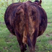
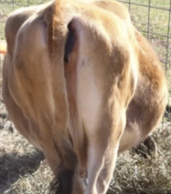
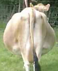
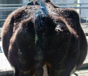
Bloat = L paralumbar fossa (distension above hips) → Backward D on LHS → Double D due to ruminal distension
Late-term pregnancy = Abdominal swelling BELOW hips (R = Uterus and L = Full rumen)
Late-term pregnancy with empty rumen
Vagal indigestion = Papple
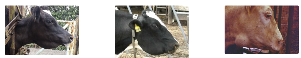
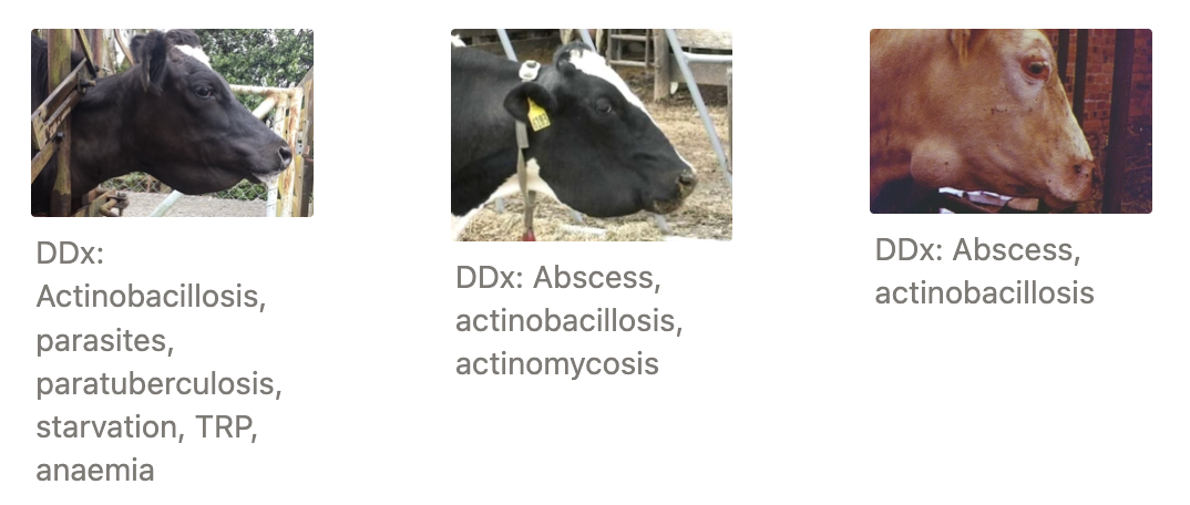
List DDx for the following jaw swellings
Requirements for transport of cancer eye
Confined to eye and eyelid (no spread to lymph nodes)
Smaller than $1 coin (3cm)
Not flyblown, discharging or bleeding
Sheep Reproduction
Cycle length
Overt oestrus length
Gestation length
Cycle: 17d
Overt Oestrus: 24hr
Longer for MA and shorter for hoggets (4hr)
Maiden hoggets do NOT seek the ram → Higher ram:ewe ratio
Gestation: 152d
Sheep Breeding Calendar
Mating/Breeding season
Mid-March/mid-April (MA)
Mate hoggets early-May
Pregnancy scan - June/July
40-45 days (MA) or 60 days (hogget) after ram removal
Lambing - Late-July/early-October (depends on mating season)
Docking - November (1 month of age)
Rings = weeks after birth (earlier)
Weaning - December (weaned at same time)
Ram preparation/soundness
NZ sheep are highly seasonal (short-day)
First = silent ovulation (not detected by ram
More fertile + higher OR @ 2nd and 3rd ovulation
Breeding too early/too later affects fertility AND fecundity
Teaser advances and synchronise mating period (get through first, silent ovulation)
17 days BEFORE ram
Ensures ewes are overtly oestrus with entire ram on 2nd cycle
Mating at same time, and shorten lambing spread
Mating early
Regional variation (dry early on)
Meet Christmas market
Mating later - Avoid poor weather conditions