human structure and function
1/25
There's no tags or description
Looks like no tags are added yet.
Name | Mastery | Learn | Test | Matching | Spaced | Call with Kai |
|---|
No analytics yet
Send a link to your students to track their progress
26 Terms
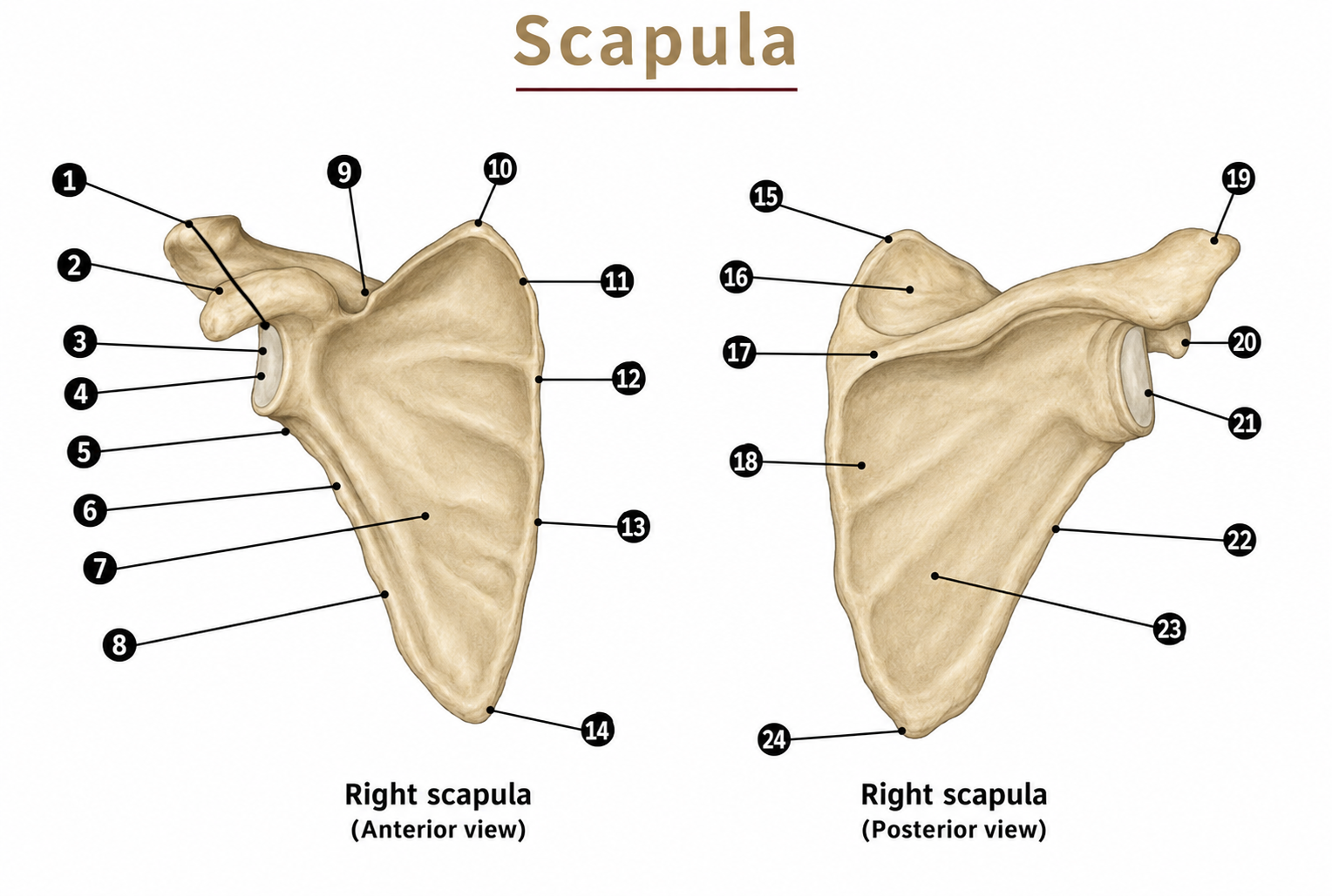
label numbers 21, 20, 17, 13, 24/14, 19, 1, 6
glenoid cavity: joints the head of the humerus at the shoulder joint
coracoid process
scapular spine: insertion of trapezius and origin for posterior head of deltoid
vertebral/medial border: insertion of rhomboids and levator scapulae
24/14. inferior angle: origin of teres major
acromion process: origin of part of deltoid, insertion of part of trapezius, attachment of coracoacromial ligament
supragenoid tubercle: origin of the long head of biceps brachii
infraglenoid tubercle: origin of the long head of triceps brachii

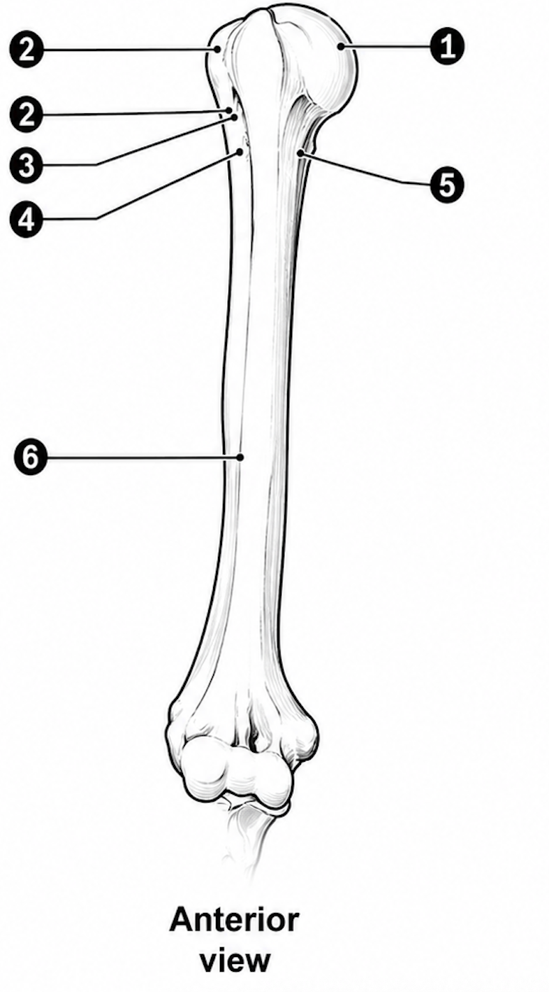
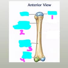
label this diagram of a proximal humerus
head of the humerus: joins the glenoid cavity of the scapula at the shoulder joint
greater tuberosity: insertion for supraspinatus, infraspinatus and teres minor (also the coracohumeral ligament)
lesser tuberosity: insertion of subscapularis
bicipital groove: between the tuberosities; for the tendon of long head of biceps
anatomical neck: every head in anatomy has a neck
surgical neck: a site named because it fractures relatively often
deltoid tuberosity: insertion of deltoid


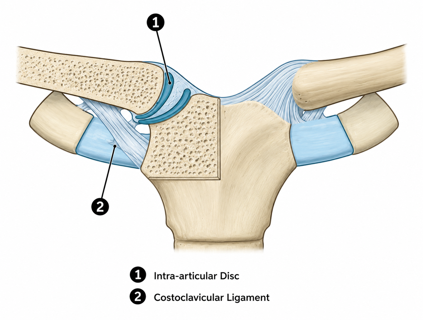
label the sternoclavicular joint ligaments
intra-articular disc: divides the joint cavity into two compartments and stops clavicle dislocating medially and acts as a shock absorber for forces transmitted along the clavicle
costoclavicular ligament: limits extreme clavicular elevation. when arm is raised overhead, this ligament becomes taut and prevents further elevation

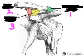
label this diagram of the acromioclavicular joint ligaments
acromioclavicular ligament: thickenings of the joint capsule above and below the acromioclavicular joint, limits elevation and depression at the acromioclavicular joint, providing horixontal stability.
coracoclavicular ligaments: this ligament complex has two parts: conoid ligament: cone-shaped, attaches medially to the coracoid process, rotates clavicle posteriorly during full arm elevation, and the trapezoid ligament: quadrilaterial shape, attaches laterally to the coracoid process, transmits compression forces from the scapula to the clavicle, thus bypassing the acromioclavicular joint.
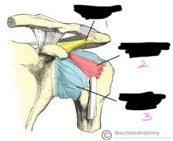
label this diagram of acromioclavicular joint ligaments
coracoacromial ligament: stops no joint movement, because it joins two parts of one bone, which never move relative to each other. protects shoulder joint from above, forming a protective arch over the humeral head. prevents extreme superior dislocation of the humeral head.
coracohumeral ligament: limits extreme humeral adduction, as well as lateral rotation, and helps support the humeral head when the arm hangs at the side.
glenohumeral ligaments: limits extreme humeral external rotation. the inferior glenohumeral ligament is an important stabilizer when the arm is abducted.

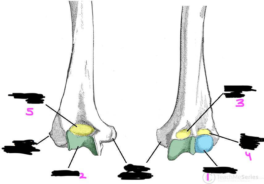
label this diagram of the distal humerus
capitulum: the rounded lateral condyle that articulates with the head of the radius
trochlea: the spool-shaped medial condyle that articulates with the trochlear notch of the ulna
coronoid fossa (anterior): receives the coronoid process of the ulna during elbow flexion
radial fossa (anterior): receives the head of the radius during elbow flexion
olecranon fossa (posterior): receives the olecranon process of the ulna during elbow extension


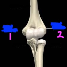
label this diagram of the epicondyles of the humerus (right arm)
medial epicondyle: origin for flexor muscles of the wrist and fingers
lateral epicondyle: origin for extensor muscles of the wrist and fingers
label this diagram of the radius
head of the radius: articulates with the capitulum of the humerus and the radial notch of the ulna
radial tuberosity: insertion of the biceps brachii tendon
styloid process of the radius: attachment for the brachioradialis muscle and radial collateral ligament
ulnar notch (distal): articulates with the head of the ulna at the distal radioulnar joint


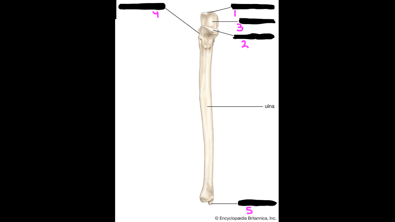
label this diagram of the ulna
olecranon process: forms the point of the elbow; insertion of the triceps brachii
coronoid process: insertion of the brachialis muscle
trochlear notch: articulates with the trochlea of the humerus
radial notch: articulates with the head of the radius at the proximal radioulnar joint
styloid proc ess of the ulna: attachment for the ulnar collateral ligament
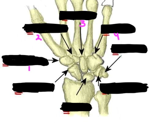
label the diagram of these carpal bones (proximal row - lateral the medial)
scaphoid: most commonly fractured carpal bone
lunate: articulates with the radius
triquetrum: articulates with the ulna via the articular disc
pisiform: a sesamoid bone within the tendon of flexor carpi ulnaris
memory aid - “Sally Left The Party”


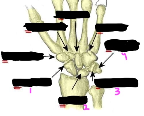
label the diagram of these carpal bones (distal row - lateral to medial)
trapezium: articulates with the base of the first metacarpal (thumb)
trapezoid: articulates with the base of the second metacarpal
capitate: the largest carpal bone
hamate: has a hook-like process (hook of hamate) on its palmar surface
memory aid - “To Take Claire Home”
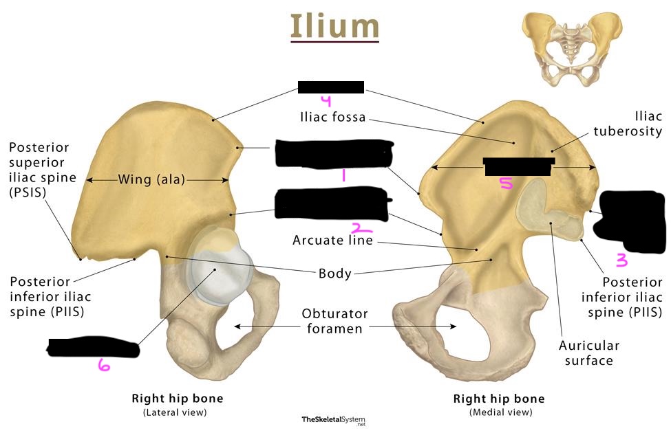
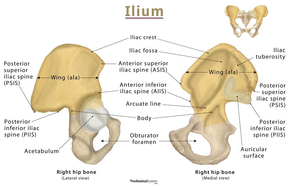
label the diagram of the illiac bone features of the pelvis
anterior superior iliac spine (ASIS): for the origin of sartorius and the attachment of the inguinal ligament
anterior inferior iliac spine (AIIS): for the origin of rectus femoris
posterior superior iliac spine (PSIS): sort-of marks the sacroiliac joint (SIJ)
iliac crest: runs between the ASIS and the PSIS - has many attachments e.g. abdominal muscles
iliac blade: iliacus originates on its front, and the gluteal muscles originate on its posterior surface
acetabulim: joins the head od the femur at the hip joint
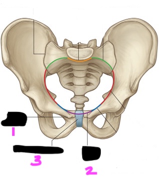
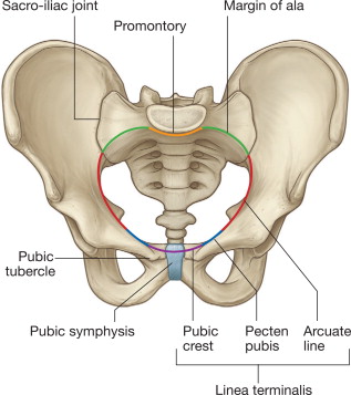
label this diagram of the pubic features on the pelvis
pubic tubercle: for the attachment of the inguinal ligament
public crest: for the origin (the lower end) of rectus abdominis
pubic symphysis: the joint between the left and right hip bones
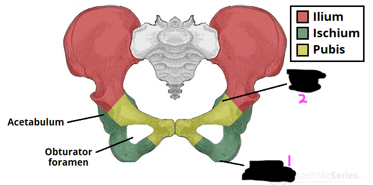
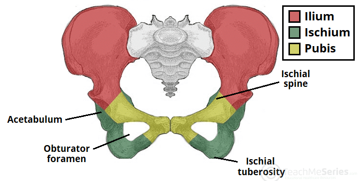
label this diagram of the ischial features on the pelvis
ischial tuberosity: origin of the hamstrings and attachment site for the sacrotuberous ligament. you sit on this bony feature
ischial spine: sacrospinous ligament attaches to it
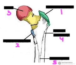
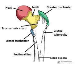
label this diagram of the femur (proximal features)
greater trochanter: insertion of gluteus medius and minimus, as well as the 6 lateral rotators of the hip joint
lesser trochanter: insertion of iliopsoas (psoas major plus iliacus)
head of femur: joins the acetabulum at the hip joint
gluteal tuberosity: insertion of part of gluteus maximus
linea aspera: insertion of the hip adductors
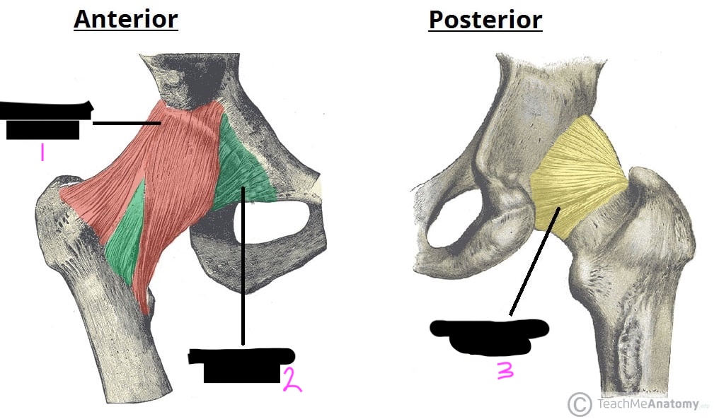
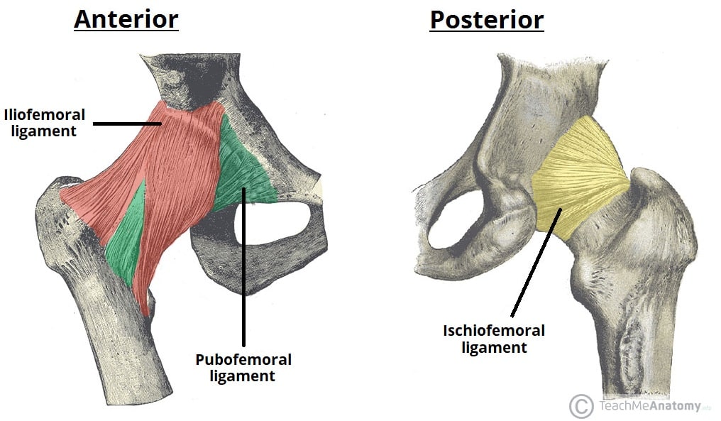
label this diagram of the hip joint ligaments
iliofemoral ligament: stops extreme hip joint extension
pubofemoral ligament: stops extreme hip joint abduction
ischiofemoral ligament: stops extreme hip joint internal rotation
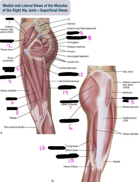
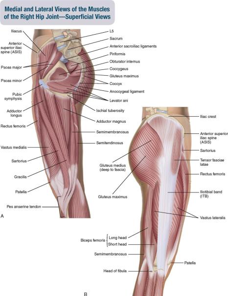
label this diagram of the hip muscles
iliacus: hip flexion
psoas major: fip flexion
sartorius: hip abduction, flexion, lateral rotation
rectus femoris: hip flexion
tensor fasciae latae (TFL): hip flexion, abduction, medial rotation
gluteus maximus: hip extension, lateral rotation, adduction
gluteus medius: hip abduction
piriformis, obturator internus: laterally rotate the hip
adductor longus: adduct and flex the hip
gracilis: adduct and flex the hip
adductor magnus: adduct and flex the hip
long head of biceps femoris: hip extension
semitendinosus: hip extension
semimembranosus: hip extension
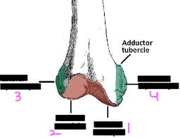
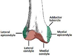
label this diagram of the femur
medial femoral condyle: join the tibia at the knee joint
lateral femoral condyle: join the tibia at the knee joint
lateral femoral epicondyle: lateral collateral ligament, lateral head of gastrocnemuis, and origin of popliteus attach
medial femoral epicondyle: medial collateral ligament and medial head of gastrocnemius attach
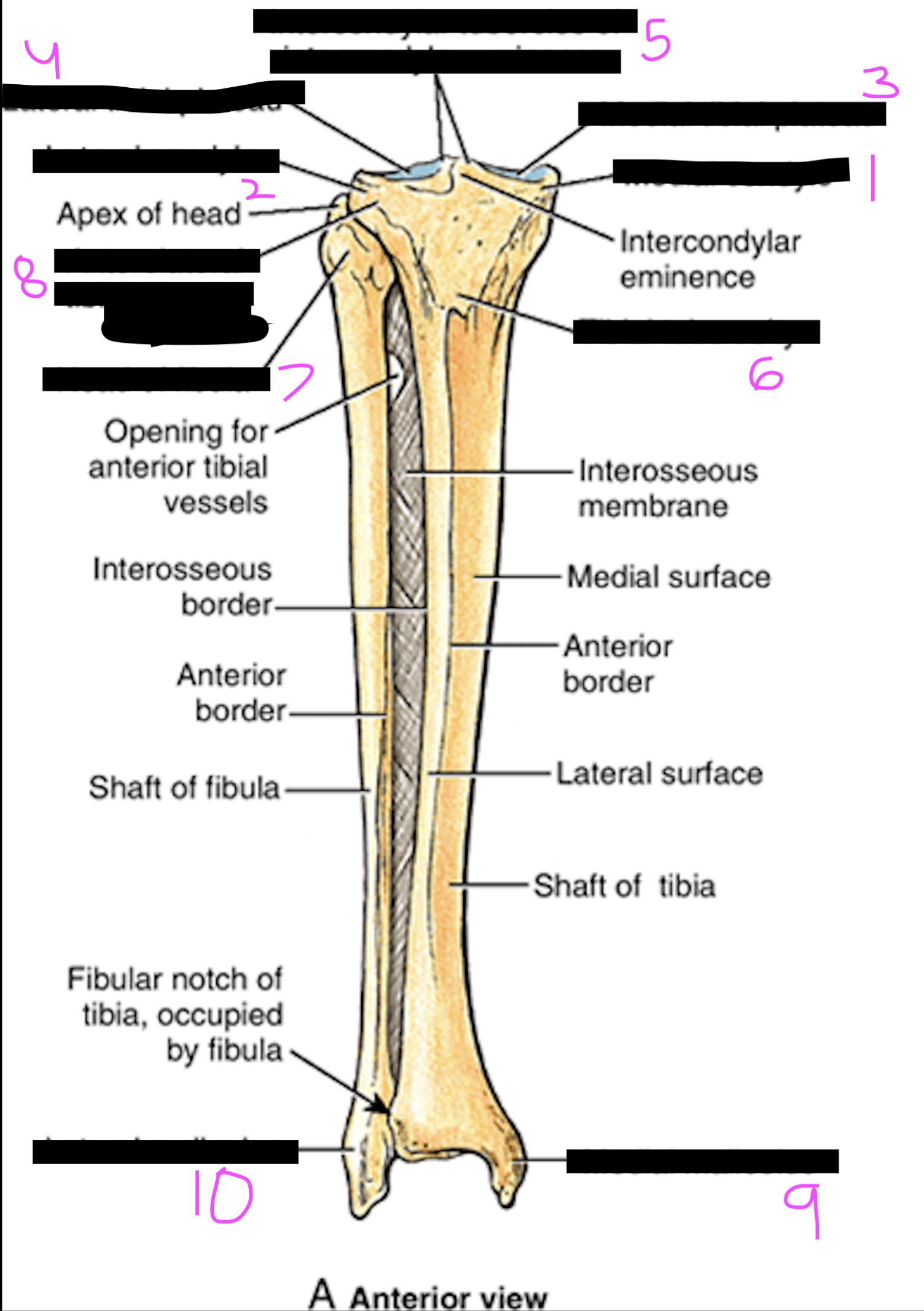
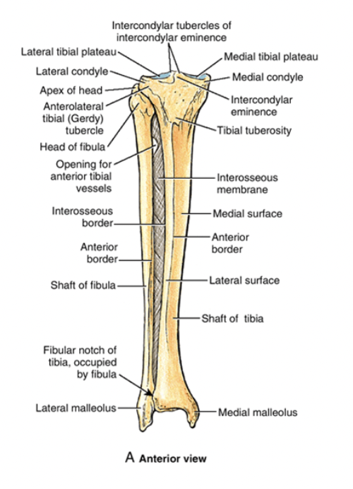
label this diagram of the tibia and fibula
medial tibial condyles: for articulation with the femur at the knee joint
lateral tibial condyles: for articulation with the femur at the knee joint
intercondylar eminence (4 tibial spines): for attachemnt of both ends of both the medial and lateral menisci
tibial tuberosity: for attachment of the patellar tendon
head of the fibula: for the attachment of the LCL and insertion tendon of biceps femoris
gerdy’s tubercle: for insertion of iliotibial band
medial malleolus: on tibia. mostly for attachment of the MCL of the ankle
lateral malleolus: on fibula. mostly for attachment of th LCL of the ankle
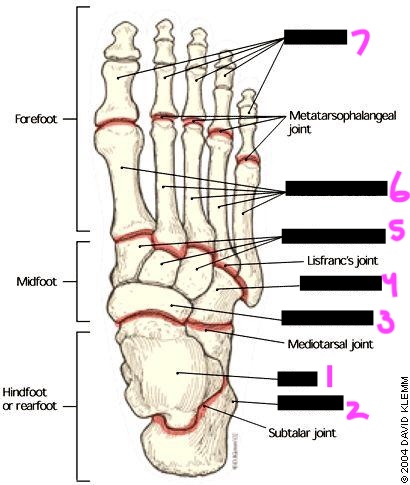
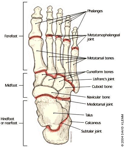
label this diagram of the foot
talus: articulates with tibia and fibula at ankle joint
calcaneus: heel bone, largest tarsal bone
navicular: boat-shaped bone on medial side
cuboid: cube-shaped bone on lateral side
cuneiforms: three wedged-shaped bones
metatarsals: five long bones of the midfoot
phalanges: toe bones
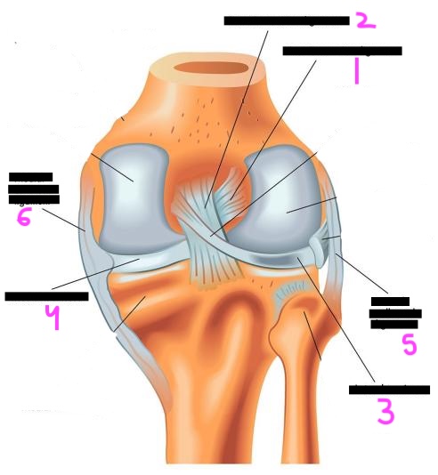
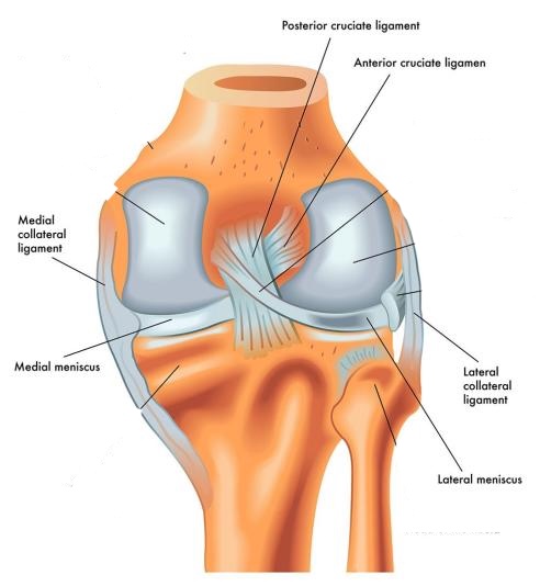
label this diagram of the knee
anterior cruciate ligament: strops the femur sliding backwards on the tibia
posterior cruciate ligament: stops the femur sliding forwards on the tibia
lateral menisci: deepen the knee joint, increasing stability; act as shock absorbers
medial menisci: deepen the knee joint, increasing stability; act as shock absorbers
lateral collateral ligament: stops the leg from adducting at the knee joint
medial collateral ligament: stops the leg from abducting at the knee joint
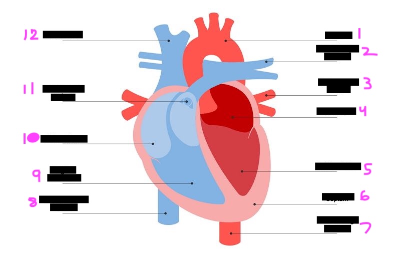
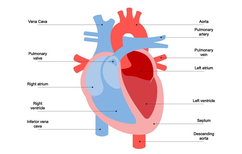
label this diagram of the heart
aorta: carries oxygen-rich blood away from the left ventricle and distributes it to the rest of the body.
pulmonary artery: branches (right and left) carrying blood to lungs for oxygenation
pulmonary vein: return oxygenated blood from lungs to left atrium
left atrium: upper left receiving chamber of the heart
left ventricle: lower left pumping chamber with thickest muscular wall
spetum: separate structures so they can perform their specific functions without interference
decending aorta: the descending portion specifically feeds the lower body
inferior vena cava: large vein bringing deoxygenated blood from lower body to right atrium
right ventricle: lower right pumping chamber, sends blood to lungs
right atrium: upper right receiving chamber of the heart
pulmonary valve: three-cusped valve at exit of right ventricle
superior vena cava: large vein bringing deoxygenated blood from upper body to right atrium
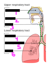
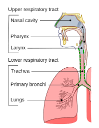
label the diagram of the respiratory tract
nasal cavity: internal space of the nose
pharynx: throat
larynx: voice box
trachea: windpipe
bronchi: main air passages to lungs
lungs
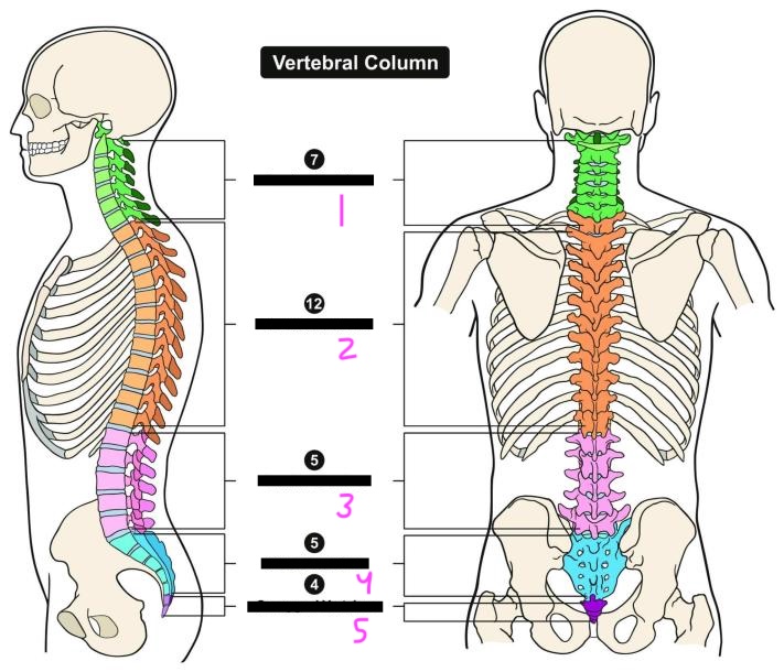
label this diagram of the vertebral column
cervical vertebra: smallest vertebrae; transverse foramina for vertebral arteries
thoracic vertebra: articulate with ribs; limited mobility due to rib cage
lumbar vertebra: largest vertebrae; bear most body weight
sacral vertebrae: fused to form sacrum; articulates with pelvis
coccygeal vertebra: vestigial tailbone
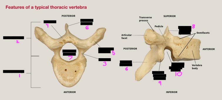
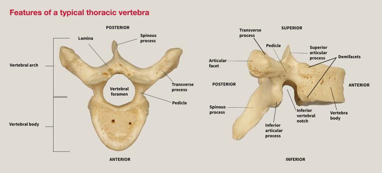
label this diagram of the basic vertebral structures
vertebral body: the anterior weight-bearing part; attaches at the intervertebral joint to the vertebra above and below it
vertebral arch: comprised of pedicles and laminae; forms the posterior boundary of the vertebral foramen
pedicle: short, thick projection protuding posteriorly from each side of the body
lamina: flat plates continuing from pedicles; attach to each other posteriorly in the midline
transverse processes: prject laterally from where each pedicle meets its lamina; for muscle and ligament attachments
spinous process: projects posteriorly from the midline junction of the laminae; for muscle and ligament attachments
vertebral foramen: the central opening within the vertebral arch; when vertebrae are joined, forms the vertebral canal
superior articular processes: project upward to articulate with the vertebra above at the zygapophyseal joints
inferior articular processes: project downward to articulate with the vertebra below the zygapophyseal joints
inferior vertebral notch: indentation on inferior surface of pedicle; when vertebrae join, forms the intervertebral foramina where nerves exit
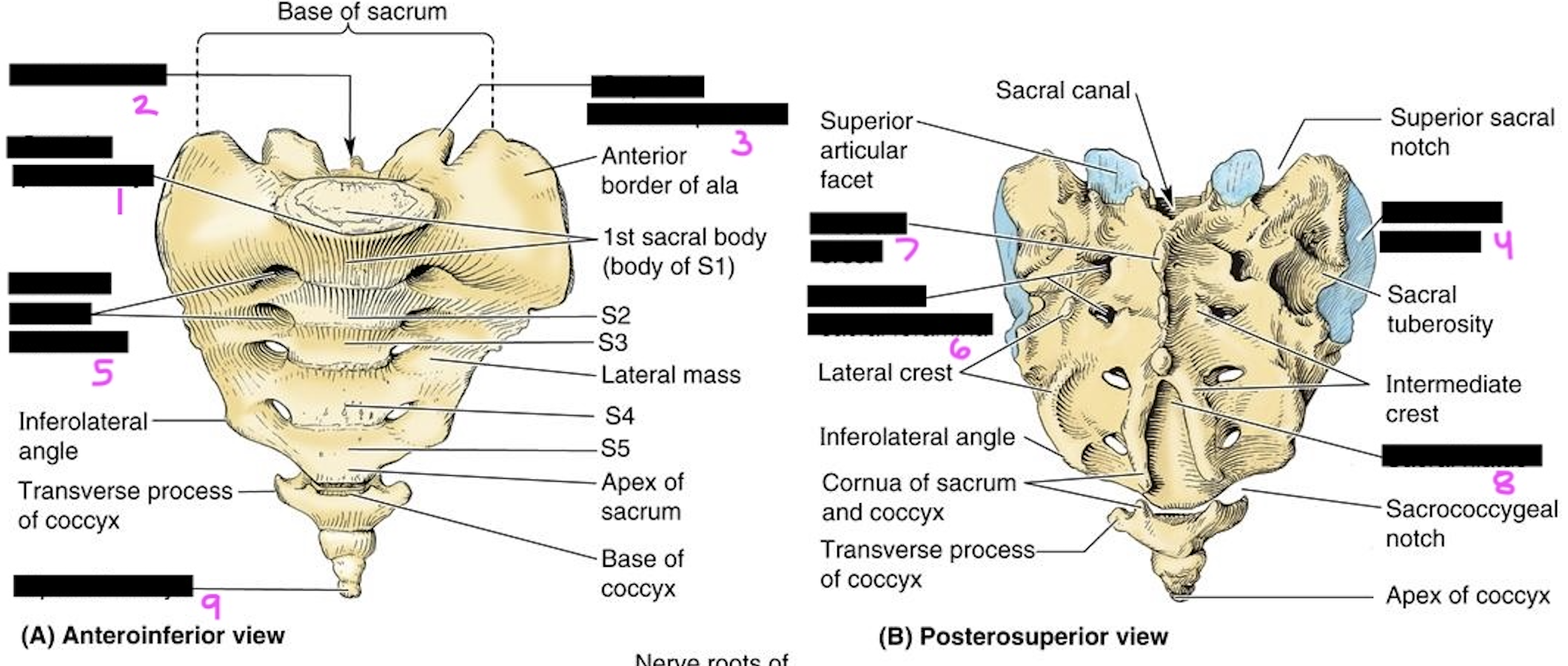
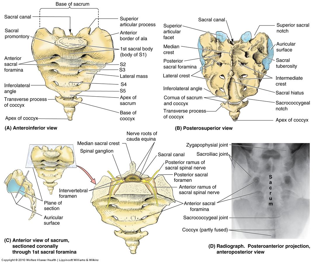
label this diagram of the sacrum
sacral promontory: Prominent anterior edge of S1 body; important obstetric landmark
Sacral canal: continuation of vertebral canal; contains cauda equina nerve roots
superior articular processes: articulate with L5
auricular surface: ear-shaped lateral surface for articulation with ilium at sacroiliac joint
anterior sacral foramina: openings for anterior rami of sacral spinal nerves
posterior sacral foramina: openings for posterior rami of sacral spinal nerves
median sacral crest: fused spinous processes along midline posteriorly
sacral hiatus: opening at inferior end of sacral canal; used for caudal epidural anesthesia
apex of sacrum: inferior tip; articulates with coccyx