Topic 30: Myosin & Actin
1/26
There's no tags or description
Looks like no tags are added yet.
Name | Mastery | Learn | Test | Matching | Spaced | Call with Kai |
|---|
No analytics yet
Send a link to your students to track their progress
27 Terms
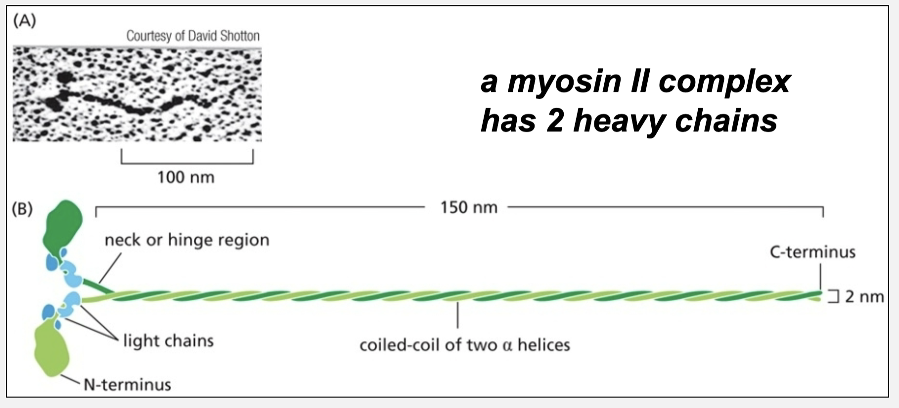
Myosin II
is a prototypical actin-based motor protein
a myosin II complex has 2 heavy chains

Myosin II: terminal head domains are what?
the motor domains

Myosin II: light chains are what?
regulatory subunits

What do the C-terminal helices in Myosin II form?
coiled-coil mediating self-assembly
Muscle myosin II self-assembles into what?
large “thick filmanets”
what are bipolar filaments made up of?
made up of many dimers oriented in opposite directions on either side of the bare zone
What is the bare zone?
portion in the middle of the filament with no myosin heads
Is myosin II a (+) or (-) end-directed actin motor protein?
plus end-directed
Which way do the myosin motor domains (heads) move/pull?
pull the (+) end of the actin filament towards themselves, sliding the actin
move/walk towards the (+) end
which end does the Myosin II walk?
walks towards the (+) end of actin filaments taking steps of defined size
If the myosin II is anchored, which way does it slide the actin filament?
slides the actin filament, pulling the plus end towards itself
Sarcomere
repeating cytoskeletal structure in myofibrils
ordered arrays of myosin thick filaments and actin thin filaments
Where do myosin thick filaments slide the actin thin filaments?
towards the center
CapZ
caps the plus end of the actin filament
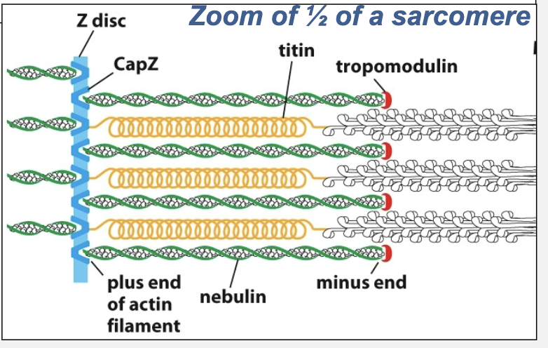
tropomodulin
caps the minus end of the actin filament

Nebulin & titan
function as molecular rulers
titan also functions as molecular spring

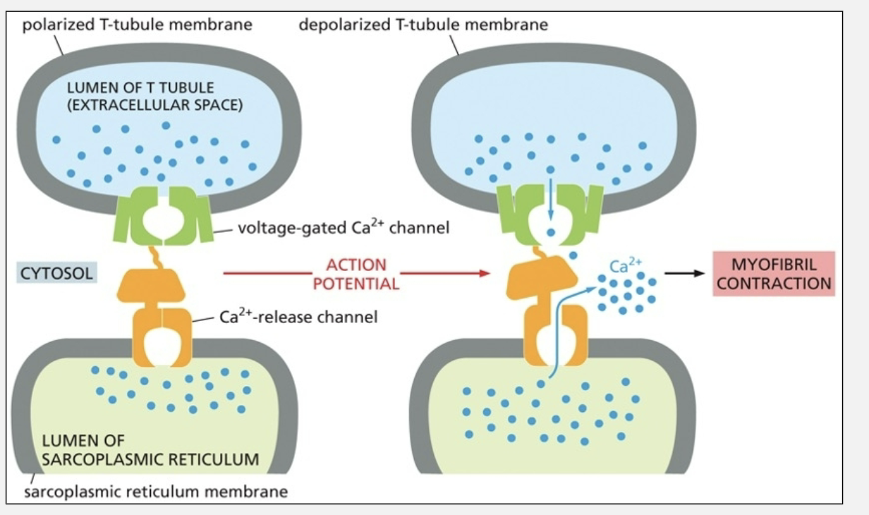
T tubules
propagate an electrical contraction signal into the interior of the muscle cell
its an invagination of plasma membrane; lumen (interior) of tubule is extracellular space
T-tubule and SR are very closely juxtaposed
what does the electrical depolarization of T-tubule membrane trigger?
triggers massive Ca2+ release
Pathways of electrical depolarization
lumen of T-tubule —> voltage-gated Ca2+ channel —> Ca2+ release channel —> huge Ca2+ release from sarcoplasmic reticulum
what happens when calcium binds to the troponin complex?
its shifts the location of tropomyosin exposing myosin binding sites
tropomyosin
binds all along the actin filaments, blocking access to myosin binding site when Ca2+ is low
Ca2+ binds to troponin C, a calmodulin-like subunit, triggering what?
a conformational shift
What does calmodulin transduce?
transduces GPCR signaling into smooth muscle contraction via myosin light chain kinase
what does myosin light chain phosphorylation activate?
myosin II
The activity of on-muscle myosin II is also regulated by what?
myosin light chain kinase
What activates Myosin light chain kinase (MLCK)?
activated by several different pathways often involving Rho GTPase
Myosin V
well adapted to carry cargo along actin filaments
has a long lever arm = long steps
each motor domain stays bound to actin filament for linger than myosin II (many steps before “falling off”)