8: nerves to the eye & nervous system
1/60
There's no tags or description
Looks like no tags are added yet.
Name | Mastery | Learn | Test | Matching | Spaced | Call with Kai |
|---|
No analytics yet
Send a link to your students to track their progress
61 Terms
where does information of the nervous system go into
information comes into the central nervous system via afferent fibres
afferent fibres have specialised nerve endings that respond to sensations as touch, pressure and temp
what 2 categories are in the nervous system
sensory ( afferent): ascending, transmits information to the cns.
motor ( efferent): descending impules from cns to the peripheral organs to cause an effect
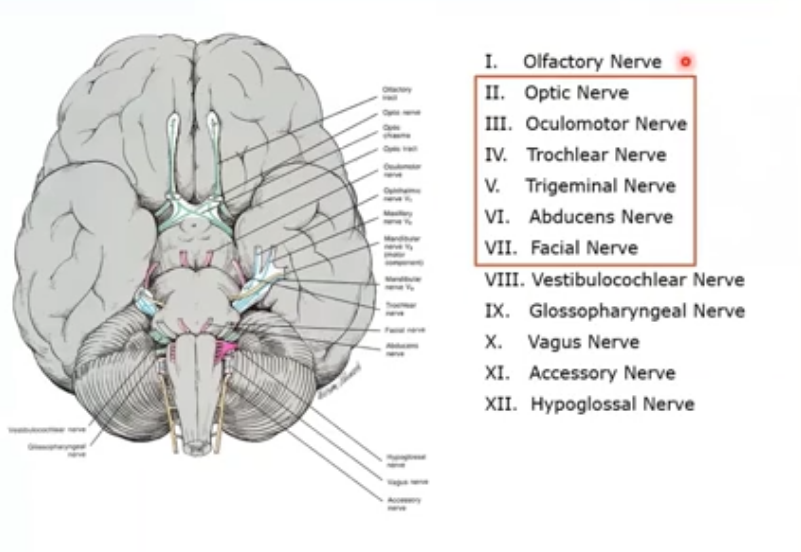
optic nerve II
origin is retinal ganglion cells
destination: LGN
function: sensory; sight
oculomotor , inferior division III
origin: midbrain
destination: medial rectus muscle, inferior rectus muscle,inferior oblique muscle and ciliary ganglion
function: motor- abduction, eleevation, extorsion , also motor to the iris sphincter and ciliary muscle in miosis
oculomotor, superior division III
origin: midbrain
destination: superior rectus muscle and superior palpebral levator muscle
function: elevation, abduction, intorsion, and elevation of eyelid
trochlear IV
origin is midrbrain
destination: superior oblique muscle
function: motor- depression, abduction and intorsion
abducens VI
origin is the pons
destination is the lateral rectus muscle
function is motor absuction
facial VII
origin is the pons
destination is the frontalis, orbicularis muscles
function is facial expressions, closure of eyelids and secretor to lacrimal gland
subdivisions of sensory afferent systems
visceral ( autonomic)
special ( somatic)
central ( comatic)
motor efferent systems subdivision
somatic motor
visceral motor: subdivided into parasympathetic and sympathetic
afferent
somatic afferent: general sensory eg pain touch and special sensory eg vision, smell , hearing
autonomic afferent: visceral sensory eg internal organs, vessels and special sensory
efferent
somatic motor
visceral motor
brachial motor - skeletal muscles, derived from branchial arteries
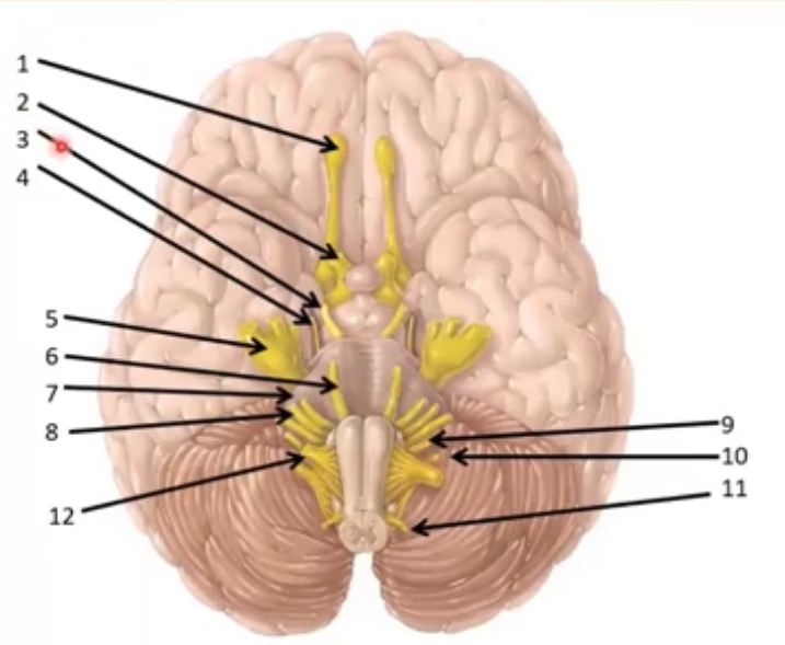
CN I & 2 emerge from the cerebrum
rest emerge from the brain stem
the optic nerve CN II
afferent - special sensory
conducts signals from the eye to the brain
made up of retinalganglion cell axons
axon bundles exit eye as optic nerve and runs nasal and posterior to the optic foramen , through canal in sphenoid bone
optic nerve visual pathway
after the 2 optic nerves pass through the canals they merge at chiams, and cross over at nasal fibres
forms optic tract where moves to LGN
follow throgh as optic radiations where enters V1
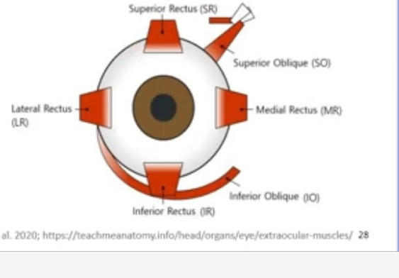
what are the extraocular muscles of the eye- muscles that move the eye: somatic
levator palpebrae superioris
superior oblique
inferior oblique
superior rectus
medial rectus
lateral rectus
inferior rectus
what nerves innervate the extraocular muscles
oculomotor: innervates all rectus, inferior palp and inferior oblique
trochlear; innervates the superior oblique
adbucens: innervates lateral rctus muscles
what does the oculomotr nerve CNIII innervate
superior rectus, medial rectus, inferior rectus, inferior oblique and superior palp levator muscles
also provides a route along which the autonomic fibres travel to innervate iris sphincter , ciliary and smoth muscles of the eyelind
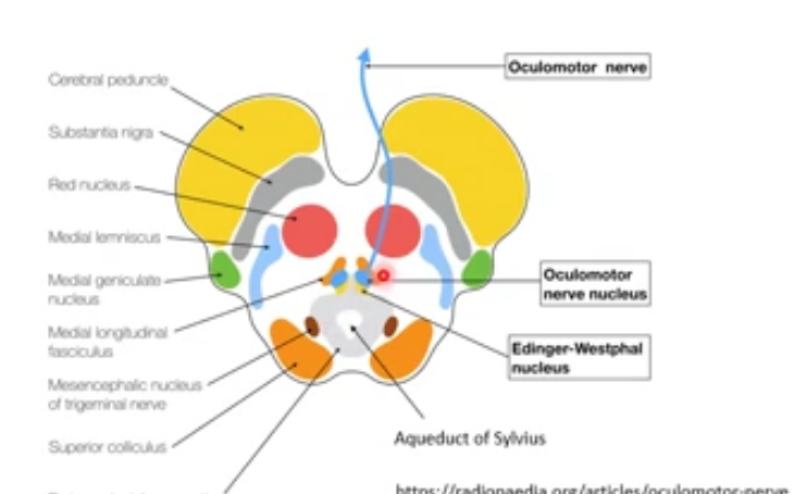
where does the paired oculomotor nerve arise from
from the large nuclei - axons arise from a subnuclesu within this nucleus
located within the midbrain at level of superior colliculus
extends in a column from posterior edge of floor of third ventricle to the trochlear nucleus
subnucleus of the oculomotor nerve
controls each muscle
nucleus of medial rectus is located toward lower border of oculomotor nucleus
nucleus of levator muscle is in the centre
oculomotor nerve pathway
fibres from each of the indv nuclei join, forming fascicular part of nerve that passes through red nucleus and decussating fibres of superior cerebellar peduncle
these fibres merge as oculor motor nerve
nerve passes between superior cerebellar and post cerebral arteries to the artery
nerves pierce roof of sinus and run within 2 dural layers above trochlear nerve
where does oculomotor nerve exit
eits the sinus and enters the orbit through superior orbital fissure havog divdided into sup and inf diivisions
where does the superior oculomotr branch move into
this ranch runs medially above optic nerve and enters the superior rectus on its inferior surface , another goes to the levator
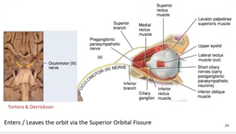
where does the inferior banch of oculomotor move to
this branch runs below optic nerve and divides into 3 branches
one branch enters medial rectus
another enters inferior rectus
third branch gives off parasympathetic fibres that form the parasymp root extending to ciliary ganglion, then runs along inferior rectus to enter inferior oblique muscle
what do the parasympathetic fibres innervate
innervates the sphincter and ciliary muscle
reaches the eye via short ciliary nerve
oculo motor nerve route from midbrain to eye
nucleus
passes posterior cerebral, sup cerebellar and posterior communicating artery
into sinus
divides into superior and inferior branches
into edinger westpal nucleus
and then either paraymp supply to iris sphincter and ciliary muscle or travels with inferior branch then inferior oblique and into ciliary ganglion
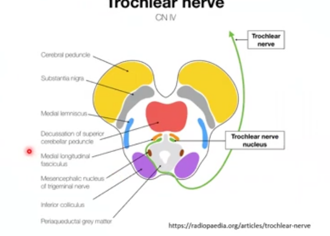
trochlear nerve CN IV
innervates superior oblique muscle
located in midbrain at level of inferior colliculus
run backwards/ dorsally to inf colliculus
only nerve to cross so innervates the contralateral superior oblique muscle
the 2 trochlear nerves cross the superior cerebellar arteries to contralateral side before emerging on posteriior side of brain stem
trochlear nerve pathway
only nerve to leave dorsal aspect of cns
as trochlear nerve emerges from dorsal midbrain, it decussates and curves around cerebral peduncle at pons , between sup and post cerebral arteries
passes between these vessels and runs to oculomotor nerve
where does the trochlear nerve go to affter passing to oculor motor nerve
enters cavernous sinus and lies between oculomotor nerve and opthalamic division of trigeminal nerve
whilst in sinus the nerve sends sensory fibres to opthalmic nerve
how does the trochlear nerve enter the orbit
enters orbit through the superior orbital fissure above the tendinous ring
runs with frontal neve to medial side of orbit and enters the superior oblique muscle
what does adbucens nerve CN VI innervate
inervates the lateral rectus muscle
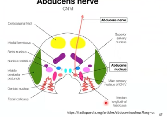
abducens nucleus
located near the inferior dorsal midline of the pons beside floor of the 4th ventricle , below cerebellum
fibres from nucleus pass through pons and lie next to corticospinal tract
exit in the groove between pons and medulla oblongata
what does the abducens nerve also contain
contains internucleur neurons that communicate with the nucleus for contralateral medial rectus muscle in oculomotor complex
this is the pathway for conjugate horizontal eye movements
this pathway recieves information from higher cns centres
abducens nerve pathway
runs along occipital bone and along temporal bone, and bends over to enter sinus
within sinus it lies near wall of internal carotid artery
small sympathetic branches leave internal carotid plexus and travel with abducens nerve
nerve carries autonomic fibres into the opppthalmic division of trigeminal nerve
how does abducens nerve enter the orbit
enters orbit through the superior orbital fissure within tendinous ring and innervates lateral rectus muscle on medial surface
what 3 nerves innervate the extraocular muscle
oculomotor
trochlear
abducens
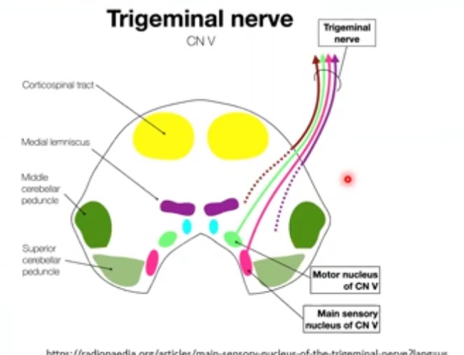
trigeminal nerve CN V
originate in the innervated structures
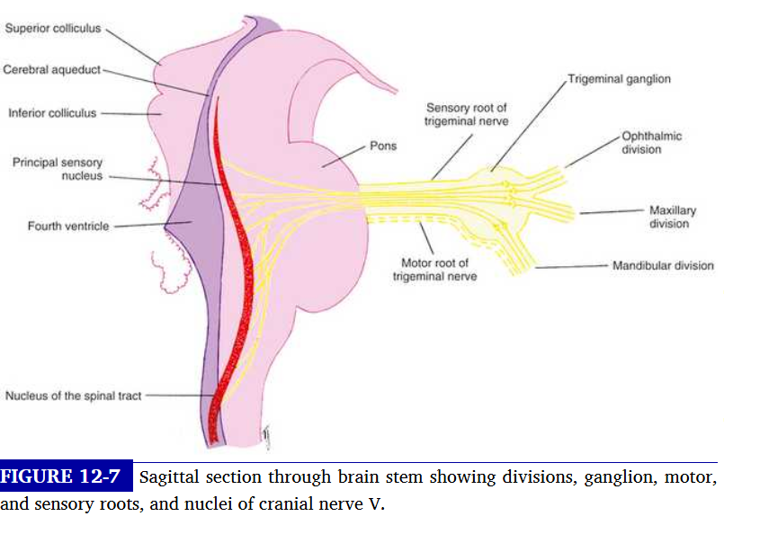
join to become larger nerves and come together in the ganglion of CN V , and then exit the ganglion and enter pons
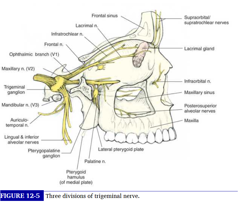
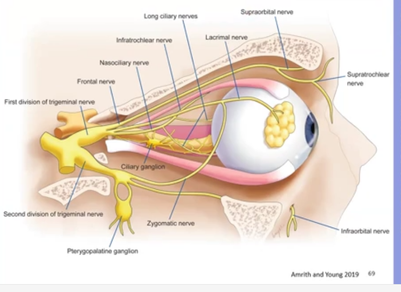
opthalmic division of trigeminal nerve: nasociliary nerve
sensory fibres form structures of medial canthal area, lacrimal sac, join to form infratrochlear nerve
this nerve penetrates orbital septum , and enters orbit below trochlea
where does motor nucleus lie in trigeminal nerve
lies medial to sensory nucleus near horizontal midline of pons where the nerve emerges from the brainstem
sensory nucleus extends veetically above and below its main section in the pons
how is nasociliary nerve formed
joining infratrochlear nerve, the anterior and posterior ethmoid nerves, long ciliary nerves and the sensory root of ciliary ganglion
where does trigeminal nerve exit
exits through 2 routes
sensory root and motor root
where does nasociliary nerve exit
exits the obit by passing through the oculomotor foramen within the common tendionous ring and superior orbital fissure and into the cranial cavity
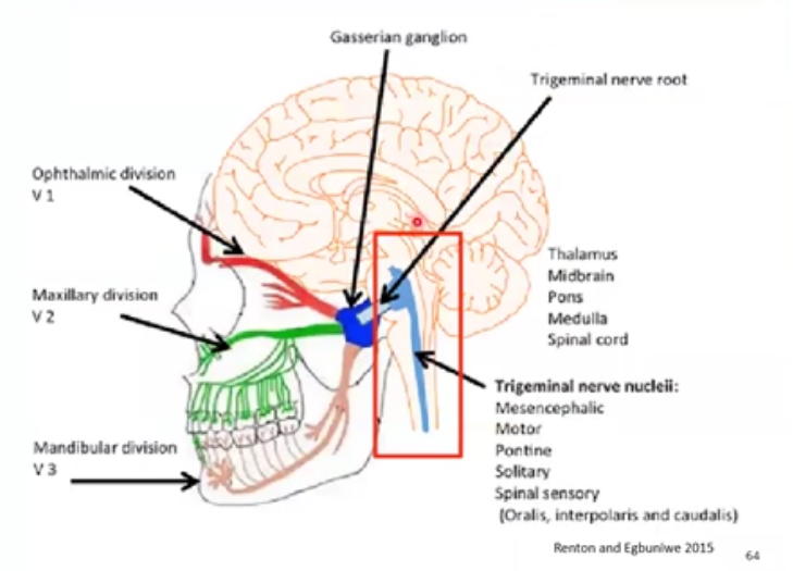
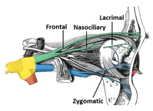
opthalmic divi of trigeminal nerve V1: frontal nerve
sensory fibres form skin and muscles and upper eyelid come to form supratrochlear nerve
this enters orbit by piercing superior medial corner of orbital septum
frontal nerve: supraorbital nerve
sensory fibres form skin and muscles form the supraorbital nerve
this nerve enters orbit as one or two branches:
one enters supraorbital notch other supraorbital artery
the supraorbital nerve joins supratrochlear and forms frontal nerve
frontal exits the orbit through superior orbital fissure above the ring
innervates skin and conjunctiva of the central eyelid
opthalmic div of trigeminal nerve : lacrimal nerve
sensory fibres from lateral aspect of upper eyelid form lacrimal gland
jon sensory fibres that serve the gland to form lacrimal nerve
runs along lateral rectus muscle and exit through superior orbital fissure
goes to lacrimal gland, and can innervate lateral eyelid
opthalmic nerve formation
after exiting orbit, nasociliary, lacrimal and frontal nerve join and form trigeminal nerve opthal div
this then enters lateral walls of sinus
maxillary V2 of trigeminal nerve
sensory, from nose, cheeks and upper jaw
infraorbital nerve and zygomatic nerve and nerves from roof of mouth form the maxillary nerve
enters the skull via foramen rotundum
innervates skin, sinuses of maxilla and mucous membrane
what is the pterygopalatine ganglion
largest of the parapsympathetic ganglia
contains post ganglionic, para and symp fibres of maxillary nerve
mainly controls secretion of glands in the face
mandibular division of trigeminal nerve V3
innervates the lower face ad contains both sensory and motor fibres
enters skull via foramen ovale
from cheeks, side of head lower haw mouth tongue and motor to lower jaw
trigeminal nerve formation
as opth and max divisions enter the skull and run within sinus
mandibular division lies below the sinus
the sensory fibres from all 3 divisions enter the trigeminal ganglion where they synapse
fibres then leave and enter the pons as either the sensory or motor root
zygomatic nerve branches provide sensory innervation of temporal and zygomatic regions
facial nerve: CN VII
has 2 roots: large motor root innervates the facial muscles
smaller root contains sensory and parasymp fibres
sensory fibres carry taste sensations from tongue
parasymp fibres supply secretomotor fibres to various glands of face
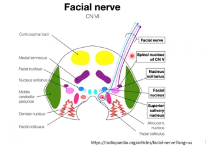
facial nucleus
motor nucleus of facial nerve is located in reticular formation of pons
upper segment supplies frontalis, orbicularis muscles and supplies remaining facial muscles
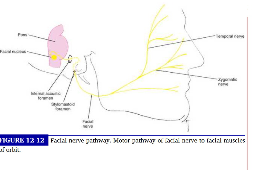
facial nerve pathway
fibres leave facial nucleus, arch around abducens nucleus and emerge as facial nerve from brain stem
enters acoustic foramen and runs through canal in temporal bone
the parasymp fibres here run to lacrimal and give off as petrosal nerve
morot fibres emerge through stylomastoid foramen, pass below external auditory canal and divide into several branches
what branches do the facial nerve divide into
upper two: temporal and zygomatic
they supply the frontalis, procerus, corrugator and orbicularis muscle
segment of the facial nerve
extratemporal: exits temporal bone via stylomastoid foramen
orbicularis muscle recieves innervation from temporal nerve and zygomatic nerve
what is the other segment of the facial nerve
labyrinthine segment goes to the greater petrosal nerve
made up of parasympathetic nerve fibres and joined by deep petrosal nerve
pupil light reflex
light shon onto the pupil, light signal stramsits via optic nerve to the chiasm
nerve fibres go to contralateral side, temp goes to ipsilateral side
at the optic tract, 90% of fibres end at LG, 10% leave V1 and follow pupil pathway
those signals go to the pretectal nucleus
from there, it send signals to the edinger wheshal nuclei
efferent: other way to the sphincter muscle
dim light
pupillary dilator muscle fibres contract and widne ppil
post ganglionic parasymp fibres from long ciliary nerve innervate the dilator muscle
pupil near response
accom, convergence and pupil constriction
pre tectal nucleus plays role in light near