cornea IV dystrophies
 Call Kai
 Learn
Learn Practice Test
 Spaced Repetition
 Match
Match Flashcards
 Knowt Play
1/61

There's no tags or description
Looks like no tags are added yet.
Name | Mastery | Learn | Test | Matching | Spaced | Call with Kai |
|---|
No analytics yet
Send a link to your students to track their progress
62 Terms
what are corneal dystrophies ()5
•Group of genetic eye disorders - not caused by outside factors such as injury or diet - no systemic associations
•Build up of abnormal material (lipids and cholesterol crystals) in the cornea - loss of transparency
•Slowly progressive
•Tend to be bilateral
More than 20 types
how do we examine corneal dystrophies (3)
•Often found incidentally
•Silt-lamp examination
•Detailed history (FH)
what are the general risk factors of corneal dystrophies (3)
•Family history (genetic)
•Any age
M=F (except Fuchs F>M)
general symptoms of corneal dystrophies (3)
•Variable effect on vision - Normal vision - non-severe sight impairment - severe visual impairment
•FB sensation/Pain
-Photophobia
what is the general management of corneal dystrophies (6)
•Depends on the severity and the type of dystrophy
•Asymptomatic - monitor
•Early stages - ocular lubricants, hypertonic eye drops (↓corneal oedema)
•Corneal erosion - antibiotics, bandage lenses, laser/scrape (phototherapeutic keratectomy - removes abnormalities)
•Ectasias - rigid contact lens
Severe cases - corneal transplant (lamellar or penetrating keratoplasty
what are the differences among corneal dystrophies (5)
•Severity and onset of symptoms
•Age of presentation
•Extraneous materials (lipid, cholesterol)
•Location of cornea affected
•Treatment
state the classifications of corneal dystrophies (4)
•Anterior - epithelium and Bowman’s membrane - superficial
•Stromal - stroma (thickest layer) - May progress to affect other layers of the cornea
•Posterior - endothelium and Descemet’s membrane - Can potentially progress to affect all layers.
•Ectasia - change of corneal shape
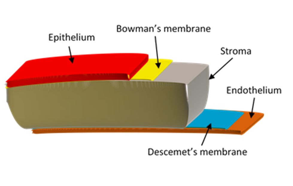
state the anterior corneal dystrophies to know (3)
Epithelial Basement Membrane
Meesmans
Reis-Buckler
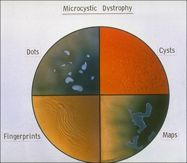
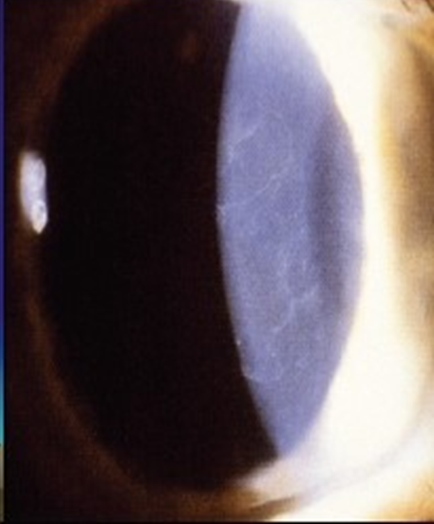
describe Epithelial Basement Membrane dystrophy (Coogan’s or map-dot-fingerprint dystrophy) (3)
•Most common anterior corneal dystrophy
•Defect in epithelium-basement membrane interaction
•Prevalence ~2.5%
what are the symptoms of Epithelial Basement Membrane dystrophy (2)
•Often asymptomatic
•Recurrent corneal erosion - pain, lacrimation, photophobia (~10% in 3rd-4th decade)
what are the signs of Epithelial Basement Membrane dystrophy (5)
•Microcysts - small cystic spaces
•Diffuse grey patches (maps) - changes in the BM
•Fine refractile lines (fingerprints) - folds in BM
Creamy white cysts (dots) - dead cells
note these are best seen in retro illumination or scleral scatter
what is the management of Epithelial Basement Membrane dystrophy (2)
Tear supplements if needed
bandage contact lenses (as per recurrent corneal erosion)
describe Meesman’s (juvenile) epithelial dystrophy (2)
•Rare, non-progressive abnormality of epithelial metabolism
•Mild, may cause recurrent epithelial erosions in adulthood
what are the symptoms of Meesman’s (juvenile) epithelial dystrophy (5)
•Unaffected until 4th or 5th decade of life
•May report:
mild irritation
sl. blurred vision
photophobia
watery eyes
what are the signs of Meesman’s (juvenile) epithelial dystrophy (2)
•Onset in infancy
•Multiple, discrete tiny uniform epithelial microcysts, maximal centrally (extend towards but do not reach the limbus)
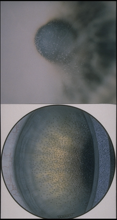
what is the management of Meesman’s (juvenile) epithelial dystrophy (1)
tear supplements
describe Reis-Bücklers dystrophy (3)
•Progressive, AD inherited dystrophy
•Degeneration of Bowman’s layer with subepithelial collagen deposition
sometimes categorised as a stromal dystrophy
what are the symptoms of Reis-Bücklers dystrophy (3)
•Severe recurrent erosions in childhood
•Pain, lacrimation and photophobia
•VA↓ later (30-40’s )
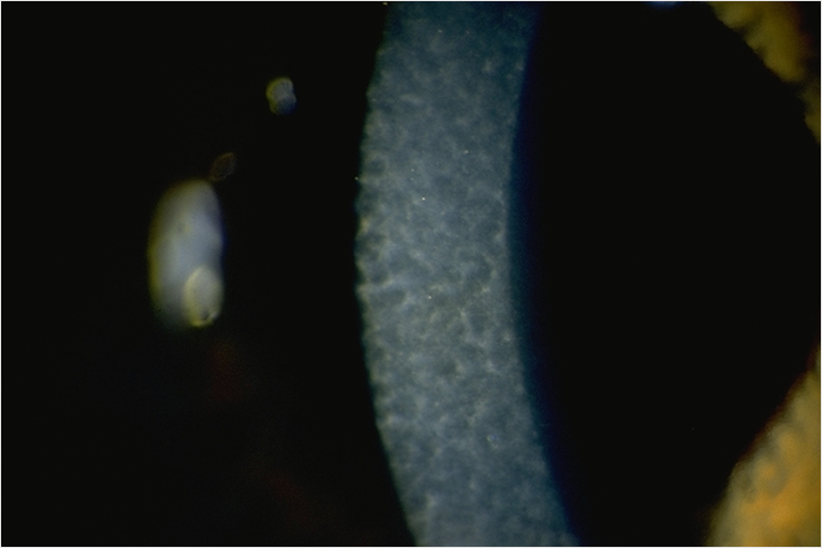
what are the signs of Reis-Bücklers dystrophy (3)
•Grey-white subepithelial opacities (reticula/honeycomb pattern)
•Most dense centrally, ↑density with age
•Corneal sensation becomes reduced (may alleviate symptoms)
what is the management of Reis-Bücklers dystrophy (2)
•Tear supplements including gels
•Surgery for advanced cases - excimer laser superficial, keratectomy or lamellar/PK if VA↓
state the stromal dystrophies to know (3)
Lattice
Granular
Macular
describe lattice stromal dystrophies (3)
•Rare, AD inherited
•Progressive deposition of amyloid (abnormal protein) in stroma
•3 types - Type I isolated to the eye, type II systemic amyloidosis (meretoja’s syndrome), type III v rare isolated to the eye & those of Japanese origin
what are the symptoms of lattice stromal dystrophies (2)
• reduced VA - especially as cornea opacifies
•Symptoms of recurrent corneal erosion - pain, lacrimation and photophobia
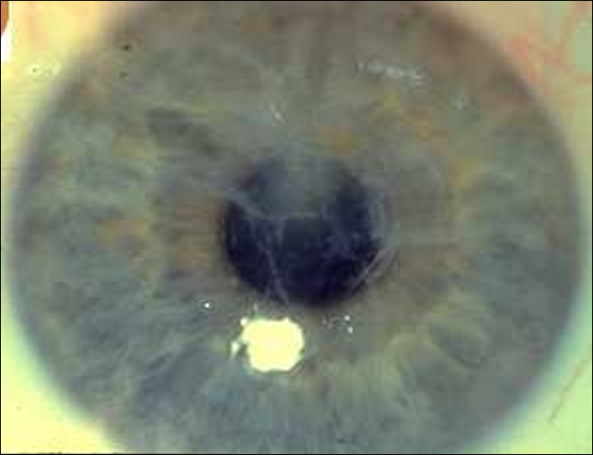
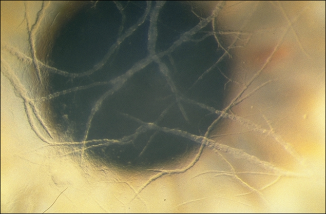
what are the signs of stromal dystrophies (3)
•Branching criss-crossed lines of amyloid
•Progressive stromal haze
•Reduced corneal sensation
what is the management of lattice stromal dystrophies (2)
•Tear supplements
•Frequently requires surgery - Penetrating or deep lamellar keratoplasty, or excimer laser keratectomy
describe macular stromal dystrophies (4)
•Rare, AR inherited
•Deposition of glycosaminoglycan in the stroma
•Most severe corneal dystrophy
relatively common in iceland
what are the symptoms of macular stromal dystrophies (3)
•VA reduced (often severe) from end of first decade
•+/- recurrent epithelial erosions & pain
Photophobia and disability glare
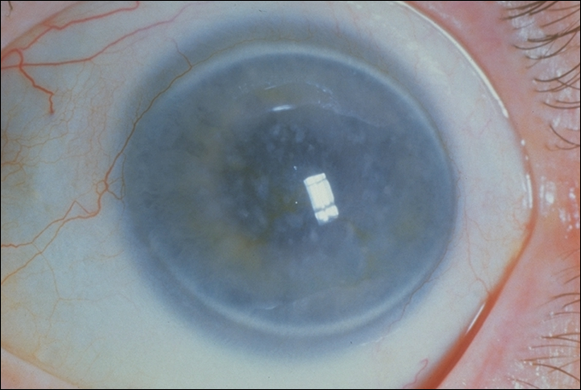
what are the signs of macular stromal dystrophies (3)
•Ill-defined greyish opacities and stromal haze - starts centrally
•Eventually involves full-thickness stroma with no limbal clear zone
•Corneal sensation diminished
what is the management of macular stromal dystrophies (1)
•Deep lamellar keratoplasty is usually required by the 5th decade - corneal transplant surgery in which the diseased corneal layers are removed down to Descemet’s membrane, while the patient’s own endothelium is preserved.
describe granular stromal dystrophies (2)
•Rare, AD inherited
•Deposition of hyaline material (usually protein rich, smooth, glassy, translucent substance in the stroma)
what are the symptoms of granular stromal dystrophies (3)
•Glare and photophobia (teens)
•VA often normal - may decrease later as cornea opacifies
•+/- recurrent epithelial erosions
what are the signs of granular stromal dystrophies (3)
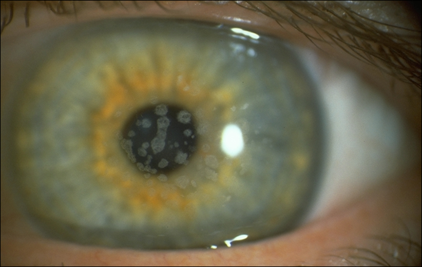
•Discrete white crumb like opacities initially in central anterior stroma (deeper later)
•Progressively coalesce and spread outwards (sparing the limbus)
•Corneal sensation diminished
what is the management of granular stromal dystrophies (1)
•Deep lamellar keratoplasty is usually required by the 5th decade
state the posterior corneal dystrophies to know (2)
Fuch’s Endothelial Dystrophy !!!
Posterior Polymorphous
describe Fuch’s Endothelial Dystrophy (4)
•Common, sporadic (AD inheritance)
•Progressive endothelial dysfunction → pump failure → corneal oedema
•F:M 3:1
•Develops middle age or later (50-70 yrs)
what are the symptoms of Fuch’s Endothelial Dystrophy (4)
•Initially asymptomatic
•Gradually ↑ blurring & glare
•Symptoms worse on waking and improve during the day (due to corneal oedema)
•General FB sensation → sharp pain++
what are the early signs of Fuch’s Endothelial Dystrophy (5)
•Bilateral with asymmetry
•Pigment dusting on endothelium
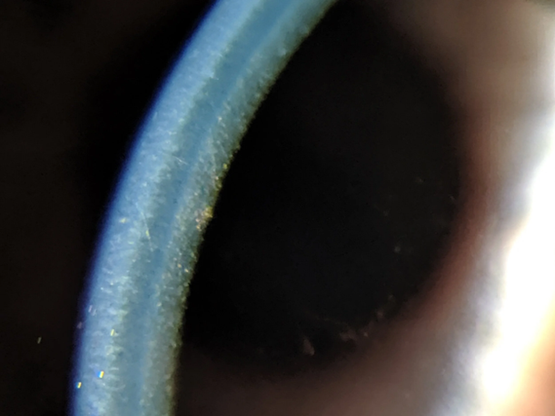
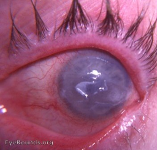
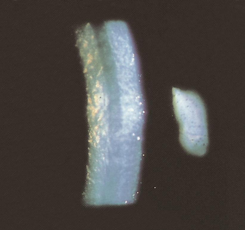
•Corneal guttata → ‘beaten metal’ appearance - irregular ‘warts ‘or ‘excrescences’ on Descemet’s membrane, secreted by anormal endothelial cells
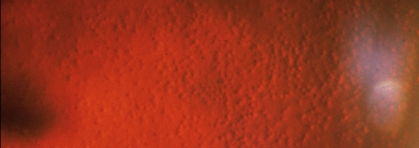
•Specular microscopy: ↓cell count, ↑cell size ↑ central corneal thickness (CCT)
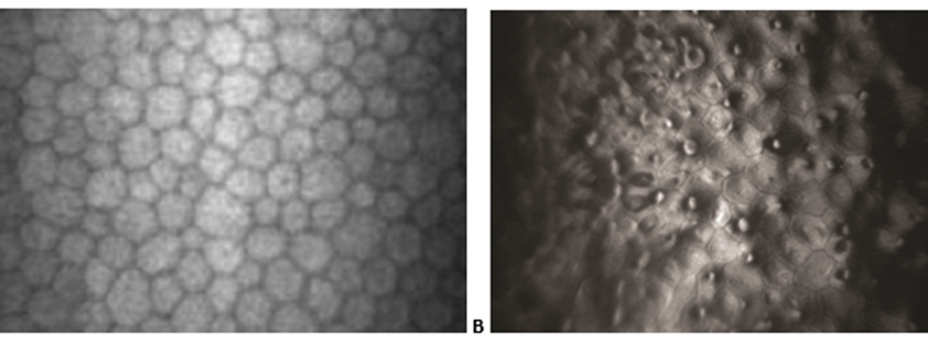
what are the later signs of Fuch’s Endothelial Dystrophy (2)
•Stromal oedema → folds in Descemet's membrane and epithelial bullae (fluid filled vesicles (blister like) in the epithelium (painful))
•Recurrent corneal erosion → vascular pannus and stromal haze
what is the optometric management of Fuch’s Endothelial Dystrophy (3)
•Monitor CCT, photography and imaging
•Ocular lubricants
•Refer routinely when symptomatic
what are the secondary care options of Fuch’s Endothelial Dystrophy (3)
•Corneal hydration - sodium chloride 5% prn (waking), hair dryer
•Ruptured bullae - bandage CL, cycloplegia, antibiotic ointment and lubricants
•May require corneal surgery - Posterior lamellar (DSEK, DMEK) / Penetrating keratoplasty
describe Posterior polymorphous (4)
•Asymptomatic corneal dystrophy
•Evident from teens onwards
•Abnormal collagenous membrane secreted by endothelium
•Occasionally associated with iridoschisis, band keratopathy & glaucoma
treatment (1) and signs (1) of Posterior polymorphous
•Asymmetrical vesicle-like lesions in Descemet’s with variable morphology
•No treatment needed
state the corneal ectasias to know (3)
Keratoconus !!!
Keratoglobus
Pellucid Marginal Degeneration
what is keratoconus (4)
•Conical distortion of the cornea
•Stromal thinning, irregular shape
•Presents teens-twenties
•Progressive – usually stabilises by mid 30s
what is the aetiology of keratoconus (6)
Unknown aetiology
Associated with:
•Atopy
•Repeated trauma (eye rubbing)
•Connective tissue disorders e.g. Marfan’s syndrome
•Systemic conditions e.g. Downs syndrome
•Prevalence 0.05-5% (1 in 2,000) - FH: 10% of offspring (may be more subclinical)
what are the risk factors of keratoconus (2)
•Atopy, connective tissue disorders
•Earlier presentation - worse prognosis
what are the symptoms of Keratoconus (2)
•Increasingly blurred vision
•Acute hydrops - pain++ and sudden loss of vision
what are the signs of Keratoconus (3:4)
Refractive:
•Progressive irregular astigmatism and myopia
•Scissoring and oil droplet reflexes (ret and ophth)
•Poor spectacle VA
Corneal steepening/thinning:
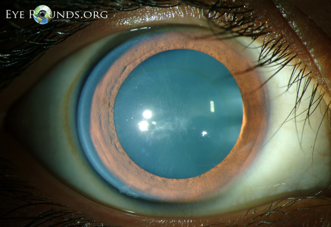
•Vogt’s striae (vertical lines in the stroma)
•Fleischer ring (iron at base of cone)
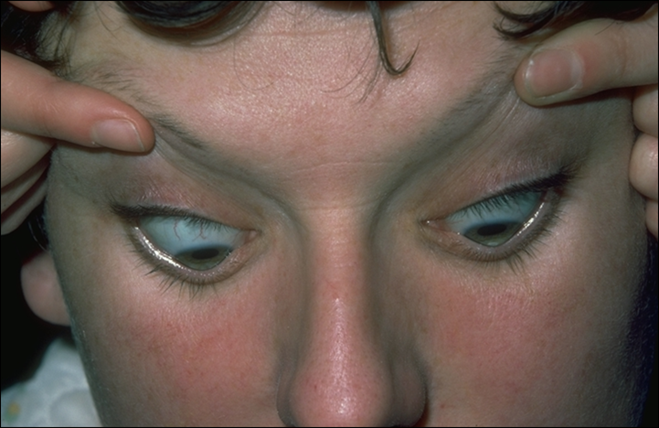
•Munson’s sing (conical distortion of lower lid on downgaze)
•Corneal topographical changes
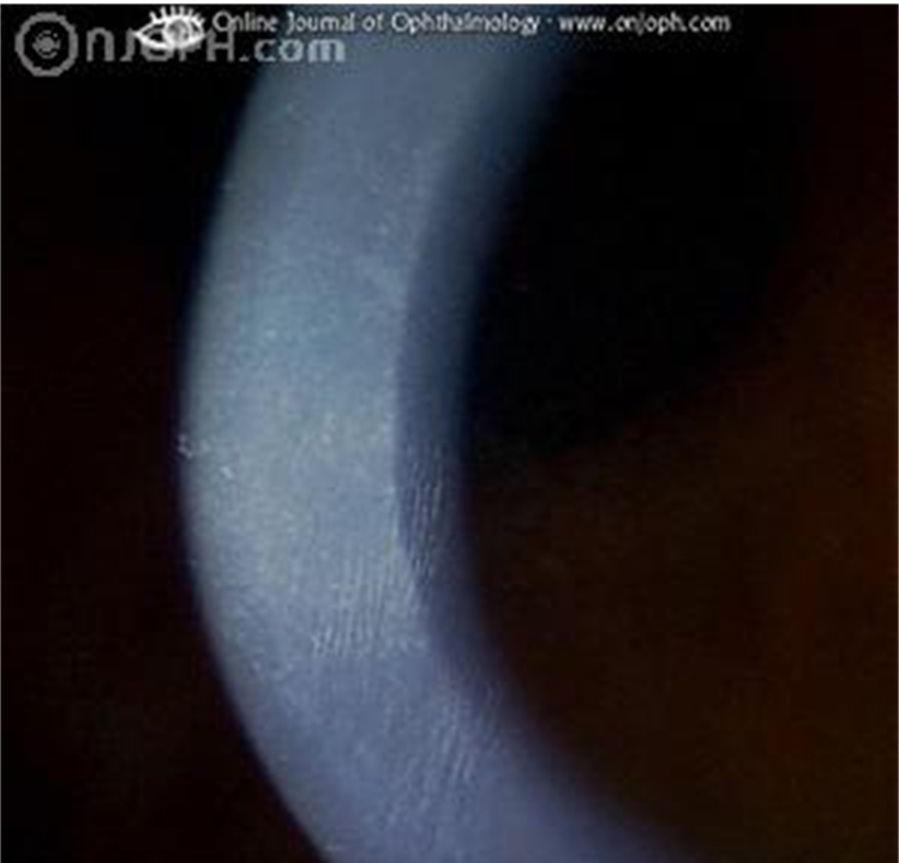
what are the complications associated with Keratoconus (2)
Acute hydrops
apical scarring
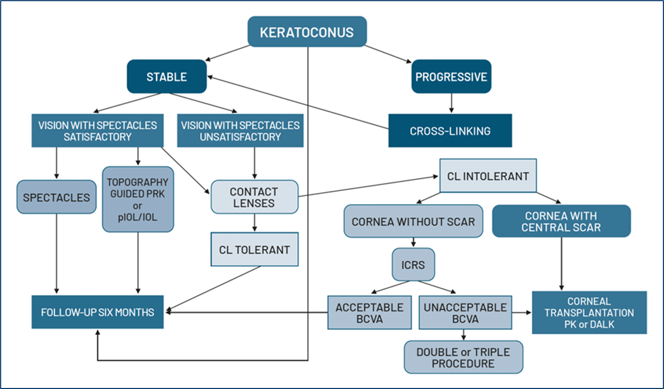
what is the management of Keratoconus - mild (1) moderate (4) severe (1) and hydrops (1)
mild - spectacles or CL correction
moderate:
RGP lens
Scleral lens
Intrastromal corneal ring segments (INTACS) also called intracorneal ring segments (ICRS) - flattens the cornea
Collagen cross linking - effective in halting progression
severe:
surgery (deep anterior lamellar keratoplasty or penetrating keratoplasty)
hydrops:
topical steroids, lubrication, cycloplegia, systemic analgesia
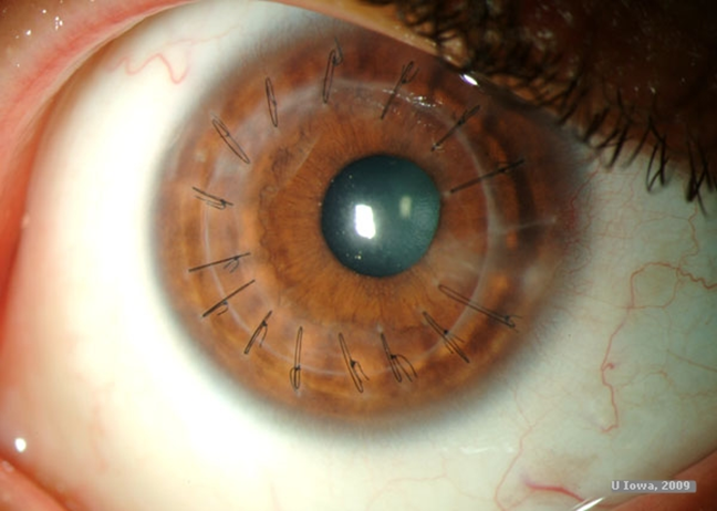
describe keratoglobus, signs, symptoms anf tx (6)
•Rare, unknown aetiology
•Global corneal thinning (entire cornea)
•Leads to gross protrusion of cornea
•Significant risk of rupture with minor trauma
•Vision is poor due to irregular myopic astigmatism
Tx: scleral lenses
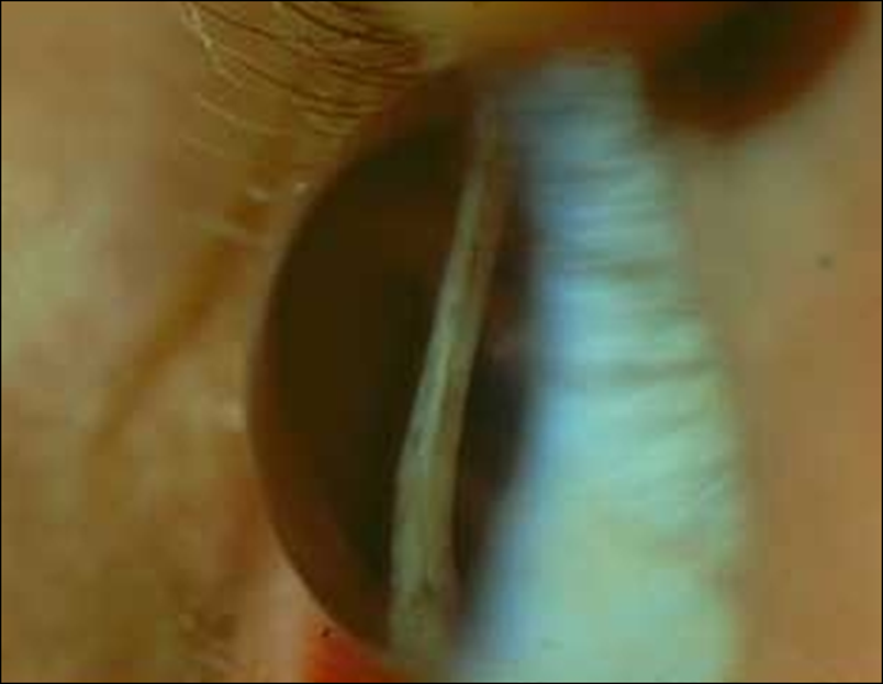
describe Posterior Keratoconus (3)
•Rare, non-progressive congenital
•Steep posterior surface (normal anterior surface)
•Treatment not usually necessary (surgery)
describe Pellucid Marginal Degeneration - symptoms, signs and tx (8)
•Rare, bilateral, progressive ectasia of the peripheral cornea - ‘beer belly’
•Inferior crescent shaped band (typically 4 & 8 o’clock) of stromal thinning
•Normal thickness central cornea
•Can cause severe deterioration in vision
•Irregular astigmatism
•Hydrops, Vogt striae & scarring are relatively rare
•Concentric folds in DM
Management - Scleral contact lenses (surgery not very successful)
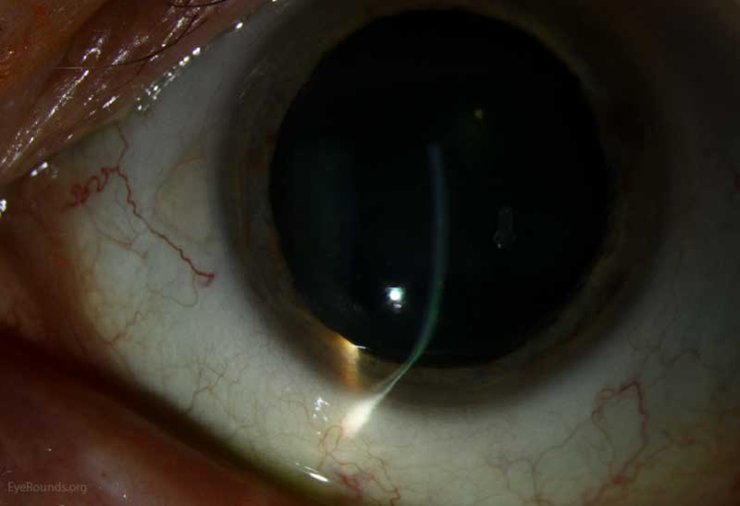
what is the aetiology of Acute Hydrops (1)
Descemet’s membrane rupture — acute leakage of aqueous into the stroma and epithelium — corneal oedema and scarring
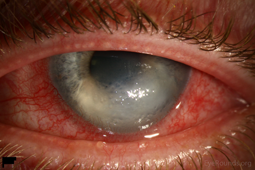
what are the predisposing factors of Acute Hydrops (2)
•Corneal ectasia (e.g. Keratoconus)
•VKC, asthma
what are the sx of Acute Hydrops (4)
•Pain++
•Sudden loss of vision
•Photophobia
Watering
what are the signs of Acute Hydrops (3)
•Stromal oedema
•+/- epithelial oedema
•Descemet's membrane rupture may be seen
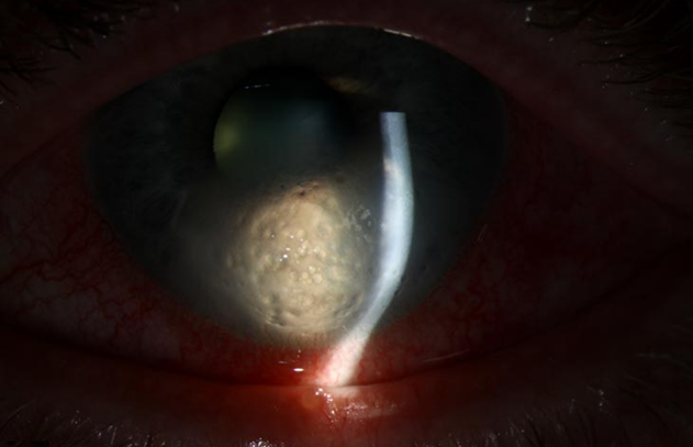
what is the management of Acute Hydrops (7)
•Acute: cease CL wear
•Monitor weekly for vascularisation/compilations
•Majority resolved 2-4 months
•Corneal topography may change (CL refit)
•Topical lubricants
•Systemic analgesia (pain relief medication)
Refer if neovascularisation
what is a corneal transplant rejection (3)
•30% of transplants experience at least one episode of immune rejection
•Maybe reversible - prompt treatment
•Lamellar and full thickness grafts
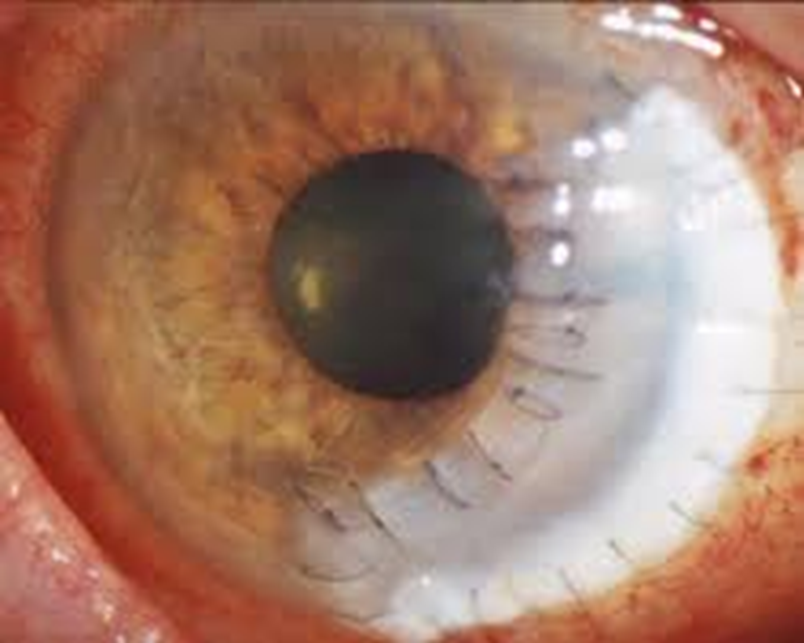
what are the sx of corneal transplant rejections ()
•Redness, watering and photophobia
•Blurred vision and pain/discomfort
what are the signs of corneal transplant rejections (8)
•Any layer of cornea - endothelium most serious
•Sub-epithelial opacities
•Anterior ciliary injection
•Transplant oedema
•Rejection line (Khodadoust ine)
•Keratic precipitates
•AC flare and cells
Raised IOP
what is the management of corneal transplant rejections (2)
•Same day referral
Any sign of problem with corneal graft - same day referral to eye clinic