New Material Review Sheet for Final - Posterior Segment & Ocular Disease Spring 2026
1/107
There's no tags or description
Looks like no tags are added yet.
Name | Mastery | Learn | Test | Matching | Spaced | Call with Kai |
|---|
No analytics yet
Send a link to your students to track their progress
108 Terms
you must either run an OCT, refer to ophthalmology, or both
What must you do when you are unsure about what is happening in the macula?
white
In the US, macular degeneration is most common in (black/white) individuals
hypoxic stress and oxidative reactions AT THE LEVEL OF THE RPE AND OUTER RETINAL LAYERS
What is the pathophysiology of AMD?
-age
-smoking
-race
-genetics
What are the most significant risk factors for AMD?
-intake of dietary anti-oxidants
-intake of omega-3 fatty acids
-amount of exercise
-UV exposure
-serum cystatin levels elevated
-blue > brown iris
-divorced > married
What are the "other" risk factors for AMD (Aside from the significant ones)?
-drusen
-pigmentary changes
-RPE clumping
-RPE window defects/dropout
-geographic atrophy (end stage)
What retinal changes does DRY AMD include?
dark brown/black
What color is RPE clumping in AMD?
dull yellow
What color is drusen in AMD?
dull yellow
What color is the RPE dropout/window defects of AMD?
loss of retinal tissue over the top of an area of RPE loss
What are effaced window defects?
orange/dark red
What color will effaced window defects be with AMD?
referral
Serous detachment w/ AMD is always a _______
leaking choroidal neovasc
There is assumed to be what associated with serous detachment?
appears as a raised, yellow/orange area under the RPE (sub-RPE) and maybe under the retina (subretinal)
What does serous detachment look like on retinal OCT?
No
Will a non-exudative choroidal neovascular membrane leak on FA?
No
Will a non-exudative choroidal neovascular membrane leak on indocyanine green?
Yes
Will a non-exudative choroidal neovascular membrane EVENTUALLY leak?
the area of the RPE detachment is filled almost entirely by the neovascularization itself
What does the OCT-A overlay of a non-exudative choroidal neovascular membrane show?
NO -- Wet AMD suggests vascular leakage
Is non-exudative choroidal neovascular membrane termed WET AMD?
-hemorrhage
-neovascularization
What does WET macular degeneration include?
darker
If there is hemorrhage, the subRPE portion is (brighter/darker)
brighter
If there is hemorrhage, the subretinal portion is (brighter/darker)
Yes -- anytime
Whenever Bruch's membrane is compromised, is there risk of choroidal neovascularization?
Yes -- there is a break in Bruch's membrane
Is choroidal neovascularization possible in AMD?
hyperreflective deposits above (retina side) the RPE
How do subretinal drusenoid deposits appear on OCT?
bad
Subretinal drusenoid deposits are a (good/bad) sign
Early AMD
What form of AMD is this pic?
Early AMD
What form of AMD is this pic?
Intermediate AMD
What form of AMD is this pic?
Advanced AMD
What form of AMD is this pic?
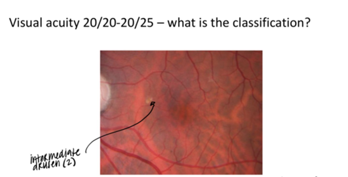
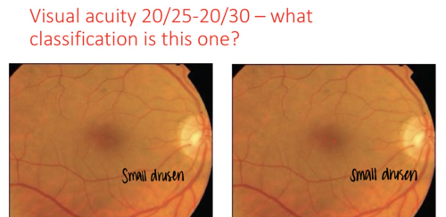
-unlimited small drusen
-less than 5 intermediate drusen
What classifies early AMD?
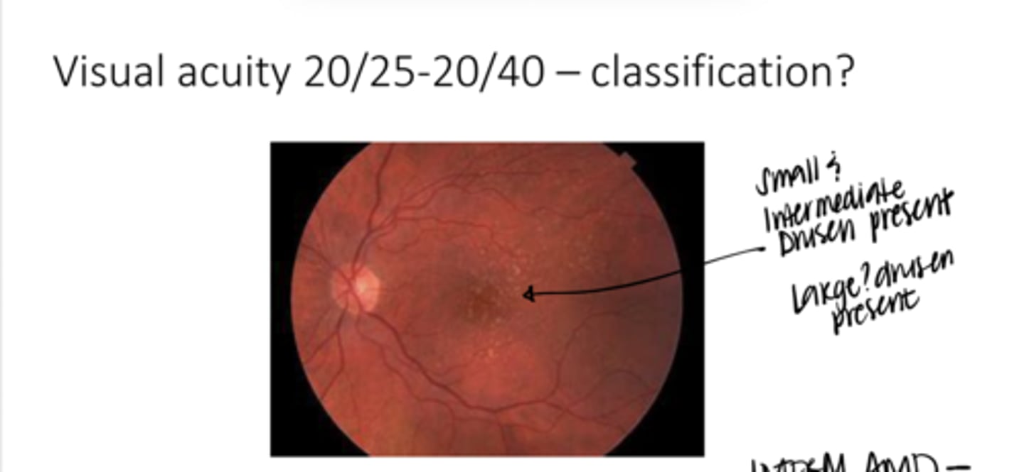
-5-20 intermediate drusen
-Intermediate RPE clumps
-less than 4 large drusen
-window defects may be present but are not geographic atrophy
What classifies intermediate AMD?
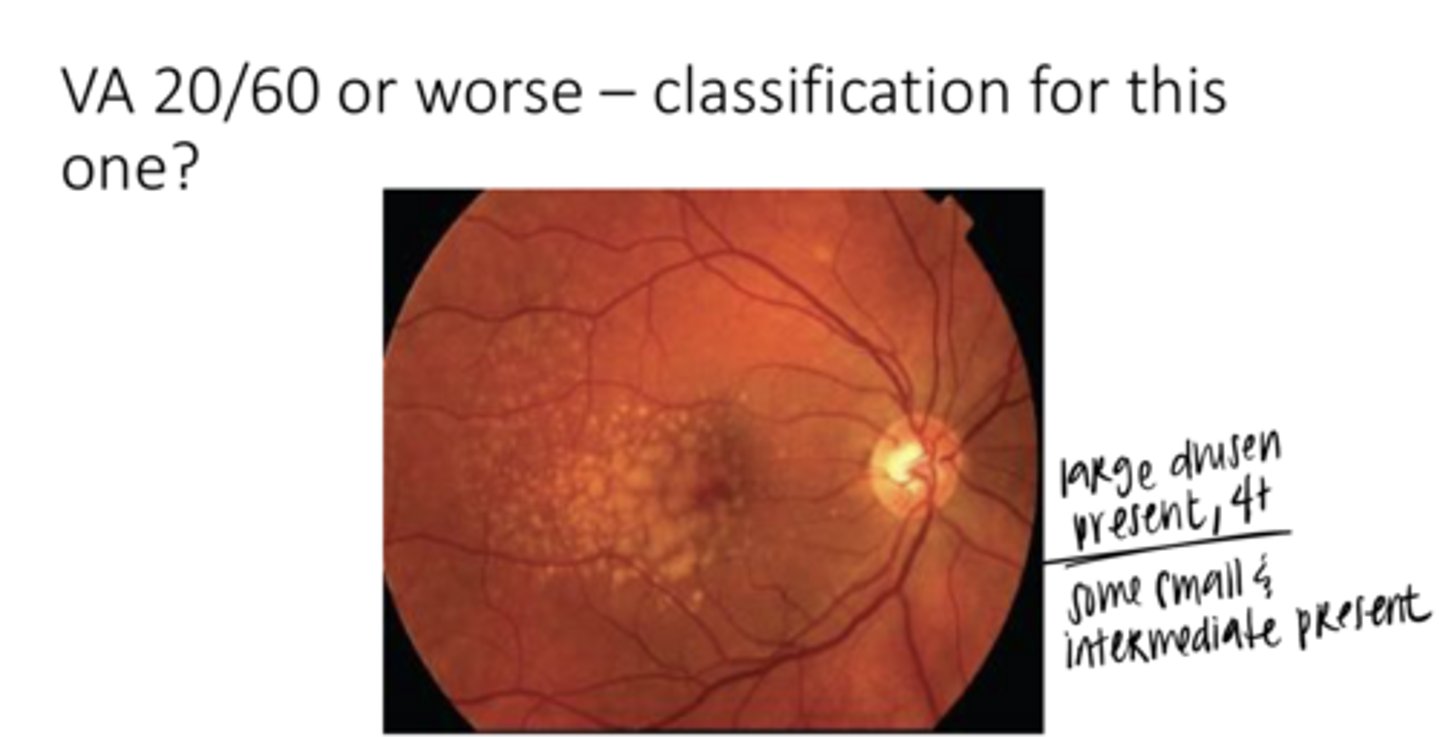
-More than 20 intermediate drusen
-Geographic atrophy
-Choroidal neovasc
-Nonexudative macular neovascularization (not Wet AMD)
What classifies Advanced AMD?
false
True or False:
Pigmentary changes are considered mild or early AMD
RPE window defects
Which are worse?
RPE window defects or RPE pigment clumping
OCT for sure in advanced AMD! FAF maybe
What do intermediate or advanced stages of AMD require?
No -- but you must at least get an OCT
Do you technically HAVE to refer a patient with no choroidal neovasc? What MUST you do in these advanced/intermediate stages?
-large numbers of intermediate drusen
-Any number of large drusen
-Any significant (intermediate or larger) pigmentary changes
What are the AMD Red flags?
-large drusen (>125um)
-Intermediate drusen (63-125um)
-Pigmentary abnormalities (hyperpigmentation, hypopigmentation, non central geographic atrophy)
-bilaterality
What are the signs of worsening AMD?
-drusen
-RPE window defects
-Serous RPE (PED) & serous retinal detachment
When imaging a patient with AMD, what should we look for on OCT?
in the RPE
Where will drusen be on OCT of a patient with AMD?
thinned/missing RPE with extra choroidal reflectivity
What are RPE window defects that can be seen on OCT imaging of an AMD patient?
dark areas of elevation
What will serous RPE/retinal detachments look like on OCT of an AMD patient?
-Lipofuscin
-Hyperautofluorescence/Hypoautofluorescence
When imaging a patient with AMD, what should we look for on FAF?
lipofuscin collects in RPE cells that are functioning abnormally
What is lipofuscin and why can we see it on FAF of AMD patients?
poorly functioning RPE
What hyperautofluoresces on FAF?
missing RPE (dropout/window defects)
What hypoautofluoresces on FAF?
Maybe -- it is highly variable
Does MPOD correlate with development and progression of AMD?
maybe
Does dark adaptometry correlate with development and progression of AMD?
No
Is genetic testing recommended for patients with AMD?
No
IS skin carotenoid testing recommended for patients with AMD?
Indocyanine green angiography (ICG) & OCT-EDI (enhanced depth imaging)
Imaging of the choroid can be improved with _________
Yes, can be used to assess the choroid/choriocapillaris and outer retina
Can OCT-A be used for testing AMD patients? How?
-vit C
-vit E
-beta carotene
-Zinc
-Copper
What did the AREDS1 supplement contain?
Intermediate or worse AMD
Who benefitted from the ARED1 supplement?
No
Did patients with mild AMD benefit from AREDS1 supplement?
higher dietary carotenoids
What benefited ALL AMD patients in any stage of the disease?
Smokers (past smokers, heavy passive/second hand smokers)
_______ had a higher risk of lung cancer with the AREDS1 formula
attributed to the beta carotene
Why did Smokers (past smokers, heavy passive/second hand smokers) have an increased risk of lung cancer with the AREDS1 formula?
False
True or False:
The AREDS2 trial included patients with mild forms of AMD
No -- none of these additions had any benefit
Did lutein, zeaxanthin, or omega-3 fatty acids being added to the original AREDS1 supplement have any benefit?
Lutein and zeaxanthin
Betacarotene in the original AREDS1 supplement was replaced with what d/t having more benefit?
No -- there was no evidence of benefitting these patients
Overall, should AREDS1 or AREDS2 supplements be prescribed to patients with mild AMD or prophylactically?
No
Is there any study that has tested the benefit of AREDS supplementation prophylactically or in mild AMD?
contact the patient's physician
What do you have to do BEFORE prescribing a supplement for AMD?
-discuss smoking cessation if relevant
-discuss diet (green leafy vegetables, colorful fruit, omega-3 fatty acids as in fish, reduction of saturated fats)
-Discuss exercise
-Discuss weight loss if relevant
-Discuss UV protection
-All patients with the AMD dx receive an Amsler grid
-Fundus photography
What needs to be done FOR EVERY PATIENT with AMD?
small drusen with a few intermediate drusen
REVIEW: What is the characteristics of early AMD?
1 year
Early AMD Follow up schedule?
No associated "dramatic findings" (MPOD, autofluorescence, dark adaptation, skin carotenoids), normal BMI, diet, non-smoker
9 months
Early AMD Follow up schedule?
One associated "dramatic" abnormal finding, normal BMI and diet, nonsmoker
9 months
Early AMD Follow up schedule?
Normal findings, BMI high or poor diet, non-smoker
6 months
Early AMD Follow up schedule?
One associated "dramatic" abnormal finding, BMI too high or poor diet, non smoker
6 months
Early AMD Follow up schedule?
Smoker
Supplement
Intermediate AMD Follow up Schedule?
At this level, all patients will get prescribed what?
Some sort of imaging (OCT/FAF)
Intermediate AMD Follow up Schedule?
You must do what for these patients?
Referral or a short (4 month) F/U
Intermediate AMD Follow up Schedule?
If the testing is dramatically worse than expected or there is a serous detachment, what should be considered?
yes
Intermediate AMD Follow up Schedule?
Should all of these patients get an Amsler grid?
Yes
Intermediate AMD Follow up Schedule?
Should you do fundus photography on these patients?
Refer them all
Advanced AMD Follow up Schedule?
How should you manage these patients?
yes
Advanced AMD Follow up Schedule?
Should you prescribe these patients supplements?
anti-VEGF meds
What is the first-line ophthalmological treatment of wet AMD?
-Lucentis
-Avastin
-Eyelea
-Vabysmo
What are the most popular anti-VEGF injections?
That Lucentis and Avastin had similar efficacy in treating WET AMD
What did the CATT study demonstrate?
targets 2 inflammatory markers (Anti-VEGF and Angiopoietin-2)
What is the MOA of Vabysmo?
maybe
Can Vabsymo be injected less often than Eyelea?
Yes -- but they do not completely stop the progression or reverse the disease
Are there now intravitreal injections that slow the progression of geographic atrophy?
OCT-A
Pachychoroidal diseases can be differentiated with what tool?
-Dilation of choroidal vessels in Haller's layer
-Thinning of choriocapillaris and Sattler's layer
-Hyperpermeable vessels in Haller's layer and choroidal filling defects (reduced perfusion)
What is the issue associated with Pachychoroidal diseases?
-RPE abnormalities (RPE pigmentary changes)
-photoreceptor abnormalities
Pachychoroidal diseases may be associated with what?
OCT-A
______ can be useful in differentiating the different pachychoroidal diseases
Some lead to neovasc and some do not
Why would we want to differentiate the different pachychoroidal diseases?
-PDT
-Anti-VEGF meds
-or both
What may be applied in order to treat pachychoroidal disease?
serous retinal detachment
Central serous chorioretinopathy will typically involve a ________
Central Serous Chorioretinopathy (Pic)
Central Serous Chorioretinopathy (Pic)

large
Pachyvessels will be (small/large) in Central Serous Chorioretinopathy?
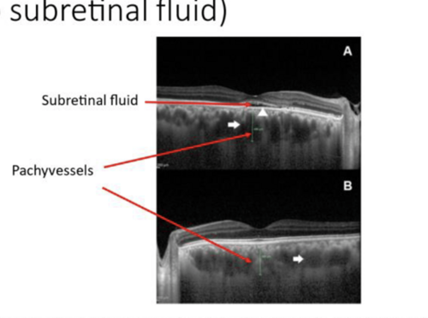
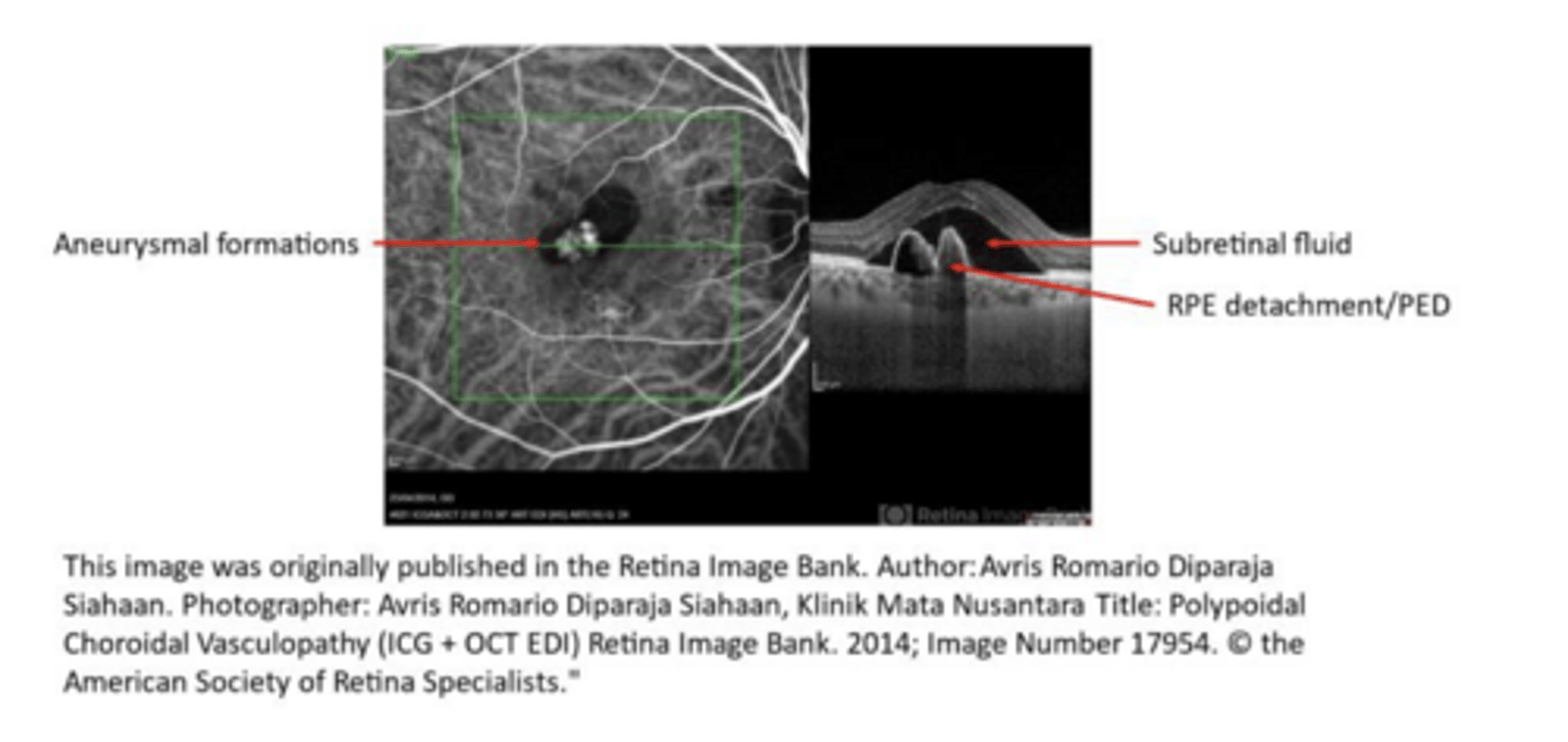
-Aneurysms on choroidal vessels
-Subretinal Fluid/RPE Detachment
What are the defining characteristics of Polypoidal choroidal vasculopathy/aneurysmal Type 1 Neovascularization (PCV/AT1)?

Polypoidal choroidal vasculopathy/aneurysmal Type 1 Neovascularization (PCV/AT1) (see pic)
Polypoidal choroidal vasculopathy/aneurysmal Type 1 Neovascularization (PCV/AT1) (see pic)
Hypoperfusion syndrome
______ is possible precursor to the ocular ischemic syndrome (OIS)
-dull ache in or around the eye
-TIA symptoms
-light induced amaurosis fugax
-carotid bruit
-decreased carotid pulse
What are the systemic findings of Hypoperfusion syndrome?
-unilateral venous dilation
-arterial narrowing
-venous beading
-non-tortuous vessels
-mid peripheral hemes
-possible retinal emboli
What are the ocular findings of Hypoperfusion syndrome?
Hypoperfusion + Signs of Hypoxia-Induced Inflammation
What is Ocular Ischemic Syndrome (OIS)?