Human Bio 2 Exam
1/143
Earn XP
Description and Tags
Lecture 9 (Slide 11) - Last Lecture
Name | Mastery | Learn | Test | Matching | Spaced | Call with Kai |
|---|
No analytics yet
Send a link to your students to track their progress
144 Terms
What does the autonomic system involuntarily control?
cardiac and smooth muscles, organs, glands
what are the 2 divisions of the autonomic NS
sympathetic and parasympathetic NS
what is used for each ANS impulse
two neurons (preganglionic and postganglionic ) and one ganglion for each impulse
when is our sympathetic division active
situations where we may be required to fight or flight
where do most preganglionic fibres (SNS) arise and what do they terminate
middle portion of spinal cord
terminate ganglia that lie near the cord
Why does the sympathetic division increase heartbeat and dilate airways?
For a ready supply of glucose and oxygen
What neurotransmitter is released by the postganglionic axon
norepinephrine (adrenaline)
Where do cranial nerves and fibers (PNS) arise from
bottom portion of spinal cord
the parasympathetic division gives us responses associated with _____
a relaxed state; our rest and digest system
what neurotransmitter is used by the parasympathetic division
acetylcholine
what cells form myelin in the PNS
Schwann cells; they contain myelin in their membranes
What decides which axons have a myelin sheath and which don’t
length
long axons usually have a sheath, short axons don’t
What happens in Multiple Sclerosis
myelin breaks down (neurons can’t transmit info)
What does myelin help with in PNS injury
nerve regeneration within the PNS (sheath serves as passageway for new fiber growth)
What is a nerve impulse
an electrochemical signal (conveys info in the NS)
What is resting potential
the potential energy (voltage) of a neuron at rest
Why does the resting potential exist
because the plasma membrane is polarized; positive charge outside cell, negative charge inside
What maintains resting potential
sodium potassium pump (NA out, K in neuron)
Nerve signals are also called ______ and occur in ____
action potentials
axons
What is a stimulus to the neuron
a change that activates the neuron
What is the neuron threshold
minimum voltage (-55 mV) that must be reached in order for an action potential to occur
What does the neuron do to convey a stronger signal (instead of changing action potential size)
It causes more action potential/ “fires more”
What happens during depolarization/ AP begins
Na+ channels open, Na+ rushes into cell
inside of cell becomes positive
What happens in repolarization / after depolarization
Na+ channels close, K+ channels open and K+ flows out of cell
inside cell becomes negative again → AP complete
How long is the entire process of depolarization to repolarization
only 3-4 milliseconds to complete
How much is an unmyelinated axon AP slowed
slowed by 1 ms because each axon section must be stimulated
What is saltatory conduction
AP’s jumping from node to node along the myelinated axon
faster than unmyelinated
what is the refractory period and why is it important
the period of time immediately after an AP when the axon is unable to conduct another AP
ensures one-way direction of signal
what are axon terminals and what structures are they near
the fine endings of axons
each terminal is close to a dendrite or cell body of another neuron
What is a synaptic cleft
small gap separating sending neuron from receiving neuron
What do neurotransmitters do for AP’s? Where are these NT’s stored?
transmit AP’s across synapse (junction between neurons)
stored in synaptic vesicles in axon terminals
What happens when an AP reaches the axon terminal
calcium ions enter the terminal and stimulate synaptic vesicles to merge with sending membrane
NT released into cleft and diffuses across to receiving membrane and bind to receptor proteins
What causes an excitatory response from the receiving neuron
if neurotransmitter causes sodium gates to open
what causes an inhibitory response from the receiving neuron
if neurotransmitter causes potassium gates to open
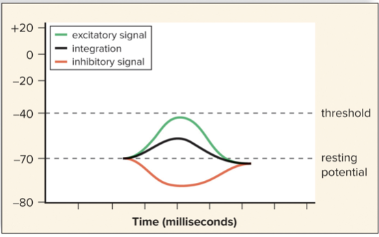
What is integration
summing up of multiple incoming excitatory and inhibitory signals
What happens when there are more excitatory signals vs inhibitory signals in integration
excitatory: it’s axon will transmit a signal
inhibitory: signals may prohibit axon from reaching threshold
After a NT has initiated a response it is removed from the synaptic cleft, why must it be removed?
to prevent continuous stimulation of receiving membranes
What 2 ways can NT’s be removed from the cleft?
Some synapses have enzymes that will inactivate the NT
Other synapses have the sending membrane reabsorb the NT
Where are ACh and NE located in the PNS
neuromuscular junctions
What are the roles of ACh and NE in the PNS
ACh: excites skeletal muscle but inhibits cardiac muscle
NE: excites smooth muscle
What does a sensory receptor do?
Conducts sensory transduction:
converts signals from environment (stimuli) into nerve impulses
what are exteroceptors
sensory receptors that detect stimuli from outside the body (taste, smell, vision, hearing etc and send to CNS)
what are interoceptors
sensory receptors that receive stimuli from inside the body (homeostasis involved)
List the 4 sensory (exteroceptor) receptor categories (based on what stimulus is detected)
Chemoreceptors, photoreceptors, mechanoreceptors, thermoreceptors
What do chemoreceptors respond to?
chemical substances (taste, smell, blood pH)
ex. nociceptors (pain receptors)
What do photoreceptors respond to?
light
What are mechanoreceptors stimulated by?
by mechanical forces (hearing, balance, touch, blood pressure)
What are thermoreceptors stimulated by?
changes in temperature; regulate body temperature
(in hypothalamus and skin)
define sensation and where it occurs
the conscious perception of stimuli
cerebral cortex
The sensation that we feel depends on what?
Which part of the brain receives the nerve signals
What happens before sensory receptors initiate nerve signals?
integration
What is sensory adaptation
a type of integration involving a decrease in response to a stimulus over time (sensory receptors send fewer impulses to the brain)
What are somatic senses associated with
skin, muscles, joints, viscera
What are the three types of somatic receptors (based on where receptors are)
proprioceptors, cutaneous receptors, and pain receptors
Where do all somatic receptors send nerve impulses
to primary somatosensory area of cerebral cortex via the spinal cord
What do free nerve endings detect
stimuli we perceive as pain and itching
What are proprioceptors/ what do they detect?
a type of mechanoreceptor involved muscle tone, equilibrium, posture
What are muscle spindles
a proprioceptor embedded in muscle fibers
Detect stretch → cause contraction
What are Golgi tendon organs?
a proprioceptor in tendoms
Detect tension → cause relaxation
What reflex involves proprioceptors(muscle spindles)?
knee jerk reflex → maintains posture
What do cutaneous receptors detect?
touch, pressure, pain, temperature
Meissner corpuscles, Krause end bulbs; all cutaneous receptors that detect….
fine touch in fingertips, palms, lips, tongue etc.
Where are Merkel disks (cutaneous receptors) found?
where epidermis meets the dermis
Root hair plexus (cutaneous receptor) detects….
hair touch
Where are the following cutaneous pressure sensitive receptors located: Pacinian corpuscles, Ruffini endings?
Pacinian: onion shaped in dermis
Ruffini: in connective tissue
What are thermoreceptors (cutaneous receptor type)
free nerve endings in the epidermis (some respond to cold, some to warm)
What happens leading up to binding of pain receptors (chemoreceptor type)?
tissue is damaged → release prostaglandin chemical → bind to nociceptors (pain receptor)
How does aspirin/ibuprofen reduce pain
by inhibiting enzymes that make prostaglandins (the pain receptor binding chemical)
What is referred pain
stimulation of nociceptors in internal organs is felt as pain in the skin
eg. pain from heart felt in left arm
what are the three layers of the eye (describe them)
sclera: outer white layer, fibrous (cornea inside is clear)
choroid: middle layer, dark pigment absorbs light
retina
what does the choroid form?
choroid → becomes iris → thickens and then forms ciliary body
iris function
regulates pupil size (hole where light enters eye)
ciliary body function
controls shape of lens for focusing
what attaches the lens to the ciliary body
suspensory ligaments
what are the two compartments of the eye made by
anterior:in front of lens, filled with clear liquid- aqueous humour
posterior: behind the lens, vitreous humor holds retina in place
When causes glaucoma and what does this result in
when the drainage ducts are blocked and the aqueous humor in the anterior compartment builds up
pressure compresses the arteries that serve retina → loss of vision
in which compartment is the third layer of the eye
the posterior compartment
What are the photoreceptors called that are within the retina layer
rods: light sensitive, no colour
cones: need bright light, detect colour
_____ is packed with cone cells
fovea centralis; when we look directly at an object it focuses on this
______ is made of sensory fibres from the retina
optic nerve; it takes nerve signals to the visual cortex in the occipital lobe
what do the cornea, lens and humor all do together
focus images on the retina (this image is inverted and reversed left to right on the retina)
when does the ciliary muscle contract vs relax, and when is lens flat vs round
relaxed muscle, flat lens: viewing a distant object
contract muscle, round lens: viewing a near object
what is eye strain, what age is this common
Fatigue from close work (contraction of ciliary muscle)
ages 40+ because lens loses elasticity
what segments do both rods and cones have
outer segment joined to an inner segment by a short stalk
where are pigment molecules located?
outer membrane of rods and cones, in the membrane of disks
where are synaptic vesicles of rods and cones located?
at synaptic endings of the inner segment of rods and cones
what is rhodopsin?
the visual pigment in rods
what happens to rhodopsin when a rod absorbs light
splits into opsin and retinal leading to a cascade of reactions → release of inhibitory NT’s
what provides us with peripheral vision
rods
what are the three types of cones?
B(blue), G(green), R(red)
combinations of cones are stimulated by in between shades of colour
what are the three layers of neurons within the retina
rod and cell cones (layer closest to choroid)
layer of bipolar cells (covers rods and cones)
ganglion cells (innermost layer, fibres = optic nerve)
what is the blind spot? how do we still have complete vision if we have blind spots?
where the optic nerve exits the eye
blind spot for right eye isn’t the same as blind spot for left eye so altogether we have complete vision
nerve impulses are carried by the optic nerves from the eyes to the ______
optic chiasma; theX shaped crossing over of optic nerve fibres
after exiting the optic chiasma, the optic nerve continues as ____ leading to ______
optic tracts
the thalamus
where does the thalamus direct nerve signals from the eye
the visual cortex in the occipital lobe
what structure is defective in colour blindness
one type of cone is defective or deficient in number (typically inability to see red and green)
what population is more susceptible to colour blindness
Males who have only one X chromosome (since gene is on the X chromosome)
affects 5-8% male population
What is 20/20 vision
you can see clearly at 20 feet what should normally be seen from that distance (good vision, not perfect)
what is nearsightedness and what causes it
can see close, cannot see far
eye is elongated; distant objects are brought into focus too far in front of retina
what solution is there for nearsightedness
wearing concave lenses (they spread light rays)