Urinary System
1/50
There's no tags or description
Looks like no tags are added yet.
Name | Mastery | Learn | Test | Matching | Spaced | Call with Kai |
|---|
No analytics yet
Send a link to your students to track their progress
51 Terms
What are the 4 major functions of the urinary system?
Removal of metabolic waste
Regulation of fluid & electrolyte balance
Regulation of blood pressure
Production, transport, storage & excretion of urine
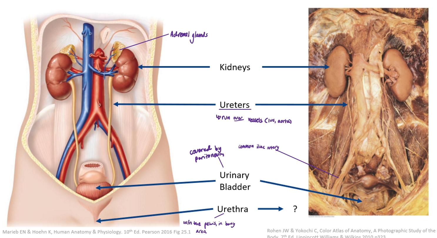
What are the 4 major components of the urinary system?
Kidneys
Ureters
Urinary bladder
Urethra
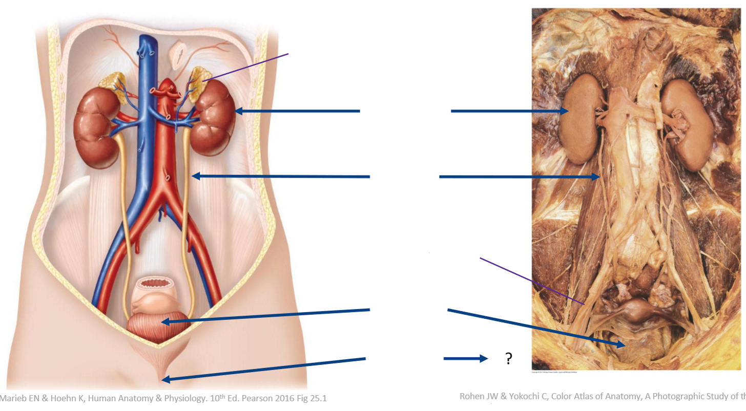
Label the following diagram
For the kidneys, state:
Which vertebra they correspond to
Where they are wedged
Which kidney is lower
Whether they are intraperitoneal or retroperitoneal
T12-L3
Wedged lateral to vertebrae in paravertebral gutter (‘either side’ of the vertebrae)
Right kidney slightly lower (but both are the same size)
Retroperitoneal
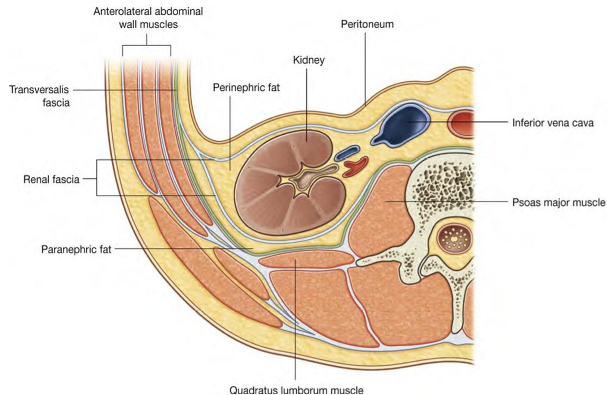
What are the kidneys surrounded by & what are the implications if this structure is removed?
Surrounded by fat
Keeps kidneys in position
Pathway for blood vessels supplying the kidneys
Kidney stripped away → kidneys start to drop → problems
Name the medial anatomical relationships of the kidneys
T12 - L3
Psoas major (but is not directly attached due to the fat)

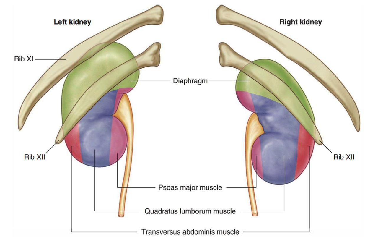
Name the posterior anatomical relationships of the kidneys
Quadratus lumborum
Thoracic diaphragm
Ribs 11-12
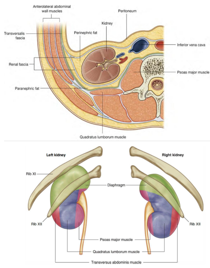
Name the anterior anatomical relationships of the kidneys
Peritoneum + abdominal viscera
Urinary viscera are retroperitoneal

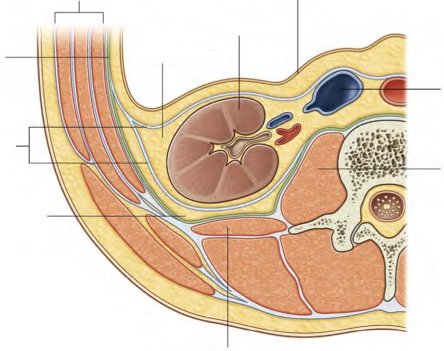
Label the following diagram
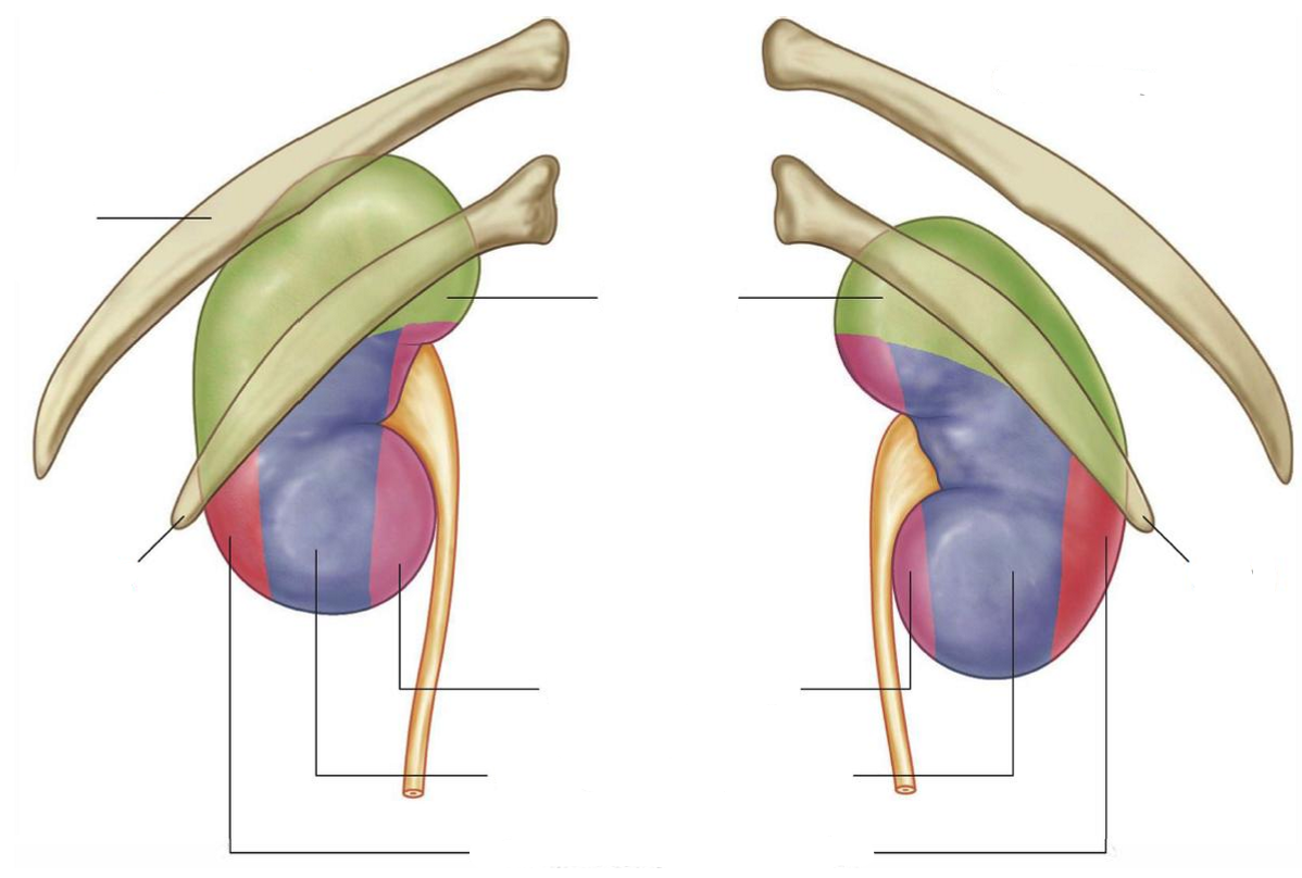
Label the following diagram of the attachments of the kidneys
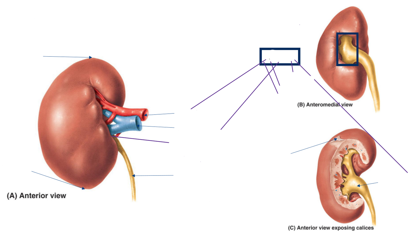
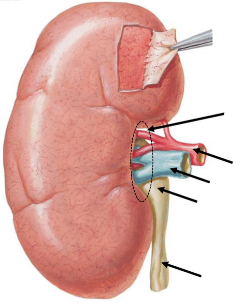
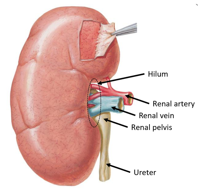
Label the following diagram of the external anatomy of the kidney
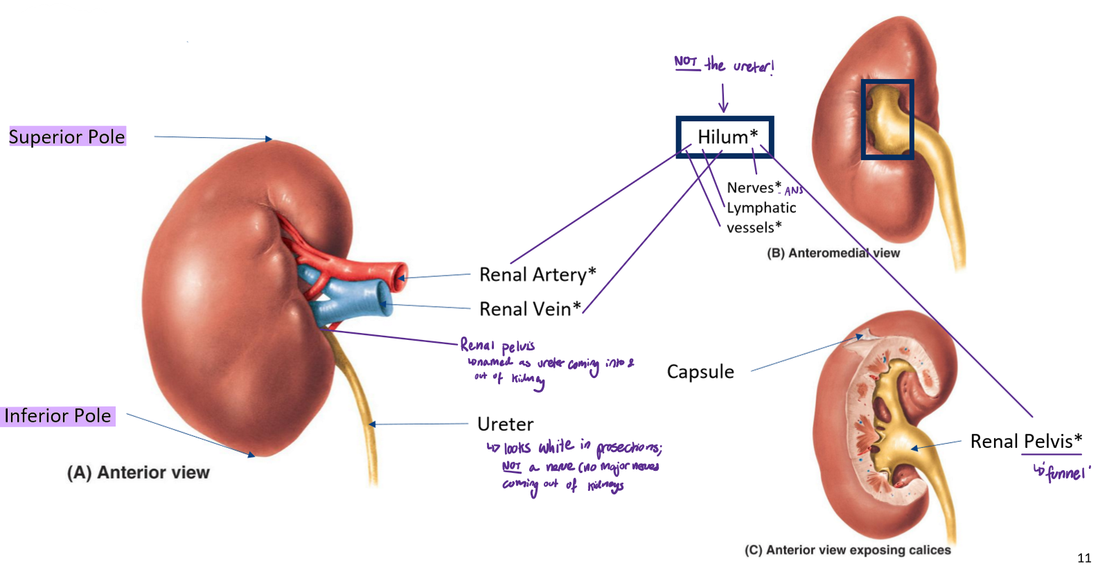
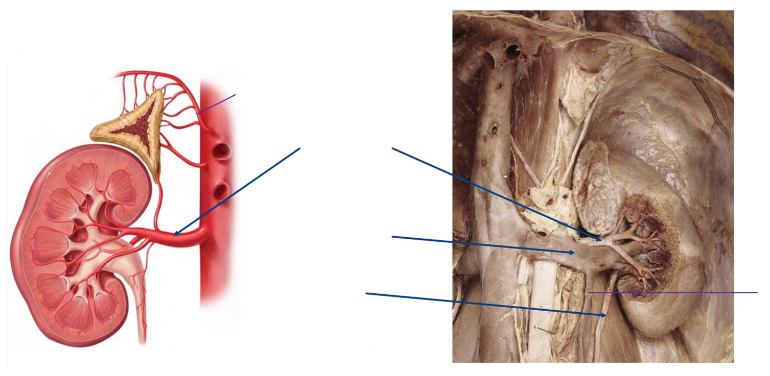
What structures make up the hilum of the kidney?
Renal artery
Renal vein
ANS nerves
Lymphatic vessels
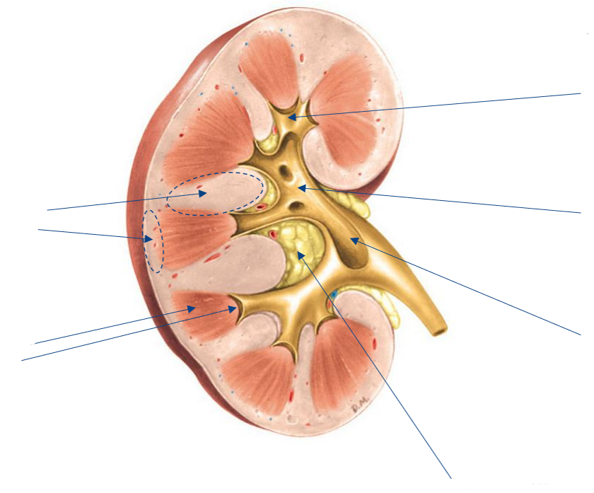
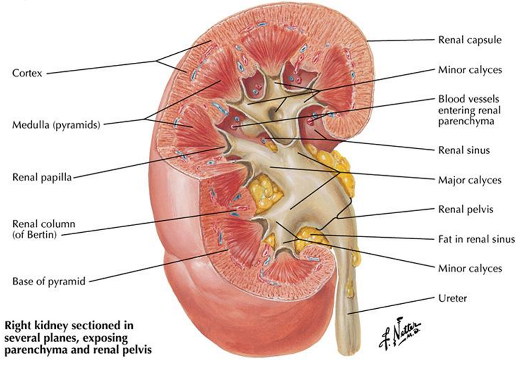
Label the following diagram of the internal anatomy of the kidney

What are the 2 solid tissues in the kidneys, and what do each of these tissues comprise?
Renal cortex
Renal columns
Renal cortex
Renal medulla
Medullary pyramid
Medullary papilla (apex of renal pyramids)
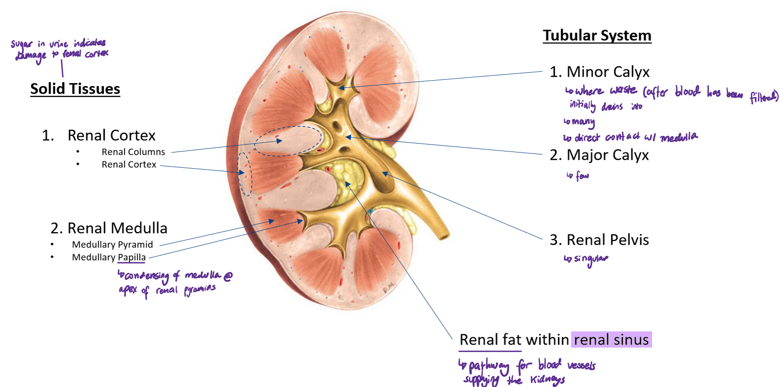
What are the 3 components of the tubular system of the kidneys? For each component, state how many there are of each
Minor calyx: many
Where waste (after blood has been filtered) initially drains into
Direct contact w/ medulla
Major calyx: few
Renal pelvis: singular

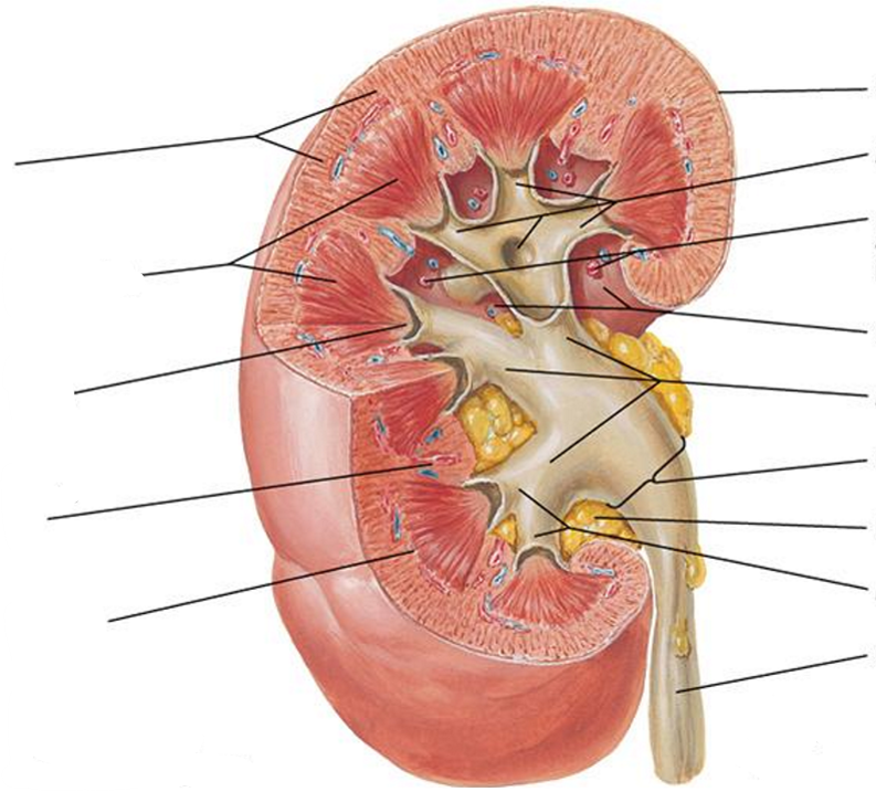
Label the following diagram of the kidney
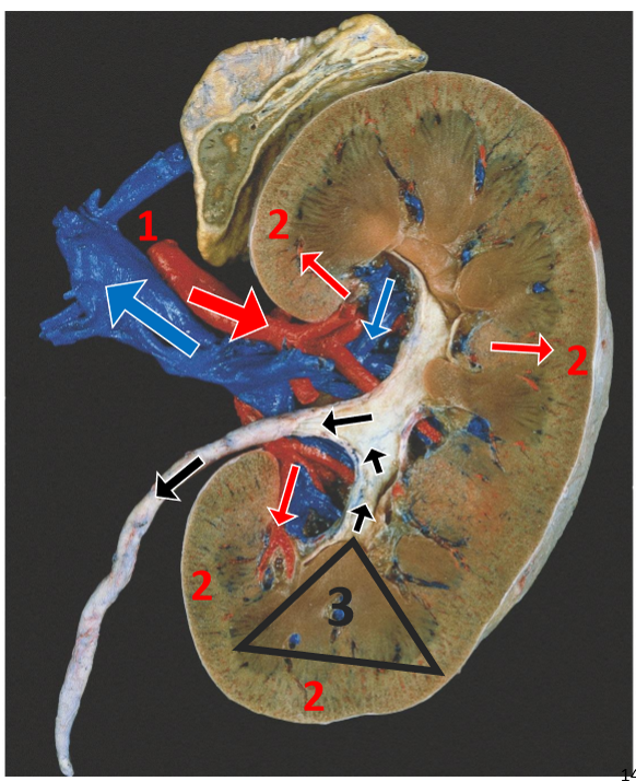
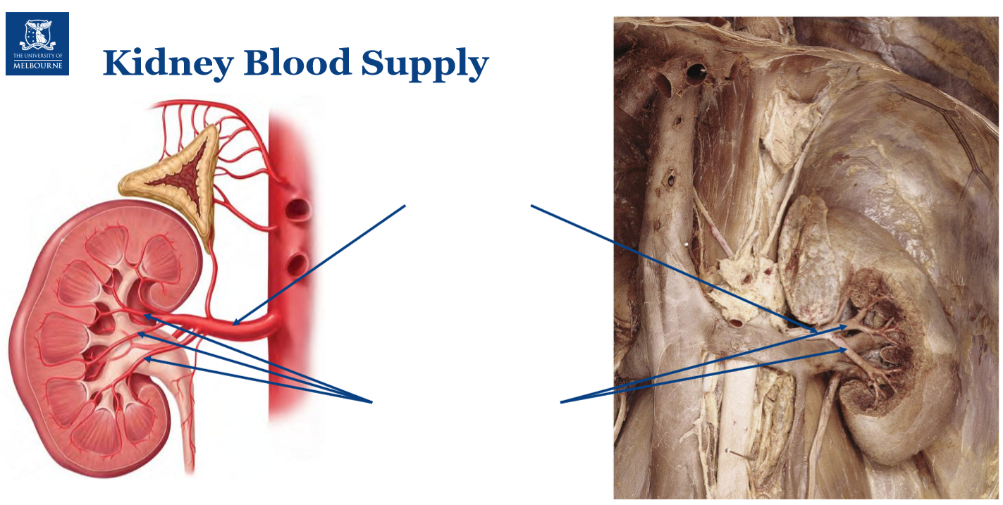
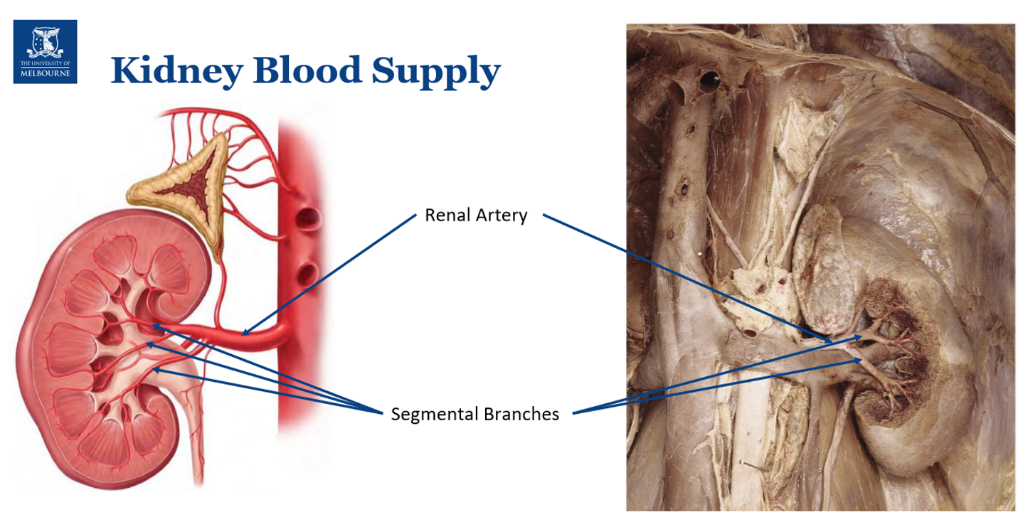
Describe the movement of blood into, throughout, and out of the kidneys
Blood enters via renal artery through hilum & progressively divides w/n kidney
Filtration occurs in glomeruli located in renal cortex
Filtrate passes through the nephron and collecting ducts w/n renal pyramids
Urine drains from minor calyx → major calyx → renal pelvis → ureter
Filtered blood returns to systemic circulation via renal vein
What is an implication of the kidneys’ location to the ribs?
Kidneys are extremely vascular; cracked rib in the back → kidney bleed (rapid blood loss)
Label the following diagram

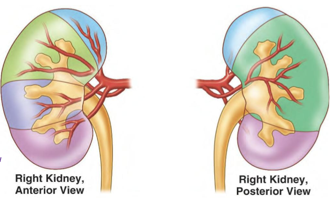
Order the following from most anterior → posterior:
renal artery
renal vein
renal pelvis
Anterior: renal vein
Middle: renal artery
Posterior: renal pelvis
Label the following image
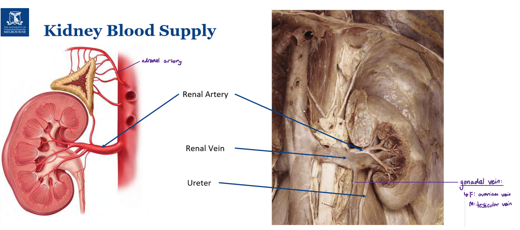
Name the gonadal veins in females vs males
Female: ovarian veins
Male: testicular veins
Where does the right gonadal vein drain into?
Inferior vena cava
Where does the left gonadal vein drain into?
left renal vein → across aorta → inferior vena cava
For the renal artery, state:
How many there are
What it branches off
How it supplies the kidneys & the relevance of this
2 renal arteries; 1 R & 1 L
Branches off abdominal aorta
Provides segmental branching of kidney; segments are surgically resectable
Artery ruptures → cannot be preserved (b/c they are too small) → ligate artery
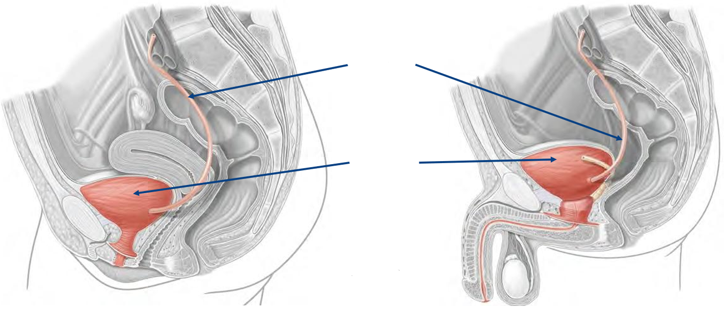
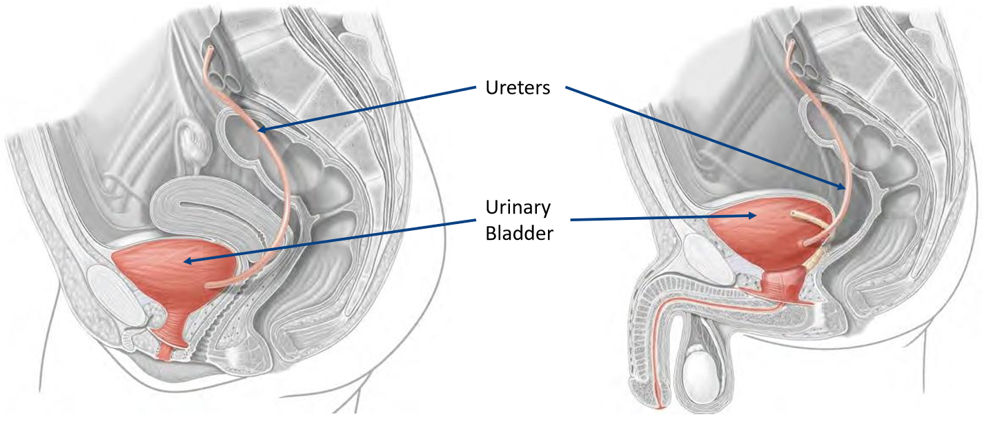
For the ureters, state:
What they are
What type of action they perform & what this facilitates
Intraperitoneal or retroperitoneal
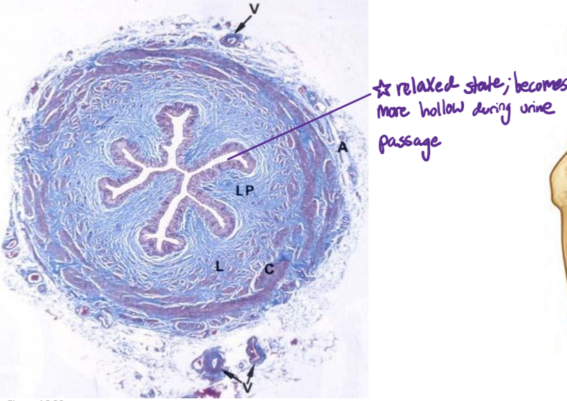
Structure during relaxed vs functioning state
Paired smooth muscle tubes
Perform peristaltic contractions → urine transport from kidney to urinary bladder
Retroperitoneal course
Relaxed state has folds, distends during urine passage
Label the following diagram
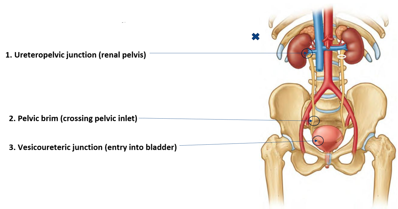
Name the 3 sites of constriction along the ureter & where they are found
Ureteropelvic junction: renal pelvis:
wide renal pelvis → narrow true ureter
‘loin-to-groin’ pain; common symptom of kidney stones
Pelvic brim: crossing pelvic inlet
Where the ureter bends as it crosses over the pelvic brim
Vesicoureteric junction: posterior entry into bladder

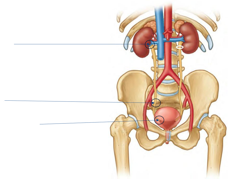
Label the following diagram of the constrictions along the ureters
What type of sphincter is the urinary bladder wall?
Functional
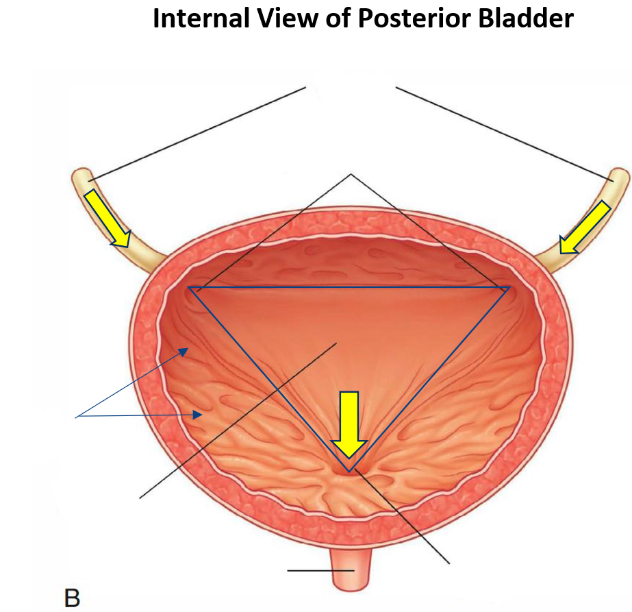
Label the following diagram of the urinary bladder

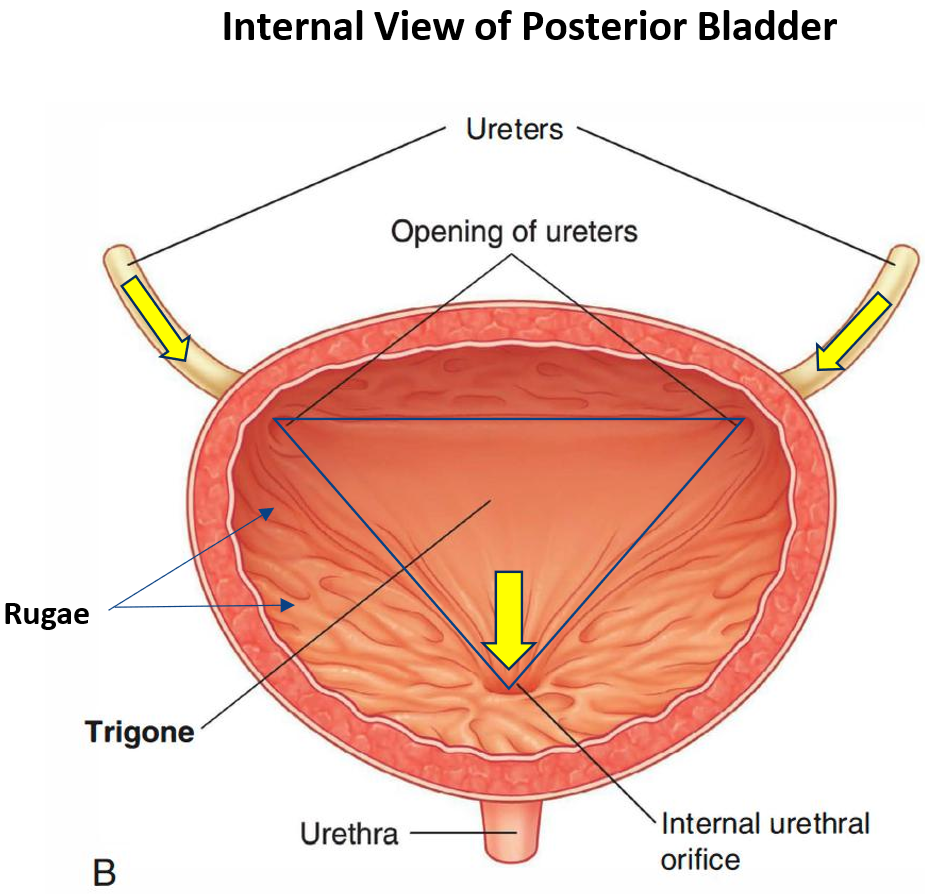
Where do the ureters enter the urinary bladder?
Ureteric opening on posterior superior surface

Describe the 3 key landmarks of the urinary bladder
Ureteric orifices: where ureters enter bladder
Trigone: smooth triangular region formed by
2 ureteric openings
internal urethral orifice
Taute; don’t want opening to be compromised as bladder fills
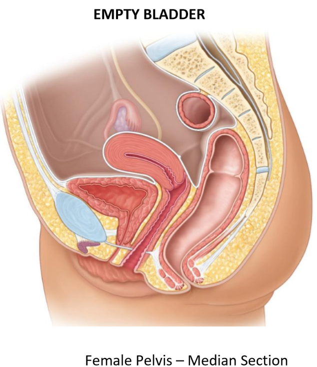
Rugae: mucosal folds when the bladder is empty
Allows bladder to fill & stretch

For the urinary bladder, state:
What type of organ it is
How it changes shape & why
Where it is located
Where it moves as it fills
Hollow (muscular) viscus
Highly distensible to store urine; changes shape from empty → expanded
Located anteriorly in pelvis
Moves into false pelvis area as it fills
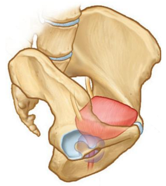
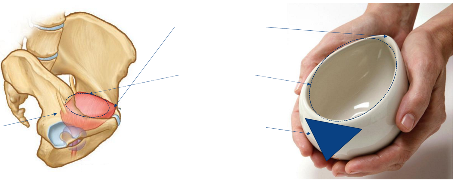
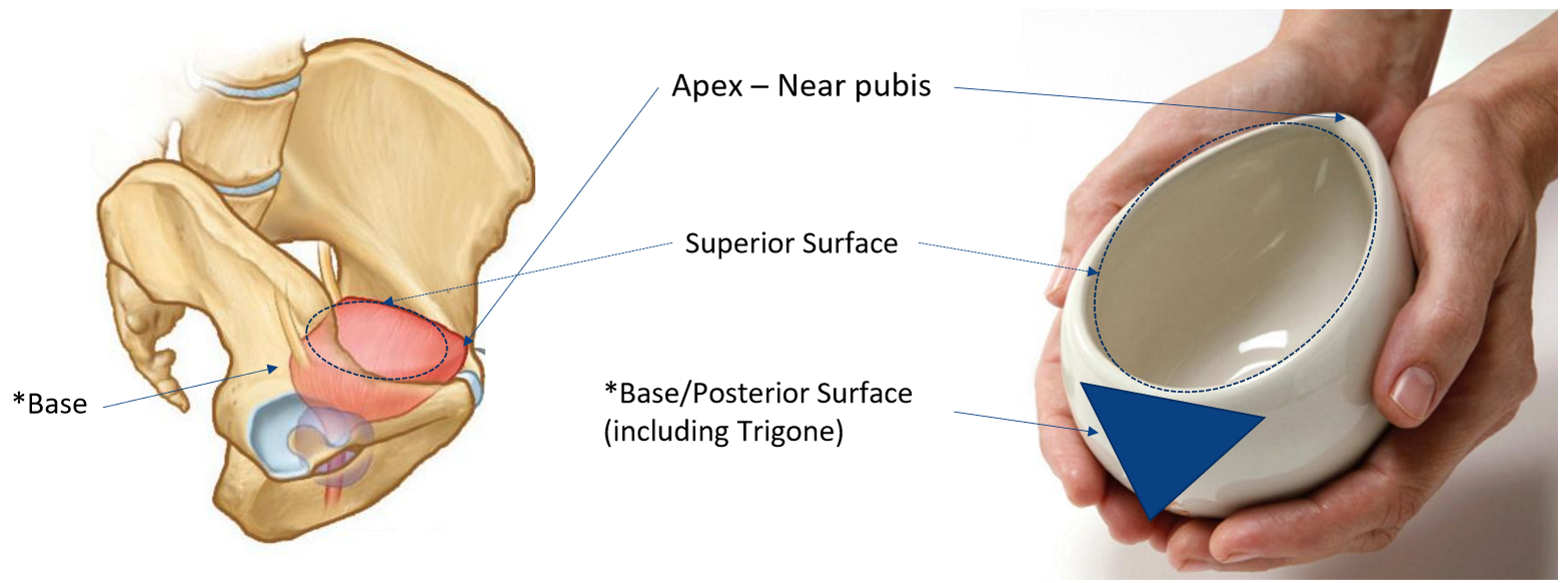
Label the following surfaces of the urinary bladder
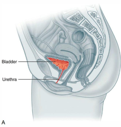
Describe the position and shape of the bladder when it is empty
Lies within pelvis, posterior to pubis
Internal surface with prominent rugae (folded mucosa)
Muscular wall contracted
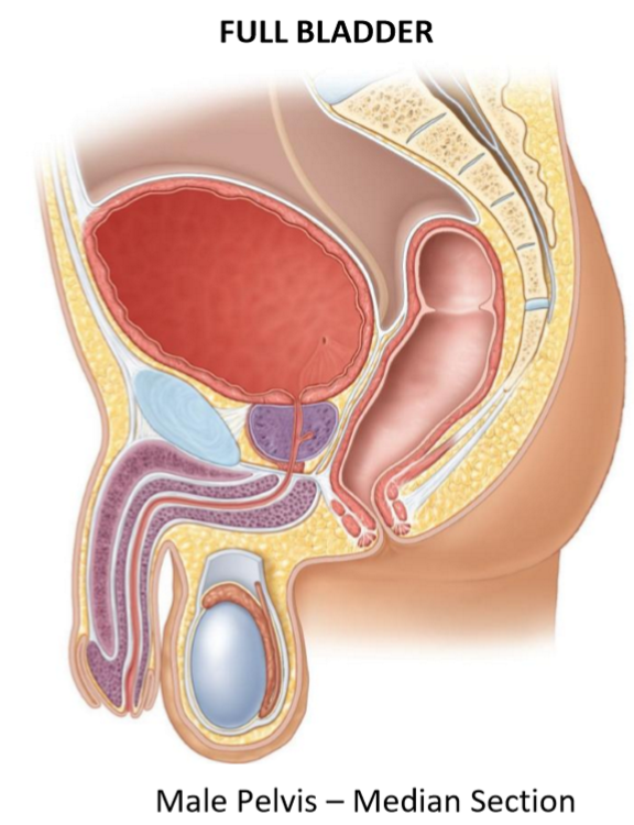
Describe the position and shape of the bladder when it is filling
Expands superiorly into abdominal cavity
Bladder neck remains relatively fixed
Rugae flatten as wall stretches
Shape becomes more rounded/expanded
Describe the process of urine outflow
Bladder stores urine until micturition
Bladder contracts → expel urine
Urine exits via internal urethral orifice
Transition from storage → excretion
Label the following image of the urinary bladder in its empty state
Label the following diagram
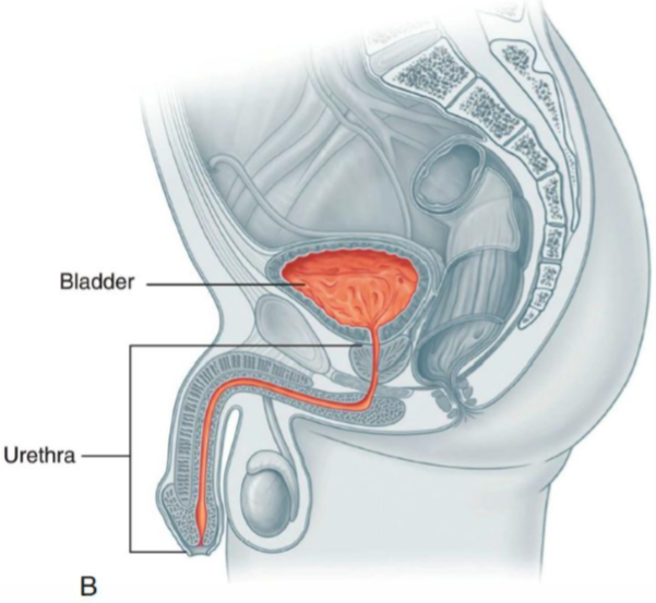
What is the urethra?
Muscular tube that carries urine from the bladder to the exterior
Terminal part of the urinary system
Where does the urethra begin?
at the internal urethral orifice of the bladder
How does the structure of the urethra differ in females to males, & what are 2 clinical implications of this?
Urethra is a lot shorter in females
Easier to get UTIs + for infections to get into bladder & spread to vag!na
Easier to insert catheters in females; no bending (which may be painful in men, also requires more skill)
Where is the external urethral orifice of the female urethra located?
Between labia minora
Anterior to vaginal opening
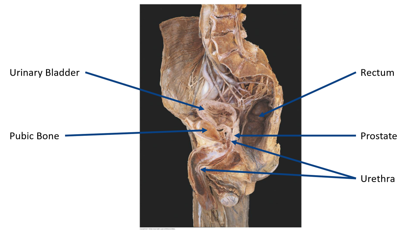
Where does the male urethra pass, & how is it distinctly different to the female urethra?
Passes through prostate & pen!s
Common pathway for urinary & reproductive systems (only urinary in females)
Name the 4 regions of the male urethra
Preprostatic (intramural) urethra
Prostatic urethra
Membranous urethra
Spongy (penile) urethra

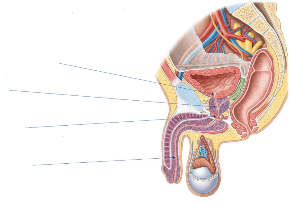
Label the following diagram of the male urethra

Where does the preprostatic (intramural) urethra pass?
Through the bladder wall

Describe the surrounding structures of the prostatic urethra, & the clinical relevance of this
Widest part of urethra
Surrounded by soft tissue, but this tissue is not stuck to the urethra
Prostatic enlargement may obstruct urine flow

Describe the structure of the membranous urethra & an implication of this
Surrounded by external urethral sphincter
Important for voluntary control of urination
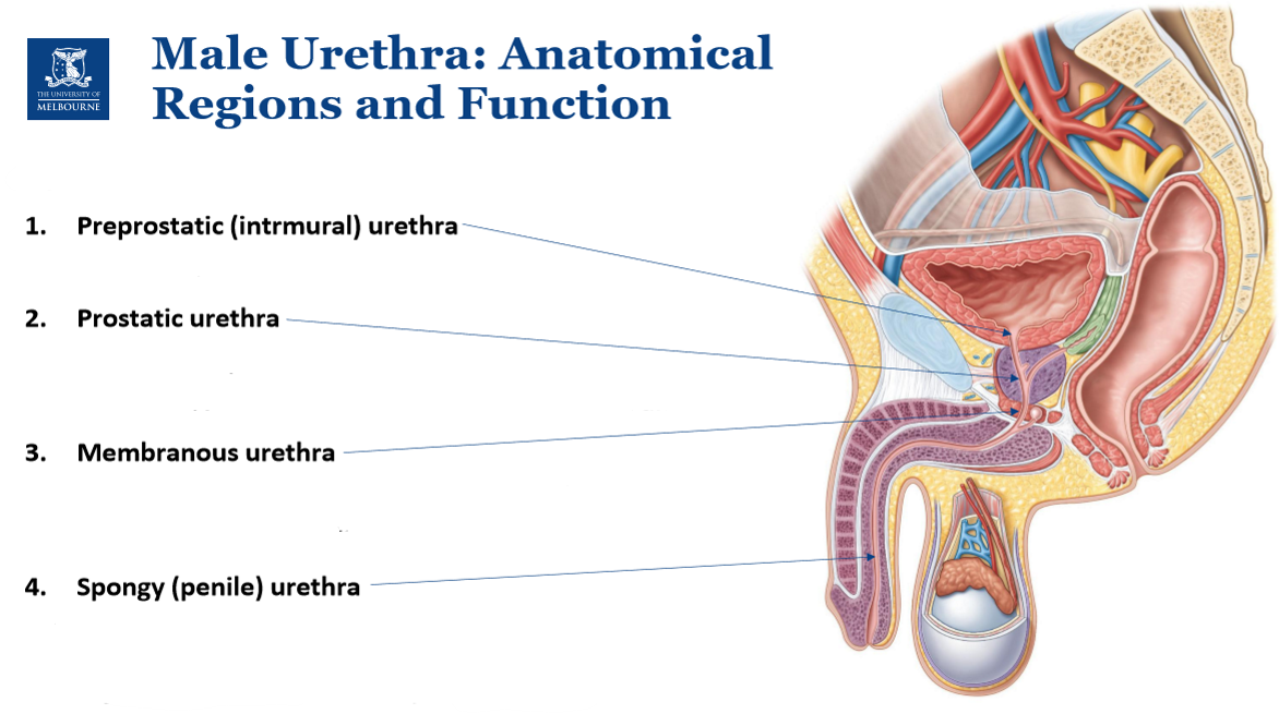
Describe the location, structure, & surrounding tissue of the spongy (penile) urethra
Located within corpus spongiosum
Structure: remains patent (open) for urination + ejaculat!on
Surrounding tissue prevents urethral collapse
