Brain Anatomy
1/23
There's no tags or description
Looks like no tags are added yet.
Name | Mastery | Learn | Test | Matching | Spaced | Call with Kai |
|---|
No analytics yet
Send a link to your students to track their progress
24 Terms
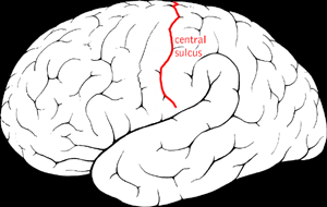
Central Sulcus
Location:
Lateral aspect of cerebral hemisphere
Description:
Groove on lateral surface of each cerebral hemisphere
Forms boundary between frontal and parietal lobes
Located between precentral and postcentral gyri
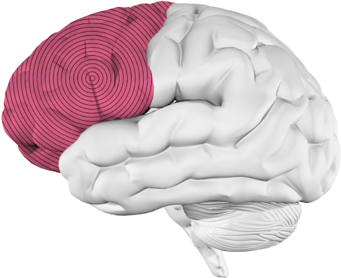
Frontal lobe
Location:
Anterior portion of cerebral hemisphere
Description:
Extends from anterior pole of brain to central sulcus
Contains precentral gyrus
Function:
Controls voluntary motor activity
Higher mental processing
Emotional behavior
Speech output (i.e., Broca's area - usually in left hemisphere)
Comment:
Named for overlying bone
Longitudinal Fissure
Location:
Midline, between cerebral hemispheres
Description:
Deep groove that separates right and left cerebral hemispheres
Comment:
Contains falx cerebri
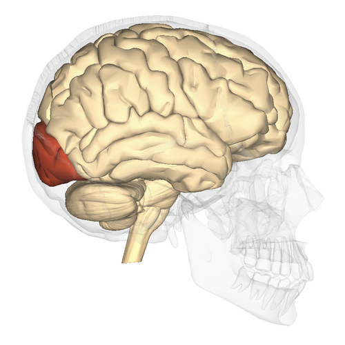
Occipital Lobe
Location:
Posterior portion of each cerebral hemisphere
Description:
Extends from parieto-occipital sulcus to posterior pole of brain
Contains lingual gyrus
Function:
Primary visual area
Comment:
Named for overlying bone
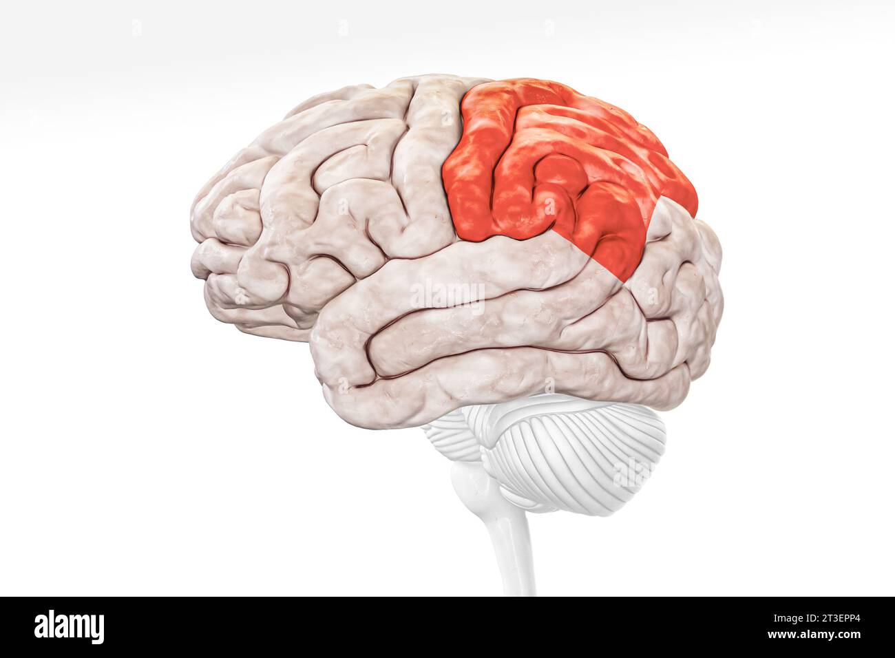
Parietal Lobe
Location:
Lateral surface of each cerebral hemisphere of brain
Description:
Extends from central sulcus (rostral) to parieto-occipital sulcus (caudal)
Includes postcentral gyrus
Function:
Reception of general sensory information from body
Tactile object recognition
Language, verbatim repetition of terms (i.e., Wernicke's area - usually in left hemisphere)
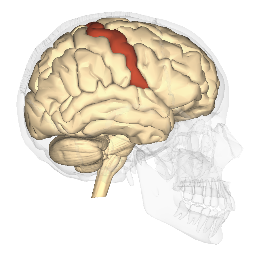
Postcentral Gyrus
Location:
Lateral aspect of each cerebral hemisphere
Description:
Distinct "fold" at anterior border of parietal lobe
Located along posterior edge of central sulcus
Function:
Receives somatosensory information from body
Comment:
Also called primary somatosensory cortex
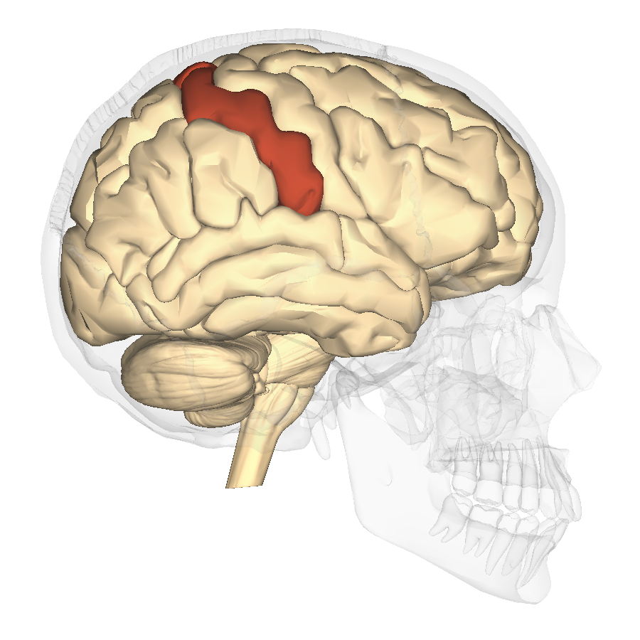
Precentral gyrus
Location:
Lateral aspect of each cerebral hemisphere
Description:
Distinct "fold" at posterior border of frontal lobe
Located along anterior edge of central sulcus
Function:
Controls voluntary movement
Comment:
Also called primary motor cortex
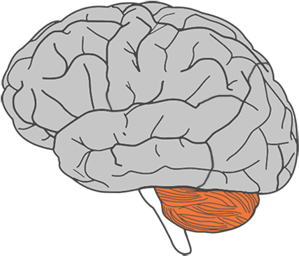
Cerebellum
Location:
Dorsal to brainstem
Description:
Composed of many lobes with highly folded cortex
Attached to pons via cerebellar peduncles
Function:
Coordinates complex movements
Monitors muscles to ensure fluid movements
Comment:
Receives extensive sensory input from body and CNS
Cerebellar cortex has folds known as folia
White matter of cerebellar lobes resembles branching tree and is called arbor vitae
Influences motor function through connections with thalamus and motor cortex
Cerebrum
Location:
Rostral portion of brain
Description:
Includes two large cerebral hemispheres separated by longitudinal fissure
Hemispheres connected by corpus callosum
Surface gray matter of each hemisphere is known as cerebral cortex
Within each hemisphere there is a core of white matter
Additional masses of gray matter located within cerebrum include basal nuclei
Comment:
Rostral = toward the nose (Latin: rostrum = beak)
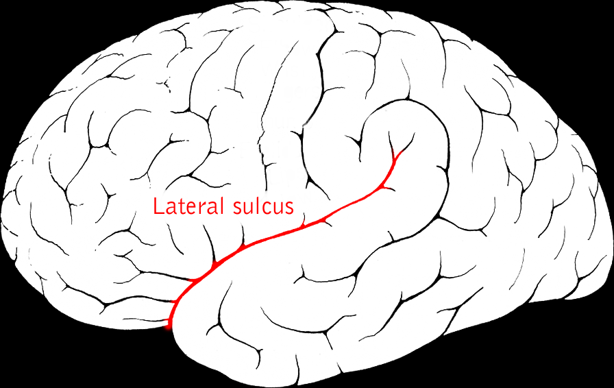
Lateral sulcus
Location:
Lateral aspect of each cerebral hemisphere
Description:
Deep groove separating temporal from frontal and parietal lobes
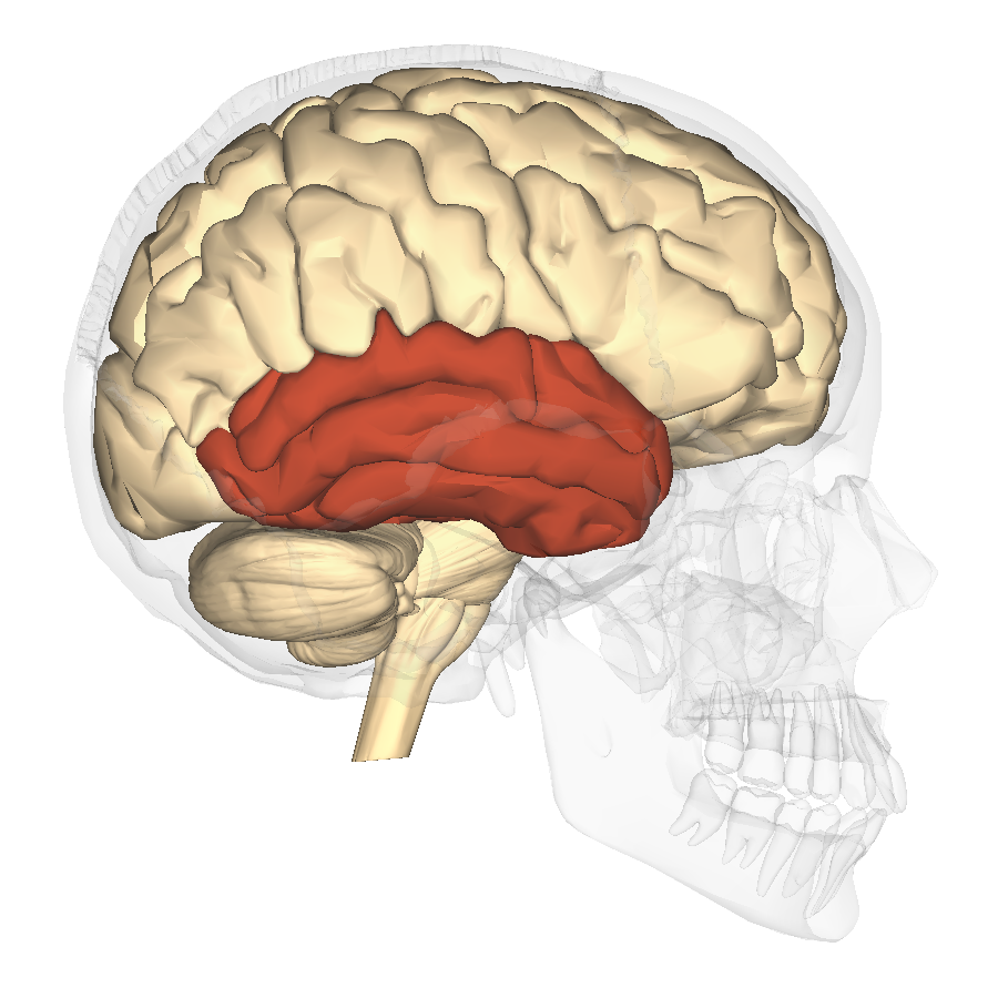
Temporal Lobe
Location:
Lateral and inferior portion of each cerebral hemisphere
Inferior to lateral sulcus
Description:
Lateral surface has three parallel gyri
Function:
Primary hearing and smell areas
Memory
Speech perception and recognition (i.e., Wernicke's area - usually in left hemisphere)
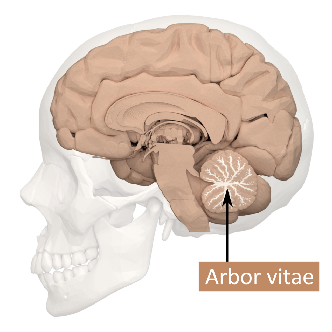
Arbor Vitae
Location:
Cerebellum
Description:
Composed of the white matter of cerebellar lobes
It's pattern resembles a branching tree
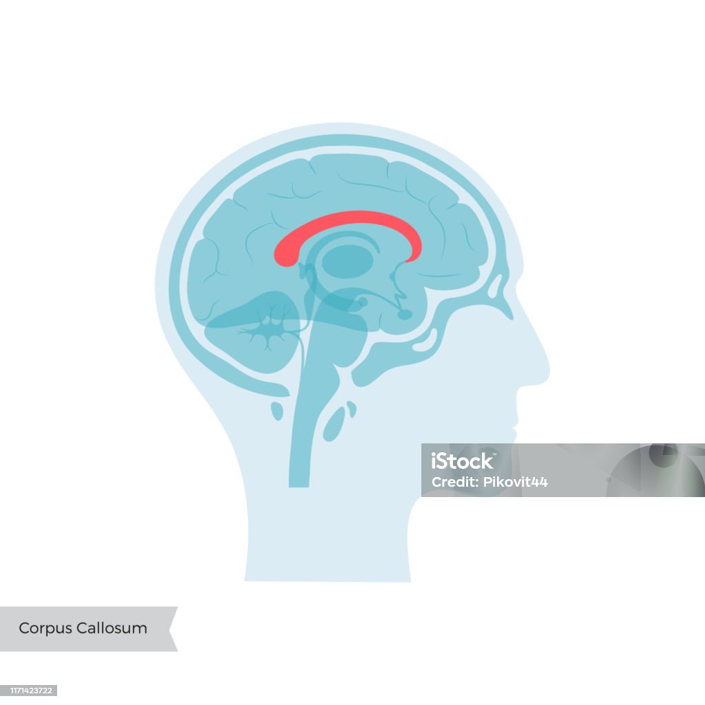
Corpus Callosum
Location:
Brain, between cerebral hemispheres
Description:
Large myelinated fiber tract connecting right and left cerebral hemispheres
Forms floor of longitudinal fissure
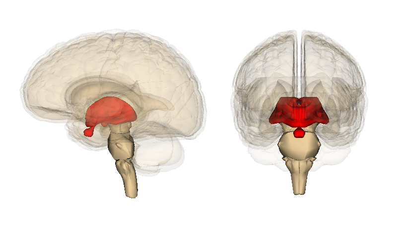
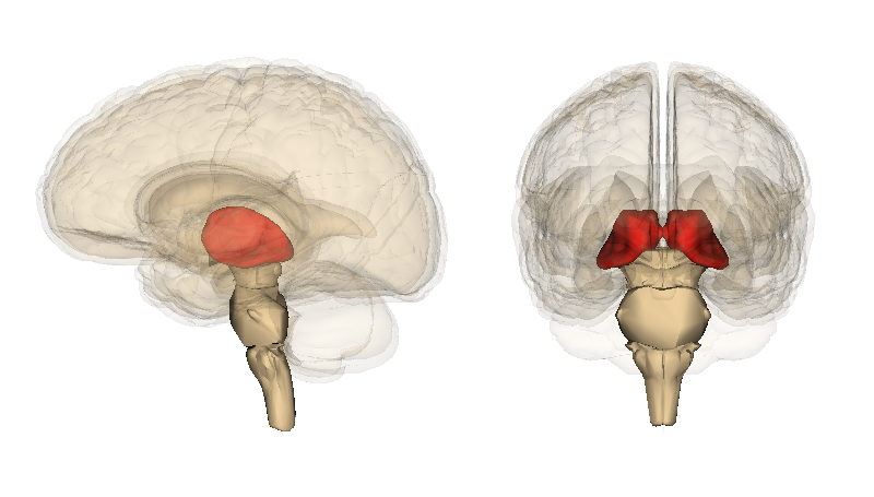
Diencephalon
Location:
Cerebrum
Description:
Composed of thalamus, hypothalamus, and epithalamus
Function:
Thalamic nuclei relay sensory information to cerebral cortex
Hypothalamic nuclei maintain homeostasis
Epithalamus includes pineal gland (produces melatonin)
Hypothalamus
Location:
Ventral diencephalon
Description:
Collection of nuclei located inferior to thalamus
Includes infundibulum and mammillary bodies
Function:
Considered master control center for endocrine system
Secretes releasing and inhibiting hormones that control anterior pituitary gland
Produces hormones that are transported to and stored in posterior pituitary gland
Controls autonomic nervous system
Regulates body temperature, food, and water intake
Regulates emotional behavior
Maintains sleep/wake cycle
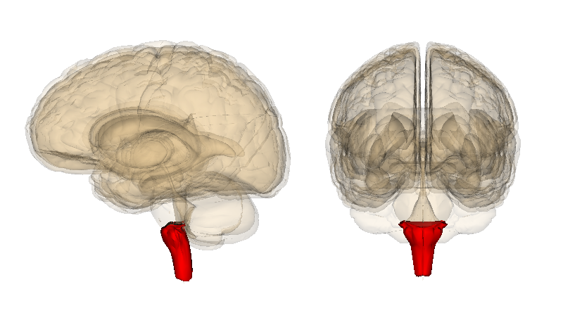
Medulla Oblongata
Location:
Most caudal portion of brain
Description:
Extends from pons to spinal cord
Associated with cranial nerves IX, X, XI, and XII
Function:
Contains respiratory, cardiac, and vasomotor centers

Midbrain
Location:
Brainstem
Between diencephalon and pons
Description:
Composed of white matter tracts and gray matter nuclei
Associated with cranial nerves III and IV
Prominent features include superior and inferior colliculi, cerebral peduncles, substantia nigra, and cerebral aqueduct
Function:
Coordinates movements in response to visual and auditory stimuli
Conveys motor information from cerebral cortex to pons
Conveys sensory information from spinal cord to thalamus
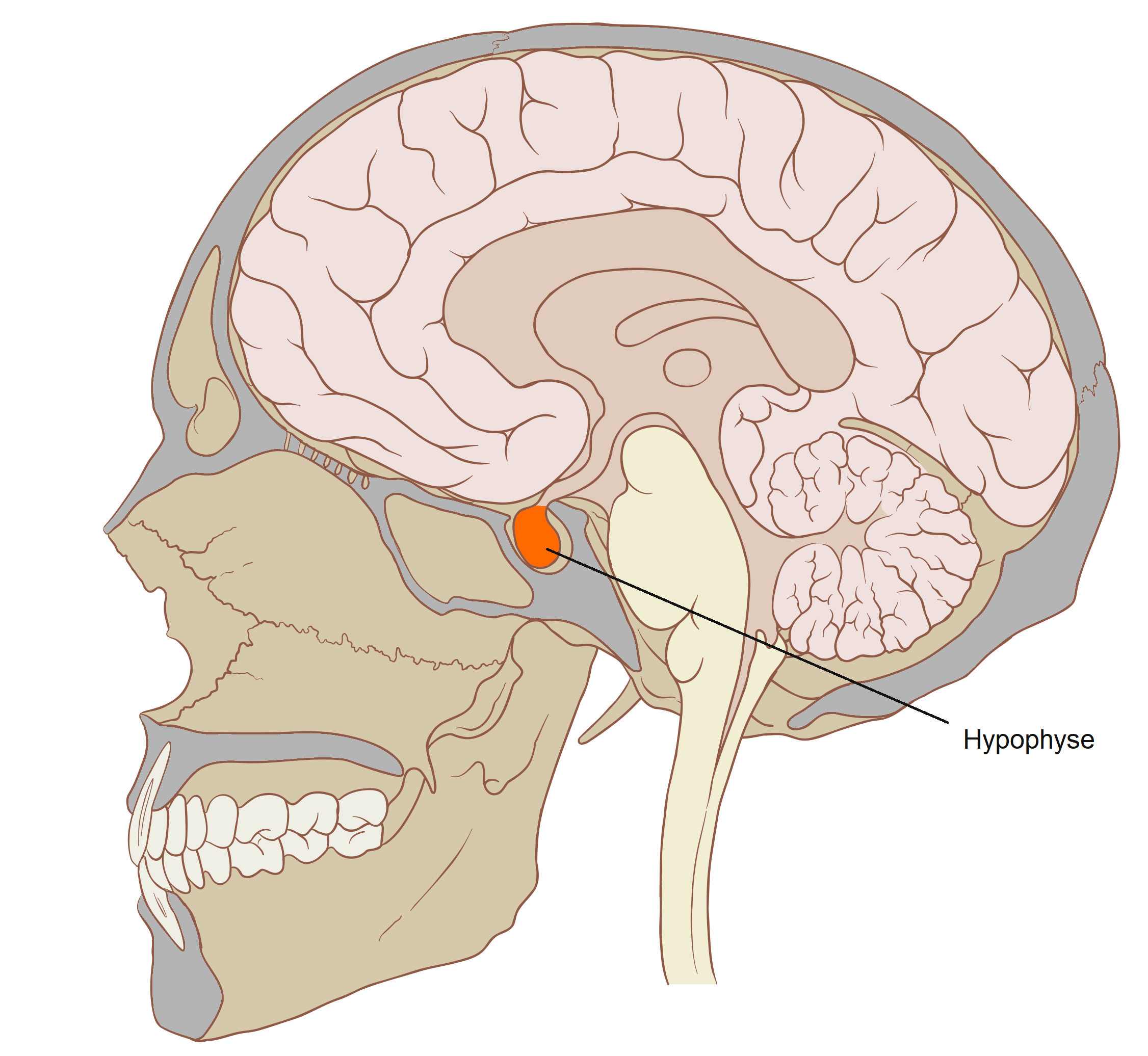
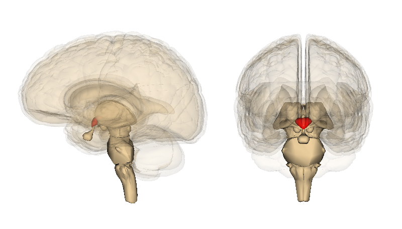
Pituitary gland
Location:
Midline of middle cranial fossa
Rests in hypophysial fossa of sphenoid bone
Description:
Small, oval bilobed endocrine gland
Two functional lobes: anterior (adenohypophysis) and posterior (neurohypophysis)
Connected by infundibulum to hypothalamus
Function:
Anterior pituitary produces the following hormones: thyroid-stimulating (TSH), prolactin (PRL), adrenocorticotropic (ACTH), growth (GH), luteinizing (LH), melanocyte-stimulating (MSH), and follicle-stimulating (FSH)
Posterior pituitary stores and releases: antidiuretic hormone (ADH) and oxytocin (OT)
Also known as:
Hypophysial gland or hypophysis
Comment:
Posterior pituitary does not produce any hormones; ADH and OT produced in hypothalamus
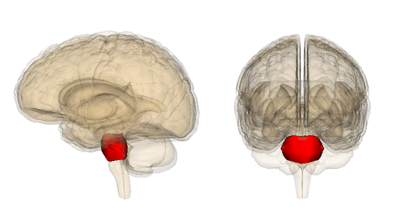
Pons
Location:
Ventral aspect of brainstem
Between midbrain (rostral) and medulla oblongata (caudal)
Description:
Characterized by distinct ventral "bulge"
Attached to cerebellum by middle cerebral peduncle
Associated with cranial nerves V, VI, VII, and VIII
Function:
Involved in control of sleep and respiration
Transfer of information to and between cerebellar hemispheres
Comment:
Latin: pons = bridge
Thalamus
Location:
Diencephalon
Description:
Paired groups of nuclei separated by third ventricle
Largest portion of the diencephalon
Composed primarily of gray matter
Function:
Primarily for relay of sensory information to cortex
Relay of motor information for movement planning
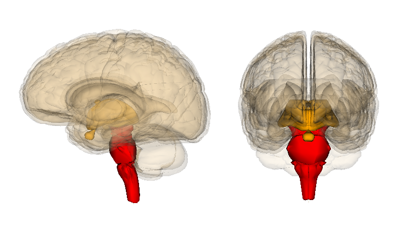
Brainstem
Location:
Caudal portion of brain
Description:
Vertical, stalk-like portion of brain
Includes midbrain, pons, and medulla oblongata
Left cerebral hemisphere
Location:
Cerebrum
Description:
Two hemispheres separated by longitudinal fissure
Composed of gray and white matter
Highly folded surface (sulci and gyri)
Four lobes visible on surface of each hemisphere: frontal, parietal, temporal, and occipital
Insular cortex (sometimes considered a fifth lobe) located in floor of lateral sulcus
Comment:
Surface gray matter is called cerebral cortex
Hemispheres connected by corpus callosum
Right crerbral hemisphere
Location:
Cerebrum
Description:
Two hemispheres separated by longitudinal fissure
Composed of gray and white matter
Highly folded surface (sulci and gyri)
Four lobes visible on surface of each hemisphere: frontal, parietal, temporal, and occipital
Insular cortex (sometimes considered a fifth lobe) located in floor of lateral sulcus
Comment:
Surface gray matter is called cerebral cortex
Hemispheres connected by corpus callosum
Parieto-occipital sulcus
Location:
Medial aspect of each cerebral hemisphere of brain
Description:
Groove that separates parietal and occipital lobes
Most obvious on medial aspect of cerebral hemisphere