Physiology Exam 3
 Call Kai
 Learn
 Practice Test
 Spaced Repetition
 Match
 Flashcards
 Knowt Play
1/69

There's no tags or description
Looks like no tags are added yet.
70 Terms
What does tetrodotoxin (TTX) do? Where does TTX come from?
TTX blocks voltage-gated Na⁺ channels, preventing action potentials and nerve signals from traveling.
It is found in animals such as pufferfish and is produced by certain bacteria associated with these animals.
What is myelin and what does myelination do for a neuron?
Myelin is a fatty insulating layer around axons. Myelination speeds up nerve impulse transmission.
What is the myelin coat and what is is made of?
The myelin coat covers most of the motor axon’s plasma membrane surface area
It is formed by shwann cells
What are nodes of Ranvier and what ion channels are found there?
They are gaps between adjacent shwann cells. Channels found in them are Voltage-gated sodium (Na⁺) and potassium (K⁺) channels.
What is saltatory conduction?
The nerve impulse/AP jumps from node to node, speeding up transmission.
What is a synapse? Are there different kinds of synapses? How do they differ?
A synapse is the gap between a neuron cell and any other cell that come very close to it to transmit an impulse.
Types:
Chemical Synapse: A synapse where a chemical neurotransmitter is secreted by the presynaptic cell.
Electrical Synapse: A synapse in which adjacent cells directly communicate through gap junctions.
Where would you find a gap junction?
The interconnecting point of two cells
What are connexins and connexons? What do these structures do?
Connexins are proteins within the plasma membranes of interconnected cells
Six connexins (the individual proteins) form one connexon.
Two connexons form one gap junction that interconnects two different cells.
What is a synaptic cleft? What happens here?
The extracellular space between the presynaptic and postsynaptic cells
Neurotransmitters travel between them to send a message
Hiarchy of skeletal muscle
Myofibrils are in myofibers, myofibers bunch up into muscle fasicles, and muscle fasicles bunch up into skeletal muscles.
What do you call the plasma membrane of a skeletal muscle cell? What about the endoplasmic reticulum?
Plasma membrane: Sarcolemma
Endoplasmic reticulum: Sarcoplasmic reticulum
Do you understand the different parts of the muscle? What is the hierarchy?
Muscle hierarchy (largest to smallest):
Muscle → Fascicle → Muscle fiber (cell) → Myofibril → Sarcomere → Myofilaments (actin and myosin)
Skeletal muscle fibers are encased in extracellular material that provides a covering, protection, and support. What is this covering?
Endomysium (connective tissue)
What supports the fascicles? The skeletal muscle?
Fascicles: Perimysium
Whole skeletal muscle: Epimysium
Each skeletal muscle fiber in an adult human is typically innervated by how many presynaptic terminals of a somatic motor neuron?
One presynaptic terminal (one neuromuscular junction).
Why study the neuromuscular junction (NMJ)? What are some unique features? What neurotransmitter is released at the NMJ?
It helps us understand how nerves communicate with muscles to produce movement.
Unique features: A specialized synapse with a motor neuron, synaptic cleft, and motor end plate.
Neurotransmitter released: Acetylcholine (ACh).
At the neuromuscular junction, the presynaptic terminal secretes an excess amount of neurotransmitter. How much excess is released, and why would this be necessary?
About 3–4 times more acetylcholine is secreted.
This ensures reliable muscle activation even if some acetylcholine (ACh) is lost or receptors are unavailable.
Once a neurotransmitter like acetylcholine is released, what happens to it? What special enzyme is present in the synaptic cleft at the neuromuscular junction?
It binds to receptors, then is broken down to stop the signal.
Special enzyme: Acetylcholinesterase (AChE).

Fill in the blanks (neuromuscular junction) Also list the steps of how this all works.
Ca2+
Na+
Na+
Gated channel in a T-tubule
Ca2+
The top-right box is the AP
The gap is the synaptic cleft
The top NT is the presynaptic cell
The bottom NT is the postsynaptic cell
Steps
Nerve impulse arrives — An action potential reaches the end of the motor neuron
Calcium ions (Ca²⁺) enter the neuron’s axon terminal.
Acetylcholine is released into the synaptic cleft
ACh attaches to receptors on the muscle cell membrane, causing sodium (Na⁺) to enter and triggering a muscle action potential.
The action potential travels along the muscle membrane and into the T-tubules, causing the sarcoplasmic reticulum to release calcium. Calcium allows the muscle to contract.
Acetylcholinesterase breaks down ACh, stopping the signal. Calcium is pumped back into the sarcoplasmic reticulum, allowing the muscle to relax.
What are myofibrils? What filaments are found in myofibrils?
They are Rod-like structures inside muscle fibers that are responsible for muscle contraction
The filaments found are thick myosin filaments and thin actin filaments
What are thick filaments made of? What are thin filaments made of?
Thick filaments: Made of myosin proteins.
Thin filaments: Made mainly of actin proteins, plus troponin and tropomyosin.
What is a sarcomere?
The basic contractile unit (the smallest part of a muscle that can shorten to produce contraction) of a muscle, extending from one Z disc to the next
What are the two binding sites on the head of myosin II molecules? What binds here?
The two binding sites are
Actin-binding site: Binds to actin.
ATP-binding site: Binds ATP for energy during muscle contraction.
What are Z disks?
Structures that mark the boundaries of each sarcomere and anchor the thin (actin) filaments.
What are I bands or A bands? What is an M line?
I band: Light band containing only thin (actin) filaments.
A band: Dark band covering the full length of thick (myosin) filaments, including overlap with actin.
M line: Center of the sarcomere where thick (myosin) filaments are anchored

Fill in the blanks
A. Z line
B. I Band
C. M Line
D. A Band
E. H Zone
F. Sarcomere
G. Z-Line
H. Myofibril
I. Z-line
What shortens during contraction? Do the filaments shorten?
The sarcomere shortens during contraction. The actin and myosin filaments do not shorten; they slide past each other.
What happens after ATP binds to a myosin head? How does it get in a “cocked” position? What is the power stroke?
ATP binds: Myosin releases actin.
Cocked position: ATP is hydrolyzed into ADP + Pi, providing energy to cock the myosin head.
Power stroke: Myosin releases Pi and pulls actin toward the center of the sarcomere.

What are the steps of the cross-bridge cycle? What is happening, and why?
Resting fiber: cross-bridge is not attached to actin
Cross-bridge binds to actin
P is released from the myosin head, causing a conformational change in myosin
Power stroke causes filaments to slide. ADP is released
A new ATP binds to the myosin head, allowing it to release from actin
ATP is hydrolyzed, and phosphate binds to myosin, causing the cross-bridge to return to its original orientation
Why? The cycle uses ATP energy to slide actin past myosin, shortening the sarcomere and contracting the muscle.
Why is calcium so important for contraction? What is troponin and tropomyosin?
Calcium (Ca²⁺): Binds to troponin, moving tropomyosin and exposing actin’s binding sites so myosin can attach.
Troponin: Protein that binds calcium and changes shape.
Tropomyosin: Protein that blocks myosin-binding sites on actin when the muscle is relaxed
Troponin is a complex of Troponin I, C, and T. What role does each one play?
Troponin I: Inhibits actin-myosin binding.
Troponin C: Binds calcium (Ca²⁺).
Troponin T: Binds to tropomyosin, anchoring the troponin complex.
Why are t-tubules so important in the cross-bridge cycle? What is a triad?
T-tubules: Carry action potentials deep into the muscle fiber, triggering calcium release so the cross-bridge cycle can begin throughout the muscle.
Triad: One T-tubule surrounded by two terminal cisternae of the sarcoplasmic reticulum.
RYR1 and Cav1.1 have important roles to play during excitation-contraction coupling.
What are these channels?
Cav1.1: Voltage-sensitive calcium channel in the T-tubule that detects the action potential.
RYR1: Calcium-release channel in the sarcoplasmic reticulum that releases Ca²⁺ into the muscle cell.
Where does most of the calcium that plays a role in contraction come from?
The sarcoplasmic reticulum (SR) stores and releases most of the calcium (Ca²⁺) needed for skeletal muscle contraction.
When is ATP used? Is it only needed during contraction? What about relaxation?
ATP is needed for both contraction and relaxation.
Contraction: ATP detaches myosin from actin and re-energizes the myosin head.
Relaxation: ATP powers pumps that move Ca²⁺ back into the sarcoplasmic reticulum, allowing the muscle to relax.
What is SERCA and is it important?
SERCA: An ATP-powered pump that moves Ca²⁺ back into the sarcoplasmic reticulum.
Importance: Yes! It lowers calcium levels in the cytoplasm, allowing the muscle to relax.
What do the tetanus toxin and botulinum toxin disrupt? What do they cause?
Disrupt the NMJ
TT: Spastic paralysis (motor neurons continue to send signals = spasms/rigidity)
TB: Flaccid paralysis (weak or paralyzed muscles)
What types molecules can be neurotransmitters? Just know the classes.
Small molecules, amines, amino acids, peptides, and lipids.
What type of neurotransmitters are acetylcholine, ATP, and GABA?
Acetylcholine (ACh): Excitatory neurotransmitter at the neuromuscular junction (Small molecule)
ATP: Purinergic neurotransmitter (Small molecule)
GABA: Inhibitory neurotransmitter (Amino acid)
Are neurotransmitters intrinsically inhibitory or excitatory or are they just chemicals?
Neurotransmitters are chemicals; their effect depends on the receptor they bind to. They can be excitatory or inhibitory depending on the receptor and target cell.
What determines if a neurotransmitter will lead to inhibition or excitation in the postsynaptic cell?
The type of receptor and ion channel on the postsynaptic cell determines whether a neurotransmitter causes excitation or inhibition.
What is an ionotropic receptor? A good example is the acetylcholine receptors in the neuromuscular junction
A receptor that opens an ion channel when a neurotransmitter binds to it, allowing ions to cross the cell membrane.
What is a metabotropic receptor? A good example of this is the receptor in type II taste cells.
A receptor that activates a G protein and triggers a signaling pathway inside the cell.
We discussed different kinds of synapses, such as excitatory cholinergic synapses and inhibitory GABAergic synapses. What is happening at each that makes it an excitatory synapse or an inhibitory synapse?
Excitatory (cholinergic): Depolarizes the postsynaptic cell, making it more likely to fire an action potential.
Inhibitory (GABAergic): Makes the postsynaptic cell less likely to fire an action potential, usually by hyperpolarizing it.
What is a central synapse? Is it the same as the NMJ? What is a bouton?
CS: A synapse between neurons in the central nervous system (brain or spinal cord).
No. A central synapse connects neurons; the NMJ connects a motor neuron to a skeletal muscle fiber.
A bouton is a small swelling at the end of an axon that releases neurotransmitters into a synapse.
What is an EPSP? What about an IPSPs?
Excitatory postsynaptic potential — makes the postsynaptic neuron more likely to fire an action potential (depolarization).
Inhibitory postsynaptic potential — makes the postsynaptic neuron less likely to fire an action potential (usually hyperpolarization).
What happens to released neurotransmitters?
They are removed from the synaptic cleft by:
Reuptake: Taken back up by the presynaptic neuron or glial cells.
Enzymatic breakdown: Broken down by enzymes.
Diffusion: Move away from the synaptic cleft.
Terminology differs from the central nervous system (CNS) and the peripheral nervous system (PNS).
What is it called when there is a grouping of neuron cell bodies in the
CNS? What about the PNS?
What is a bundle of axons in the CNS? What about the PNS?
CNS: Neuron
PNS:Ganglion
CNS2:Tract
PNS2:Nerve
What is the central nervous system composed of? How is the peripheral nervous system different? (What are efferent neurons? Is that different from afferent? Where are interneurons?)
CNS: The brain and spinal cord.
PNS: All nerves and ganglia outside the brain and spinal cord.
Efferent: Carry signals away from the CNS to muscles and glands (motor)
Afferent: Carry sensory signals toward the CNS.
Interneurons: In the CNS, where they connect and process signals between neurons
How many neurons are in the nervous system? What are neuroglia cells?
About 86 billion neurons in the brain alone.
Support cells that protect, nourish, and insulate neurons and maintain their environment.
What types of neuroglia cells are in the central nervous system(4)? What about the PNS (2)?
CNS: Astrocytes, oligodendrocytes, microglia, and ependymal cells.
PNS: Schwann cells and satellite cells.
Do you understand the difference between the central nervous system and the peripheral nervous system?
CNS: Brain and spinal cord; processes information.
PNS: Nerves and ganglia outside the CNS; carries signals to and from the CNS.
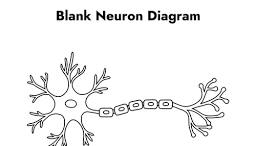
Do you remember the different parts of the neuron from earlier in the semester?
(dendrites vs. soma vs. nerve fiber vs axon hillock)

How do multipolar neurons differ from bipolar or unipolar neurons?
Multipolar: One axon, many dendrites; most common type.
Bipolar: One axon and one dendrite.
Unipolar: One process extending from the cell body that branches into two.
Can you give an example of where you find bipolar neurons? What about anaxonic neurons?
BP: The retina of the eye and olfactory epithelium (smell).
ANX: The brain and retina; they lack a distinct axon.
What is gray matter? What about white matter? Why the different colors?
Gray: Mostly neuron cell bodies, dendrites, and synapses.
White: Mostly myelinated axons that carry signals.
Myelin is fatty and white, making white matter lighter. Gray matter has less myelin, so it appears gray.
What does the nervous system develop from in the embryo (which tissue layer)?
The ectoderm (outer germ layer).
Very early on in development a neural plate forms and then a neural tube. Do you understand how the tube forms and then changes as we develop (general understanding not anything detailed)? Do NOT memorize the terms (Prosencephalon, Mesencephalon, Rhombencephalon, and etc.)
The neural plate folds inward and closes to form the neural tube. The tube develops into the CNS:
Anterior (front) end: Brain
Posterior (back) end: Spinal cord
What are neural crest cells?
Cells that separate from the developing neural tube and form many structures of the PNS, including sensory neurons and Schwann cells.
How much blood flow goes to the brain? Why would there be such a need for so much
blood flow?
About 15% of the body's blood flow.
To supply oxygen and glucose for energy and remove waste, since the brain has a high metabolic demand.
The brain is protected by the skull, membranes, and fluid. What are these membranes?What is the fluid? What is the benefit to having our brain surrounded by fluid?
Membrane: The meninges: dura mater, arachnoid mater, and pia mater.
Fluid: Cerebrospinal fluid (CSF).
CSF cushions the brain, absorbs shock, and provides buoyancy to reduce pressure on the brain.
The gap between the dura and bone is the _______________ space
Epidural space
What is the choroid plexus? What does it do?
A network of blood vessels in the brain's ventricles that produces cerebrospinal fluid (CSF)
What is the cerebrum? What is the outer layer called? Is it gray or white?
Cerebrum: The largest part of the brain; responsible for thought, memory, sensory processing, and voluntary movement
Outer layer: The cerebral cortex
Gray matter
What are the different lobes of the cerebrum? Do you remember some of the functions?
Frontal lobe: Planning, decision-making, and voluntary movement.
Parietal lobe: Processes touch and other sensory information.
Temporal lobe: Hearing (auditory experiences), memory, and language.
Occipital lobe: Vision.
What lobe deals with auditory experiences and storage? What lobe plays a role in
voluntary motor control of skeletal muscles?
Temporal Lobe.
Frontal Lobe.
What is the primary motor cortex? Why were the body parts drawn at different scales (why were some images bigger than others)?
PMC: The area in the frontal lobe that controls voluntary skeletal muscle movements.
Size represents how much brain area controls that body part. Body parts requiring precise movements (like hands and lips) have larger areas.
What is the motor circuit? Why would GABA play a role here? GABA is usually used to
calm down neurons and make them less excitable.
A network of brain areas that work together to plan, control, and coordinate movement.
GABA inhibits neurons, helping regulate movement and prevent unwanted or excessive muscle movements.
What is lateralization? Which hemisphere deals with speech and writing? What about
spatial concepts?
Lateralization: When certain brain functions are primarily controlled by one hemisphere.
The left hemisphere is speech and writing.
Right hemisphere is spatial concepts.
What is the corpus callosum? Why would anyone want a split brain?
A thick bundle of nerve fibers connecting the brain's left and right hemispheres, allowing them to communicate.
To reduce severe epilepsy by cutting the corpus callosum, preventing seizures from spreading between hemispheres.