oral anatomy permanent teeth chapter 16
 Call Kai
 Learn
 Practice Test
 Spaced Repetition
 Match
 Flashcards
 Knowt Play
1/103

There's no tags or description
Looks like no tags are added yet.
104 Terms
•What is the primary mineral in enamel, dentin, and cementum?
• In which of these hard tissues contains the highest concentration of this mineral?
primary mineral calcium hydroxiaptie
enamel has highest concentration
•Enamel is a crystalline structural unit, and is made up of millions of these what?
enamel rods
•What does the neonatal line of the enamel and dentin represent?
at birth defines tissue created before and after birth
one in dentin (line of owen)
secondary dentin
forms after the apical foramen (continues throughout life of the tooth)
teriary dentin
response to truama
formative cells of dentin
odontoblasts
•What structure within dentin allows nerve sensation to travel to the pulp?
dentinal tubules (hydodynamic theory
When the pulp is injured by a cavity preparation, extensive caries or traumatic injury, it may undergo inflammation. This pulpal inflammation is termed _________
pulpitis
reversible and irreversible
•What is the term to describe the layered formation (process) of cementum?
cementogenesis
made by cementoblasts
necrotic pulp they would feel what?
nothing the pulp has died
hypercementosis
roots growing in abnormal places
hard tissue loss of the enamel by erosion is caused by
acidic food
GERD
eating disorders
•All permanent anterior teeth are
succedaneous replace baby teeth
•All anterior teeth are composed of four developmental lobes
•3 labial lobes: mesiolabial, middle labial, distolabial
•1 lingual lobe
•All anterior teeth have two vertical labial depressions that separate the labial developmental lobes
mesiolabial and distolabial developmental depressions
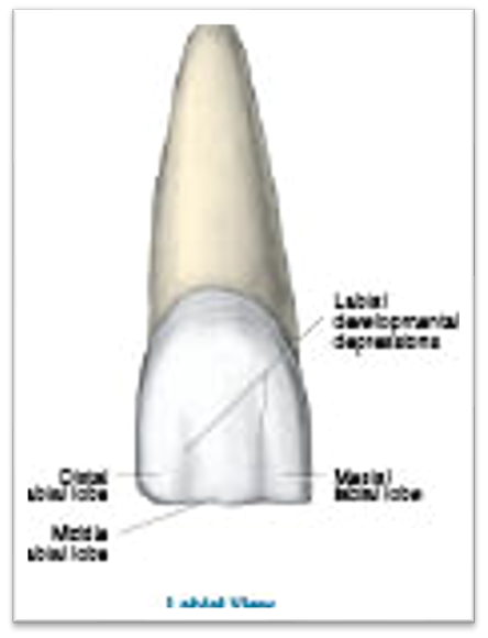
lobes of the anterior perm teeth
mesiolabial and distolabial developmental depressions
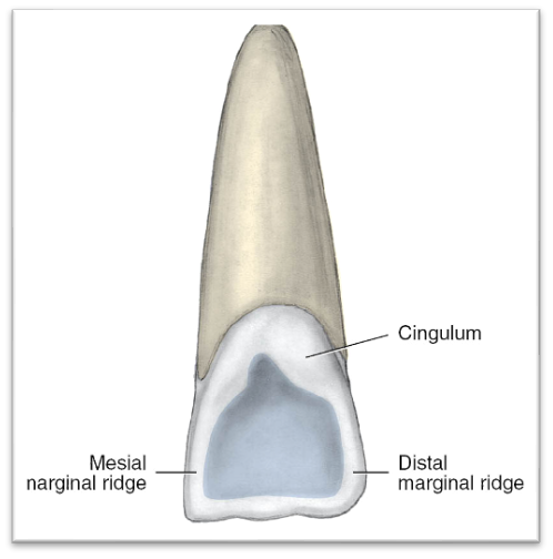
The lingual surfaces of all anteriors have a cingulum
•A raised, rounded area on the cervical third of the lingual surface in varying degrees of prominence or development
•The cingulum corresponds to the lingual developmental lobe
•Ridges are present on the lingual surface.
•The lingual surface on anteriors is bordered mesially and distally on each side by a rounded raised border, the marginal ridge.
marginal ridge

fossa
some anteriors have shallow wide depressions on the lingual surface called fossa or fossae
developmental pits
•Some anteriors have developmental pits
•Located in the deepest part of each fossa

On their lingual surface, some anteriors may have
•a developmental groove, (or primary groove): a sharp, deep, V-shaped linear depression that marks the junction among the developmental lobes
supplemental groove
•may be present on the lingual surface of anteriors
•a shallower, more irregular linear depression than the developmental groove
•branches from the developmental groove, but are not always present in the same pattern on each different tooth type.
anterior perm teeth Usually have a _____ root, with some exceptions
single
•Each root of the maxillary anterior teeth has great ___ and slight ___inclination
great lingual and slight distal
•Each root of the mandibular anterior teeth varies in angulation from nearly vertical to????
great lingual inclination, with the canines possibly having a slight distal root inclination.
•When newly erupted, each incisor has three
mamelons
•rounded enamel extensions on the incisal ridge from the labial or lingual view
•enamel extensions from the three labial developmental lobes


•____ the only permanent teeth with two incisal angles (formed from the incisal ridge and each proximal surface)
• both types are the only permanent teeth with a nearly straight___
incisors
stright incisal ridge

permanent maxillaRY INCISORS :
general features
•Each has a crown that is larger in all dimensions, especially MD, compared with a mandibular incisor
•The labial surfaces are rounder from the incisal aspect, with the tooth tapering toward the lingual
•All lingual surface features, including the marginal ridges, lingual fossa, and cingulum, are more prominent on the maxillary incisors than on the mandibular incisors
•Each root is usually shorter compared with those of other maxillary teeth and usually is without root concavities.
Maxillary incisors can also have an accentuated cingulum, with deepened
developmental grooves
•If a maxillary incisor has increased prominence of the lingual marginal ridges and a deeper lingual fossa, it may be considered a ____ incisor
shovel shaped incisor
MAXILLARY INCISORS:
CLINICAL CONSIDERATIONS 4 things
•Supragingival tooth deposits, such as dental biofilm and stain, can collect in the prominent lingual surface concavities of maxillary incisors
•Caries risk
•Periodontal risk if the groove extends to the root
•Accessibility
perm maxillary central incisors
•Universal Tooth Designation System: #8 and #9
•International Standards Organization Designation System: 1-1 and 2-1
•Palmer Notation Method: 1| and |1
Maxillary centrals:
specific features when they erupt
•They erupt between 7 and 8 years of age (root completion at age 10)
•Thus, they usually erupt after the mandibular central incisors
The maxillary central incisors are the most prominent teeth in the permanent dentition because of both their large
size and their anterior arch position
MAXILLARY CENTRALS:
Shape
•The outline of the crown from the labial or lingual view is trapezoidal
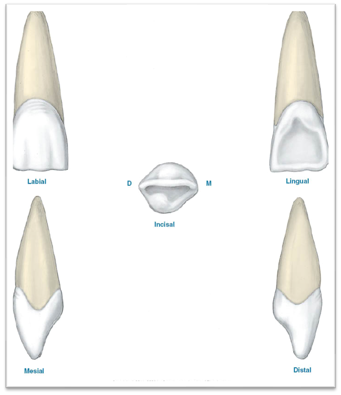
MAXILLARY CENTRALS:
root
smooth, stright conical root usually with rounded apex
crown to root ratio is usually 1: 1.5
MAXILLARY CENTRALS:
Labial view features
•The difference in sharpness of the central’s mesioincisal and distoincisal angle helps to distinguish the maxillary right central incisor from the left.
MAXILLARY CENTRALS:
Lingual view features
•The cingulum is wide and well-developed in size, as well as being slightly off-center toward distal
•The single lingual fossa is wide yet shallow; it varies in depth and diameter

MAXILLARY CENTRALS: Incisal view features
•Overall, the shape of the crown is triangular from this view, with the labial outline broadly rounded.

Maxillary central incisor:
clinical considerations
•The incisal edge or even the entire tooth is especially at risk for traumatic fracture (position, eruption)
•Because of these two factors and without full root completion, the entire tooth in a child may undergo avulsion (complete displacement of the tooth from the tooth socket)
•An open contact, or diastema can exist between the maxillary central incisors. It can be a wide, and to some patients, unattractive space.
•Incisors may appear winged when looking at the patient’s smile.
Max lateral incisors
•Universal Tooth Designation System: #7 and #10
•International Standards Organization Designation System: 1-2 and 2-2
•Palmer Notation Method: 2| and |2
maxillary lateral incisor: features
•Permanent maxillary lateral incisors erupt between 8 and 9 years of age (root completion at age 11)
•Thus, these teeth usually erupt after the maxillary central incisors have erupted
Maxillary lateral incisors: Specific features
•Maxillary lateral crowns have the second greatest degree of variation in form of any permanent tooth
•It usually resembles a maxillary central incisor in all views of the tooth but has a smaller and slightly rounder crown.
•The distal outline is rounder than the central
Maxillary lateral incisors: root
•It has a conical root that is relatively smooth and straight but may curve slightly to the distal.
•Usually, the root is as long but thinner MD and wider LL than the max central incisor
•Crown to root ratio is 1:1.5
maxillary lateral incisors:
Labial view
•The crown is smaller than that of a central incisor and less symmetrical.
•Both incisal angles are rounder than on a central incisor.

Maxillary lateral incisors:
Lingual view
•The lingual surface of the crown is narrower than the labial surface, as is the case with a central
•It has a prominent yet centered and narrower cingulum than does a central incisor, with a deeper lingual fossa.

MAXILLARY LATERAL INCISORS: Lingual view
•The marginal ridges are pronounced.
•Longer mesial marginal ridge and a shorter distal marginal ridge
The linguoincisal ridge or edge is also noticeably well-developed in size.
•A ____pit is more common on a lateral than on a central
lingual
Vertical linguogingival developmental groove originates in the
lingula pit and extends cervically (lateral incisors)
max lateral incisors
•May have a vertical distolingual marginal developmental groove originating on the distomarginal ridge which can extend to the root
Max laterals: clinical considerations
•The developmental groove can be considered a clinically adverse factor because deposits can accumulate in the groove
•Perio risk factor if it extends to the root
Perm mandibular incisors
•Permanent mandibular incisors are the smallest teeth of the permanent dentition and the most symmetrical.
•The lateral and central incisors of the mandibular arch resemble each other more than do the similar types of incisors of the maxillary arch
•Generally, a mandibular lateral is slightly larger than a central
mandibular incisors: General features
•Each mandibular incisor has a crown that is wider LL than MD,
•The root is extremely narrow on the labial and lingual surfaces and wide on both proximal surfaces.
•Proximal root concavities are also present on both types of mandibular incisors
mandibular incisors: Clinical considerations
•Supragingival tooth deposits such as dental biofilm, calculus, and stain tend to collect in the fossa
buildup of deposits is aided by the mandibular incisors position in the oral cavity near the duct openings of the submandibular salivary gland and sublingual salivary glands in the floor of the mouth
mandibular incisors: Clinical considerations
•Dental professionals must be careful to check for deposits in any mesial and distal root concavities
with attrition mand incisors can???
•With attrition, the incisal edge can change on the mandibular incisors.
•Prolonged hand instrumentation can cause further narrowing of the already narrow root surfaces of the
mandibular incisors
•Universal Tooth Designation System: #24 and #25
•International Standards Organization Designation System: 3-1 and 4-1
•Palmer Notation Method: |1 and 1
perm mandibular central incisors
•They erupt between 6 and 7 years of age (root completion at age 9)
•Thus, these teeth usually erupt before the maxillary central incisors
mandibular centrals
•They are the smallest and simplest teeth of the permanent dentition
•It has a simple root, which is wider LL than MD
•The root has pronounced proximal root concavities, which vary in both length and depth
mandibular centrals specific features
Mandibular centrals: Labial view
Both the mesioincisal angle and distoincisal angles are
•sharp or only slightly rounded; the mesioincisal angle is slightly sharper than the distoincisal angle
•The crown is quite symmetrical from the labial view, having a slight fan shape
Mandibular centrals:
Lingual view
•The crown is narrower on the lingual surface than the labial
•The single lingual fossa is barely noticeable; therefore, the mesial marginal ridge and distal marginal ridge are also barely noticeable
•Because the cingulum is centered, the faint mesial and distal marginal ridges have the same length.
Mandibular centrals: Incisal view
it has nearly symmetrical crown outline on the incisal view

Permanent mandibular lateral incisors
•Universal Tooth Designation System: #23 and #26
•International Standards Organization Designation System: 3-2 and 4-2
•Palmer Notation Method: I 2 and 2 I
Mandibular laterals: Specific features
•Erupt between 7 and 8 years of age with root completion at age 10
•Thus, these teeth usually erupt after the mandibular central incisors
features of mandibular laterals
•Slightly larger overall than a central incisor
•More variation in form than a central
•From the incisal view, the crown appears tilted or twisted distally in comparison with the long axis of the tooth

feat of mandibular laterals
•The single root is usually straight, slightly longer and wider than that of a central incisor
•The root, like that of a mandibular central, has pronounced proximal root concavities, especially on the distal surface
•The crown is not as symmetrical
•The MI angle of the incisal edge is sharper than the DI angle, which helps to distinguish the right from the left.
•The DI angle is lower than the MI angle
labial view of mandibular laterals
•labial developmental depressions are deeper than on the central incisors
mandibular laterals: Lingual view
•lingual surface has more prominent features compared to the lingual surface of a central incisor
•The small single cingulum lies just distal to the long axis of the root (not bilaterally symmetrical).
Mandibular laterals: Proximal view features
•A rounder appearance is noted both labially and lingually as compared with that of a central incisor

lingual and incisal view of mand laterals
name 1-6

Permanent canines (mandible and maxilla)
•Permanent canines are the four anterior teeth located at the corners of each quadrant for each dental arch
• The longest and most stable of teeth in the dental arch
•They are the only teeth in the dentition with a single cusp
When viewed from the proximal surface, the canine is what in shape
triangular
•When viewed from the labial or lingual, however, the crown of canine outline appears____ with five sides, similar to the premolars
pentagonal
canines are wider ___ than the incisors
LL

Similar to the other anteriors, each of the canines has an
incisal ridge
•Different from the incisors is the ___ which is in line with the long axis of the root for both maxillary and mandibular canines when first erupted
– cusp tip
may change due to attrition
canine form and function
•Pointed, tapered shape aids in piercing and tearing food
•Prominent cusp contributes to esthetics
•The incisal edge of canine is divided into two _____ rather than being nearly straight across like the incisors
cusp slopes, or ridges,
canine
•The ____cusp slope is usually shorter than the distal cusp slope for both maxillary and mandibular canines when they first erupt
mesial cusp slope
The canines are the only teeth in the permanent dentition with a vertical and centrally placed
labial ridge
the labial ridge of the canine is the result of what
•of greater development of the middle labial developmental lobe in comparison with the mesial and distal labial developmental lobes
mamelons
•are not normally present on the incisal edge as they are on incisors, but a small notch may be seen on either cusp slope
•canines
Proximal ____ are located on both proximal root surfaces
root concavities
•Each canine has a ___ and _____ on its lingual surface, like the incisors.
A vertical, centrally placed ______ extends from the cusp tip to the cingulum
cingulum and marginal ridges
lingual ridge
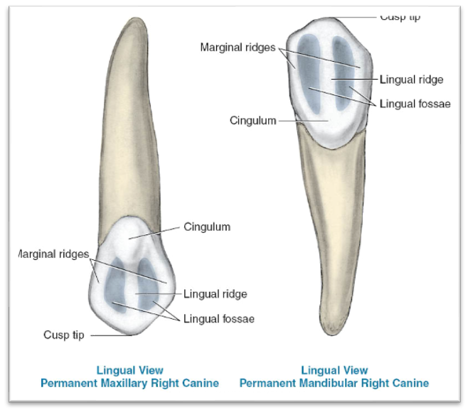
Canine eminence
•Each has a particularly long, thick root, 1:1.5 crown to root ratio

•The long and large root is externally manifested by the vertically oriented and labially placed bony ridge called the ____ of the alveolar bone, noted especially in the maxillary arch
canine eminence
Clinical considerations canines
•Changes can occur in the length of each canine cusp slope and in the position of the usually centered cusp tip.
•Proximal surfaces of the canines are more accessible from the lingual than the labial during instrumentation
Permanent maxillary canines
•Universal Tooth Designation System: #6 and #11
•International Standards Organization Designation System: 1-3 and 2-3
•Palmer Notation Method: 3| and |3
Maxillary canines: Specific features
•They erupt between 11 and 12 years of age (root completion between ages 13 and 15)
•Thus, these teeth usually erupt after the mandibular canines, after the maxillary incisors, and possibly after the maxillary premolars
Maxillary canines: Specific features
resembles a mandibular canine but the cusp is____ and more developed, and the cusp tip is ____than a mand canine
larger
shaper
•Features are more prominent than mand canine
•Crown is similar in length to a central incisor, but wider labiolingually, and narrower mesoidistally

maxillary canines: Labial view
•The distal outline is shorter than the mesial outline and usually has a depression between the distal contact area and the CEJ, which helps to distinguish the right from the left.
•The single cusp is rounded and the mesial cusp slope is shorter than the distal cusp slope, which helps to distinguish the right from the left.
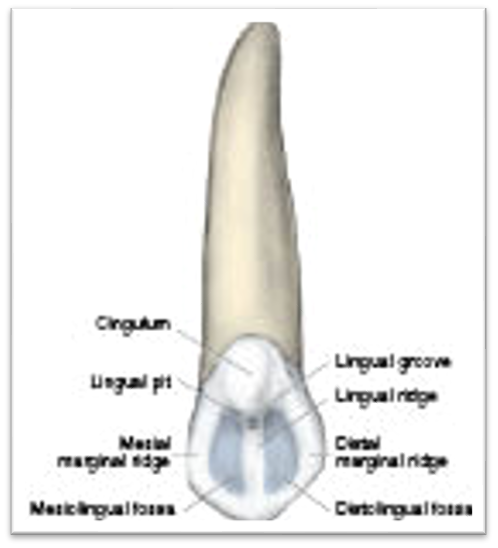
•The entire lingual surface features of the maxillary canine are _____ than a mandibular canine
more prominent
lingual of max cannines
•The cingulum is large and is centered mesiodistally on the lingual surface
•A centrally placed vertical lingual ridge is present from the cingulum to the cusp tip
•The lingual surface is sometimes separated by a shallow lingual groove, and this groove may contain a lingual pit near its center

Clinical considerations for maxillary canines
•They may fail to erupt fully, remaining impacted within the alveolar bone
•Dentigerous cyst is possible
•Possible dilaceration of root
•The cingulum may exhibit tubercles that are located near the most incisal level of the cingulum
mandibular canines
•Universal Tooth Designation System: #28 and #22
•International Standards Organization Designation System: 4-3 and 3-3
•Palmer System 3| and |3
when do mand canines erupt
•erupt between 9 and 10 years of age (root completion between ages 12 and 14).
•Thus, these teeth usually erupt before the maxillary canines and after most of the incisors have erupted

mand canines specific features
•Closely resembles a maxillary canine.
•The single cusp is not as well developed in size
•it is narrower LL and MD than a maxillary canine.
Mandibular canines: Lingual view
•The lingual surface of the crown is smoother than that of a maxillary canine
• Has a less- developed cingulum and two marginal ridges and two fossae

Mandibular canines: Labial view
•The labial surface is not as rounded as that on a maxillary canine.
•In contrast, however, it is generally rounder than a mandibular incisor
