Anatomy Practical 1
 Call Kai
 Learn
Learn Practice Test
 Spaced Repetition
 Match
Match Flashcards
 Knowt Play
1/186

There's no tags or description
Looks like no tags are added yet.
Name | Mastery | Learn | Test | Matching | Spaced | Call with Kai | Chat |
|---|
No analytics yet
Send a link to your students to track their progress
187 Terms

Ocular Lens
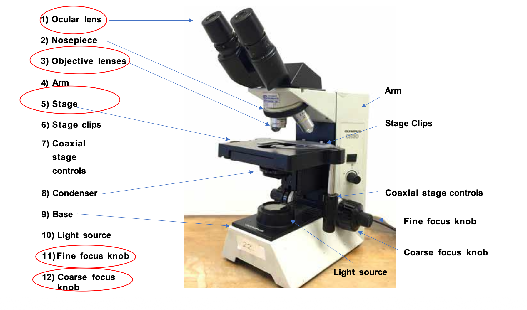

Nosepiece
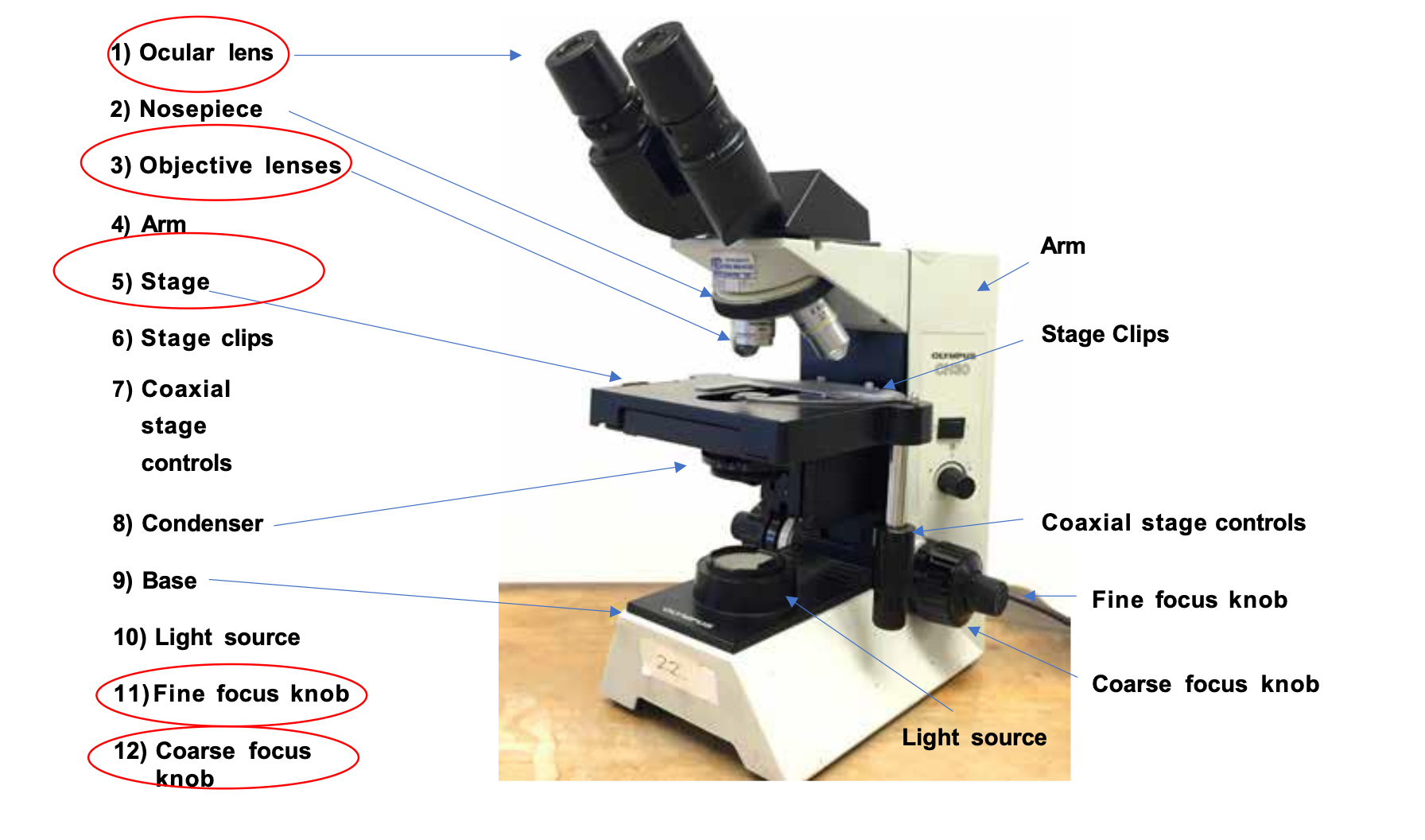
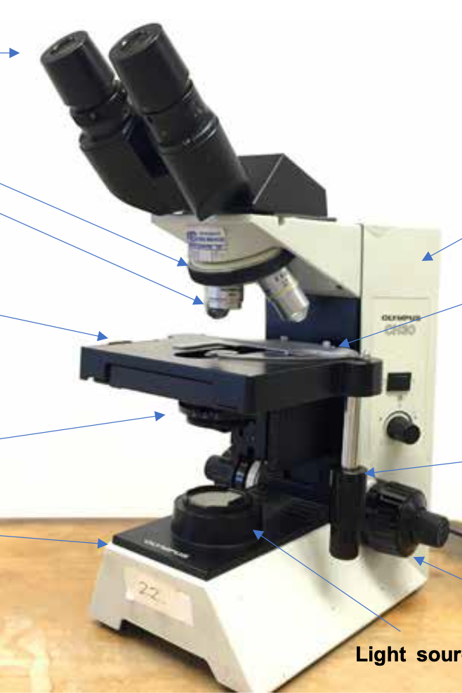
Objective Lenses (Types)
Scanning power objective lens, Low Power Objective Lens, High/Dry Power Objective Lens, Oil Immersion Objective Lens (if available)


Arm
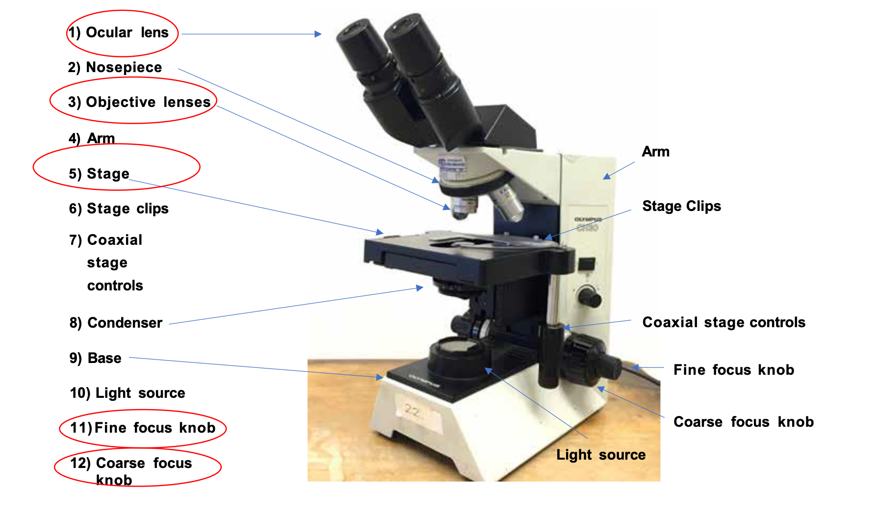

Stage


Stage Clips
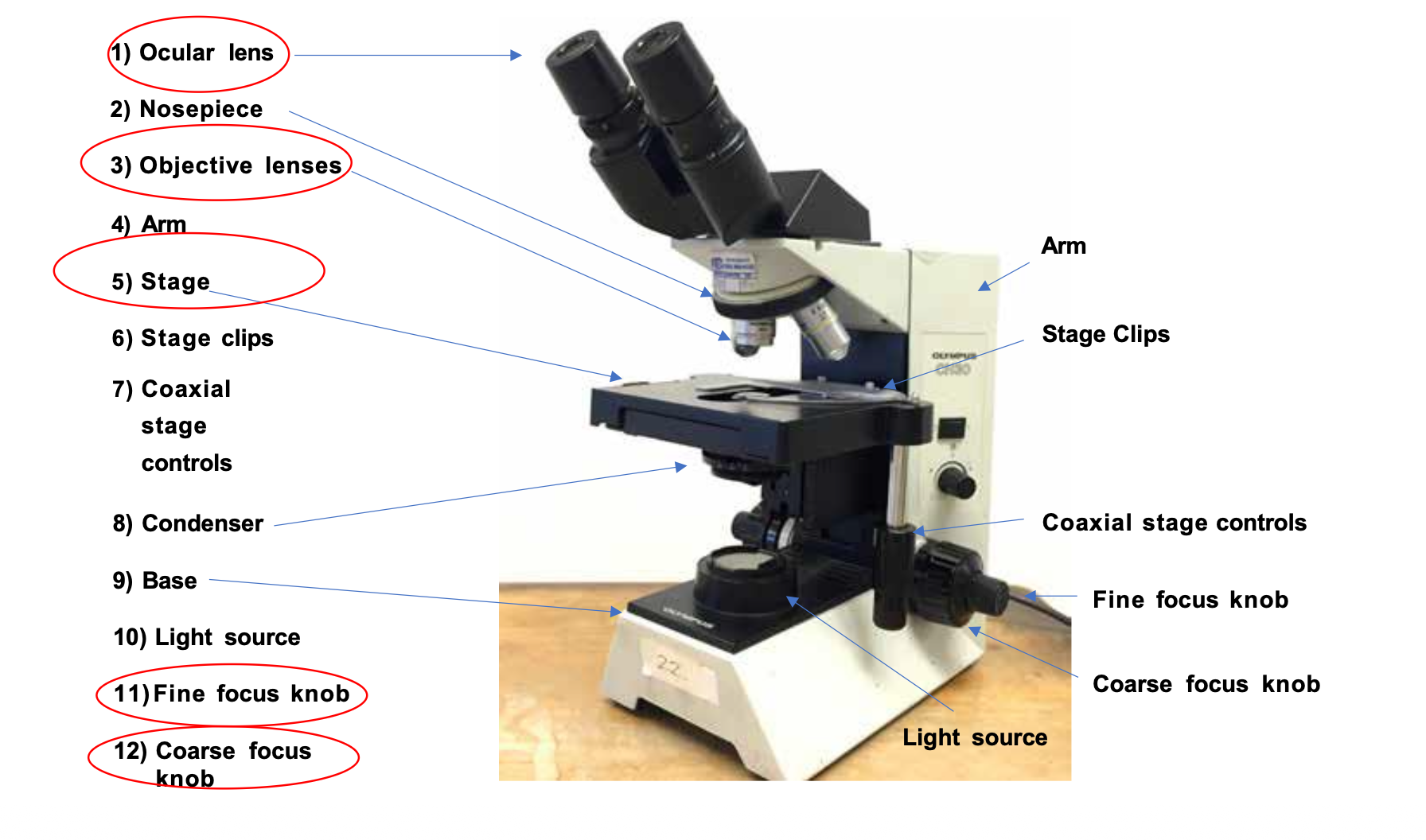

Coaxial Stage Controls


Condenser


Base
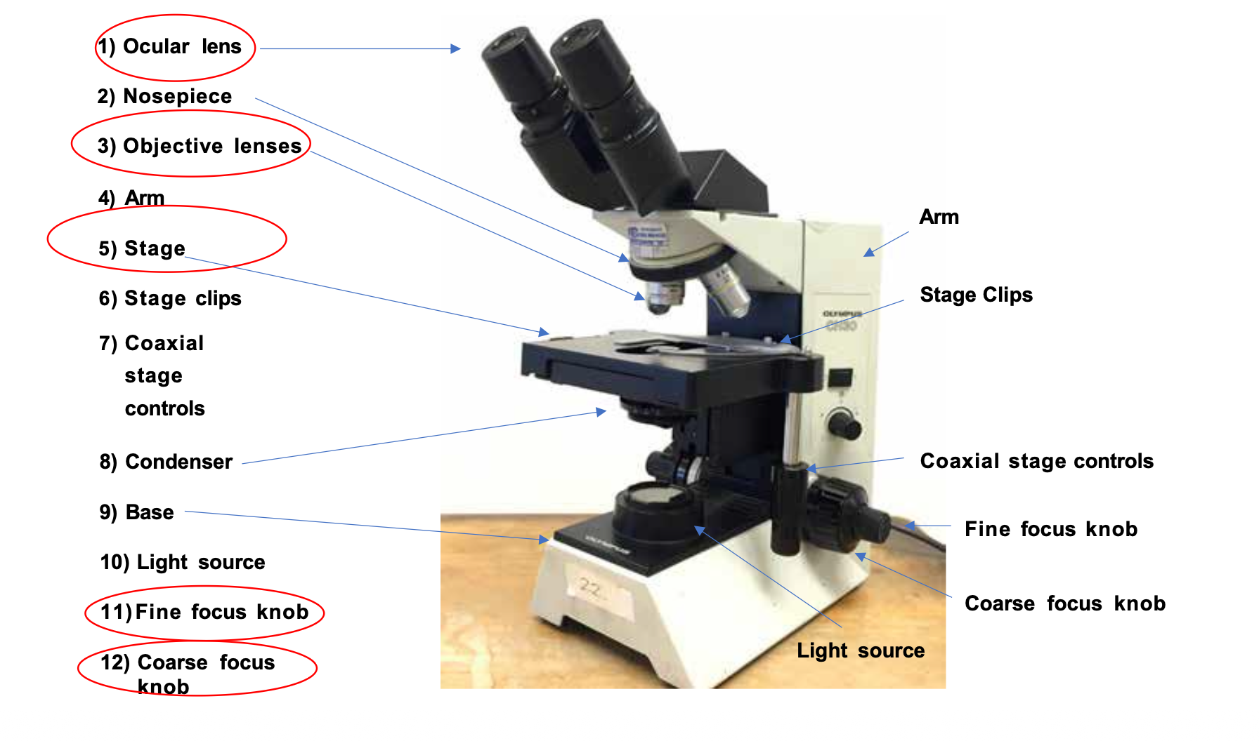

Light Source


Fine Focus Knob


Coarse Focus Knob

Ocular Magnification + Scanning Power Lens (Red)
Ocular magnification (10x) combined with the scanning power lens (4x) results in a total magnification of 40x

Ocular Magnification + Low Power Lens (Yellow)
Ocular magnification (10x) combined with the low power lens (10x) results in a total magnification of 100x

Ocular Magnification + High/Dry Power Lens (Blue)
Ocular magnification (10x) combined with the high/dry power lens (40x) results in a total magnification of 400x

Ocular Magnification + Oil Immersion Lens (Black and White)
Ocular magnification (10x) combined with the oil immersion lens (100x) results in a total magnification of 1000x


Cell Nucleus
Cell Nucleus

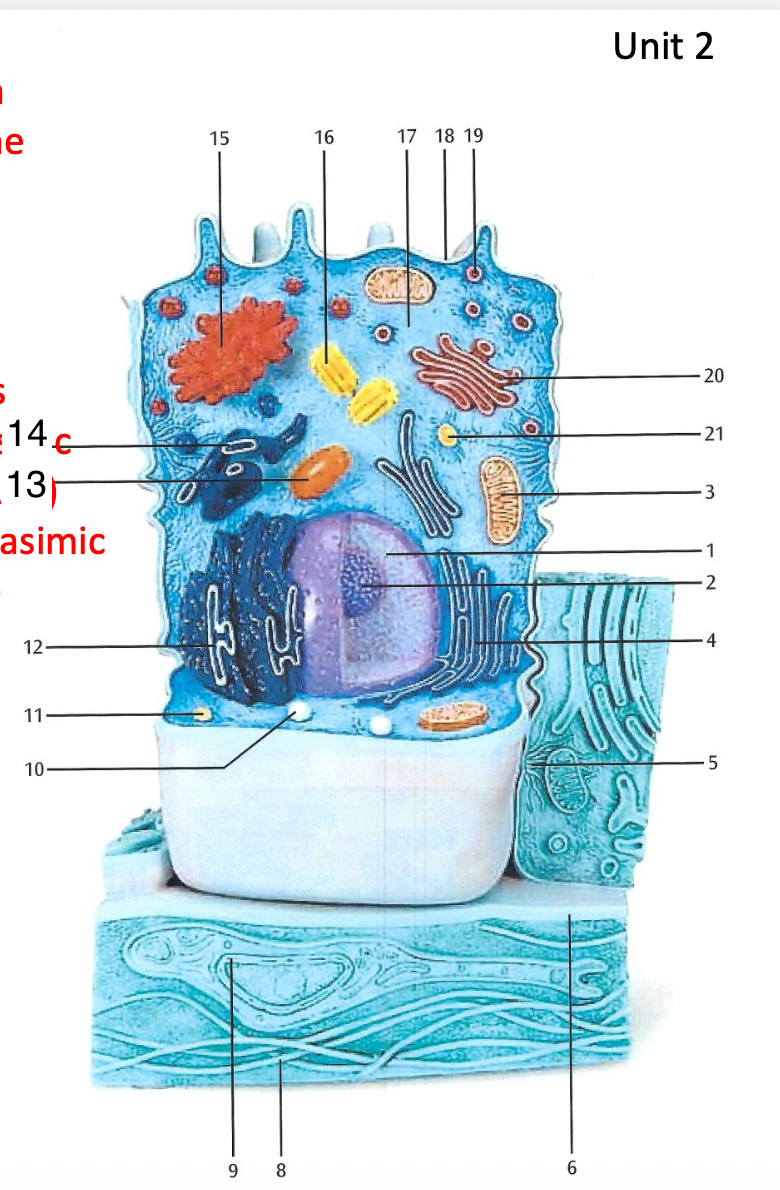
Nucleolus
Nucleolus


Mitochondria
+ 13. Mitochondria

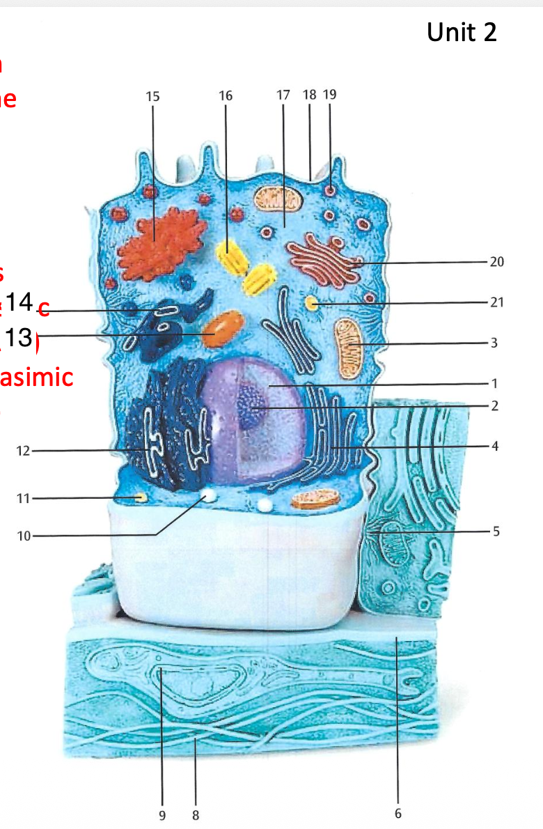
Smooth endoplasmic reticulum
+ 14. Smooth endoplasmic reticulum

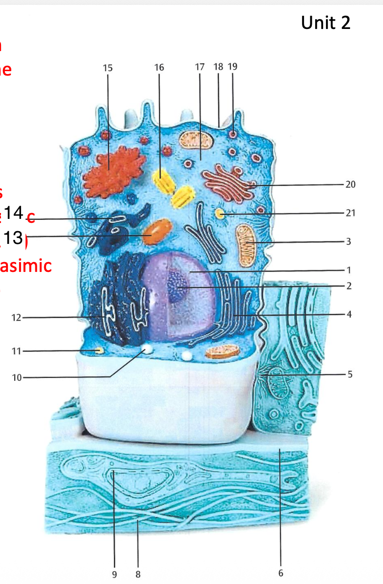
Desmosome
Desmosome


Basal Membrane
Basal Membrane


Hemidesmosome
Hemidesmosome


Collagen Fibers
Collagen Fibers


Fibroblast
Fibroblast


Peroxisome
Peroxisome

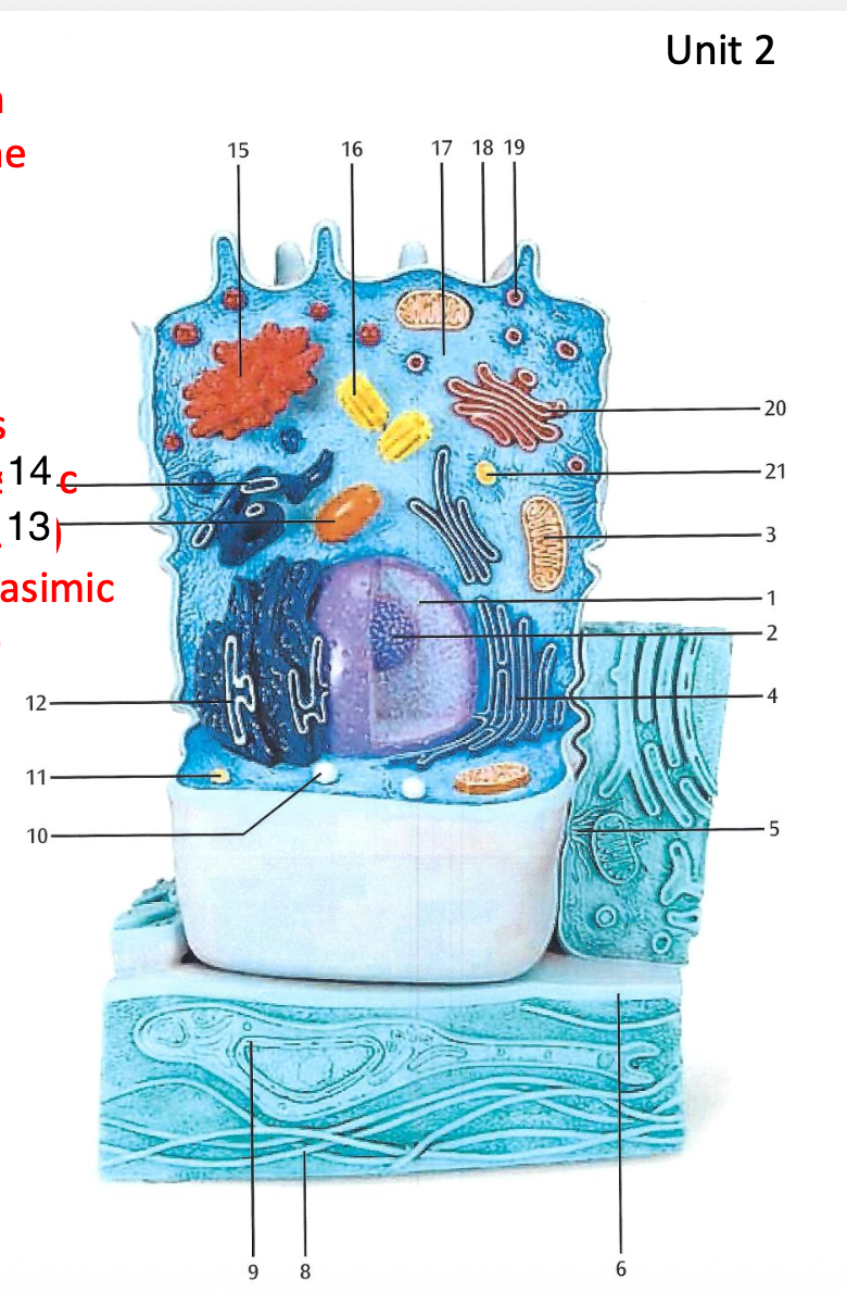
Lysosome
Lysosome


Rough Endoplasmic Reticulum
Rough Endoplasmic Reticulum
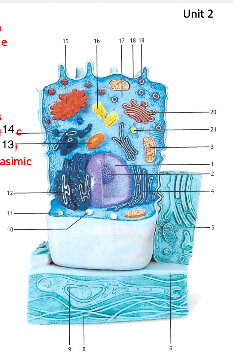

Golgi apparatus
+ 20. Golgi Apparatus


Centriole
Centriole


Cytosol
Cytosol


Microvilli
Microvilli


Secretion Vesicle
Secretion Vesicle


Identify this photograph.
Actin Filaments of the Cytoskeleton


Identify the photograph.
Microtubules


Identify the structures in this photograph.
Mitocondria


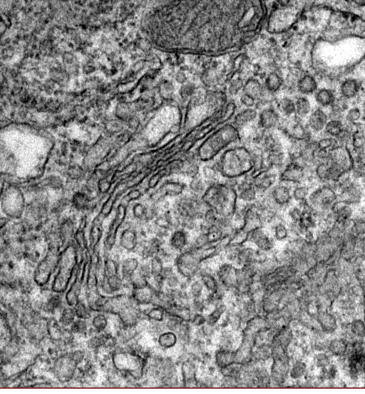
Identify the structure in this photograph.
Golgi apparatus
Define HISTOLOGY.
the study of microscopic structures of tissues
Define TISSUE.
A group of cells that have similar structure and that function together as a unit.
Define MATRIX.
the fluids and secretions found around cells in a tissue.
What are the 4 types of Tissue
Epithelial, Connective, Muscular, and Nervous.

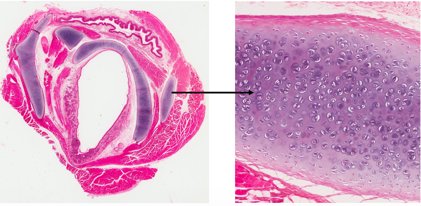
What is structure in this photograph? Name the type of cartilage that the arrow is pointing to.
1) Cross-section of the Trachea 2) Connective Tissue - Hyaline Cartilage

What type of tissue is this? Where can it be found in the body?
1) Stratified Squamous Epithelial Tissue 2) Occurs in the skin and in tissues lining the mouth and vagina.

Difference between Ligaments and Tendons
Tendons: connect muscle to bone
Ligaments: connect bone to bone
Both are made up of dense regular connective tissue

Tendon or Ligament? Why?
Ligament, connects bone to bone


Tendon or Ligament? Why?
Tendon, connects muscle to bone


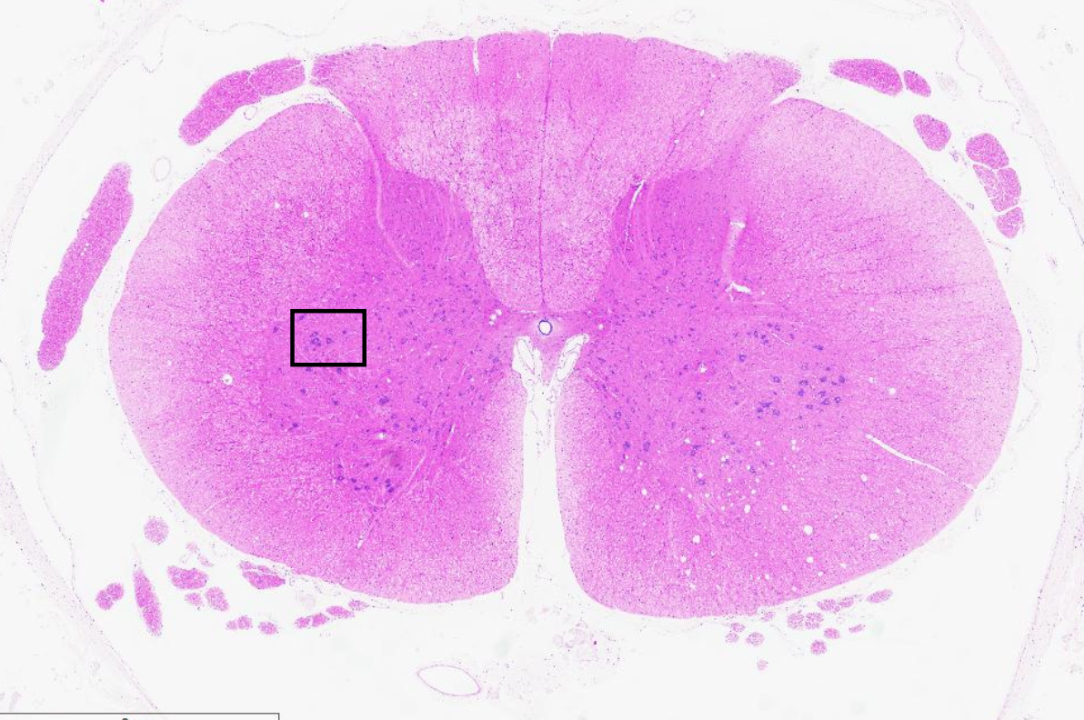
Identify the image. What type of tissue is this?
Cross-section of spinal cord, nervous tissue


Name the tissue type. Name the use of cells that the arrows are pointing to.
Nervous tissue
Spinal cord neurons
Identify the 3 types of Muscle Tissue Types
Skeletal
Cardiac
Smooth

Name the type of muscle
Skeletal Muscle


Name the type of muscle.
Cardiac Muscle


Name the type of muscle.
Smooth Muscle

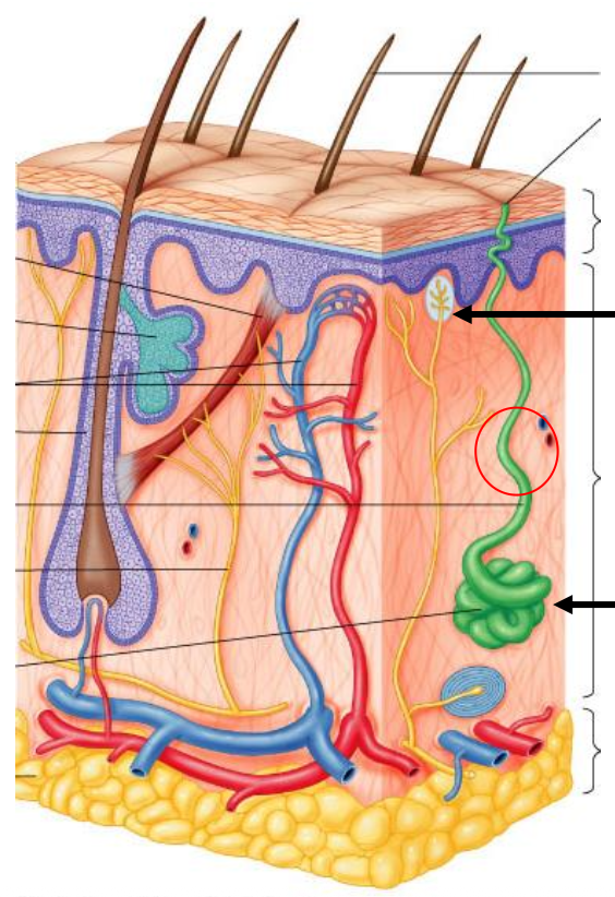
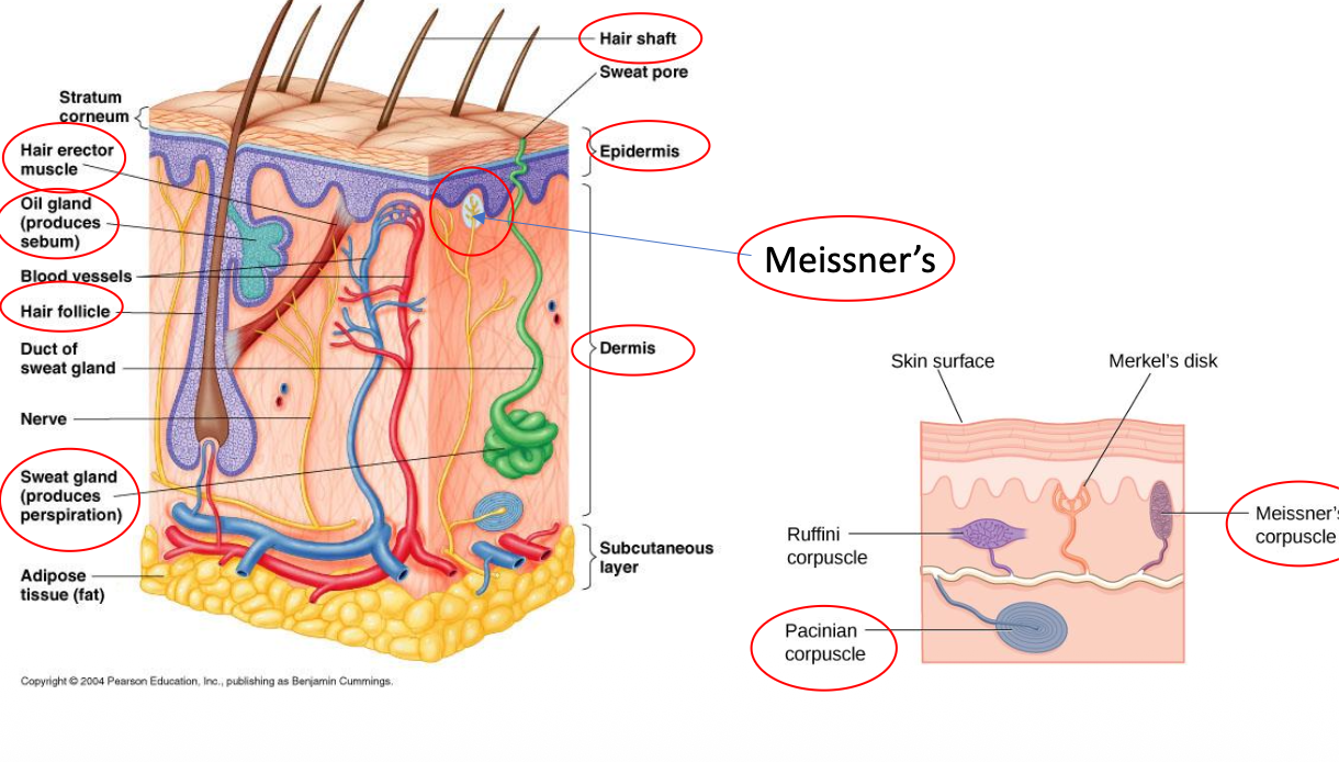
Arrector Pili Muscle
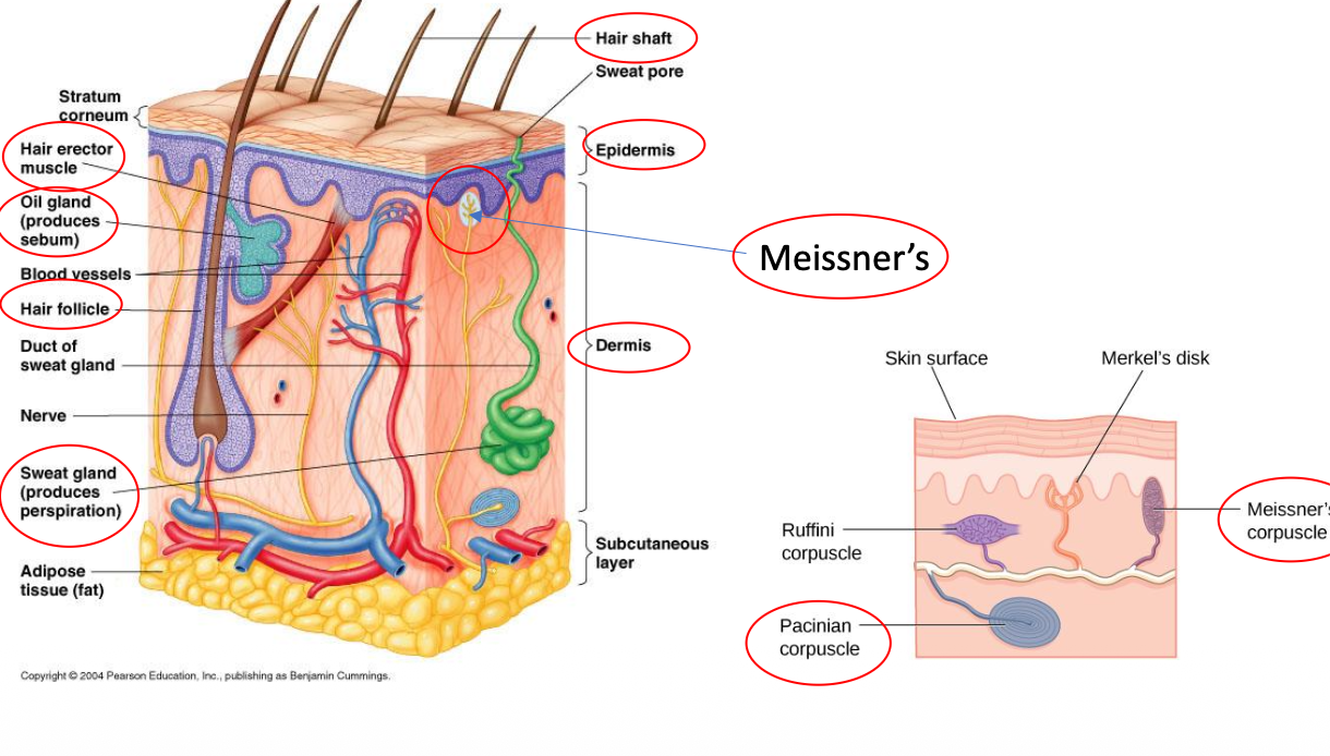
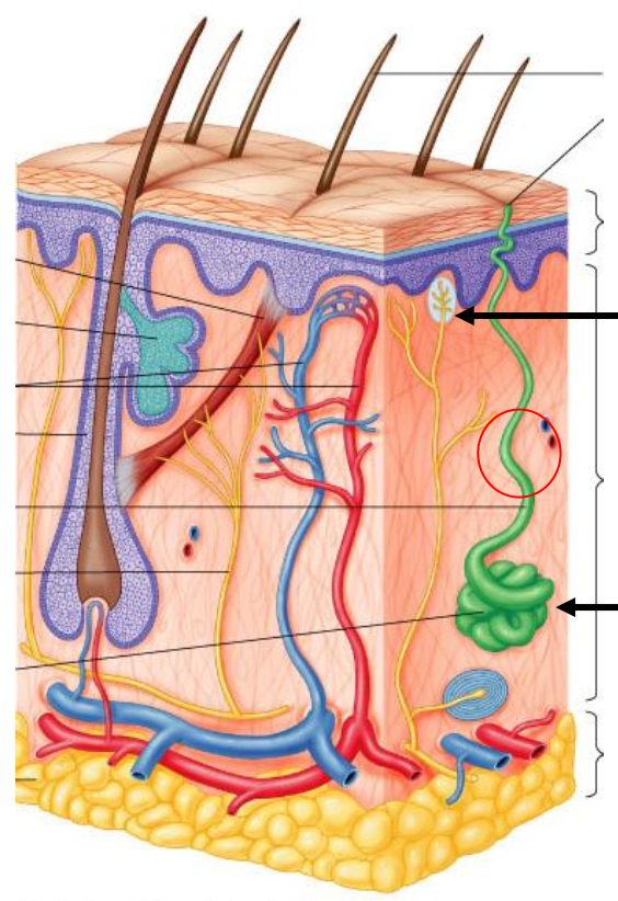
Sebaceous gland


Sweat Gland


Hair Shaft
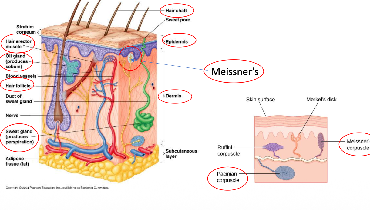
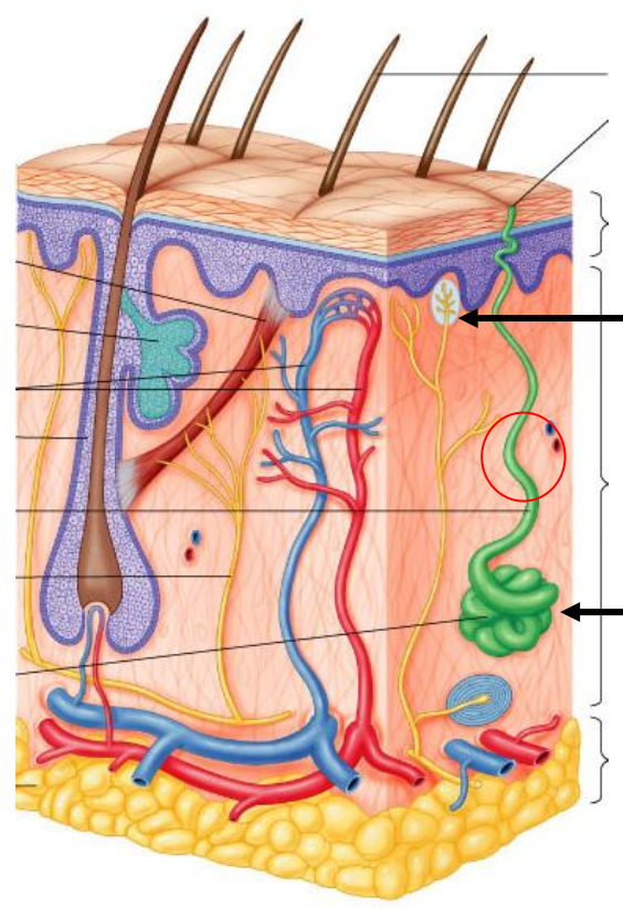
Blood Vessels
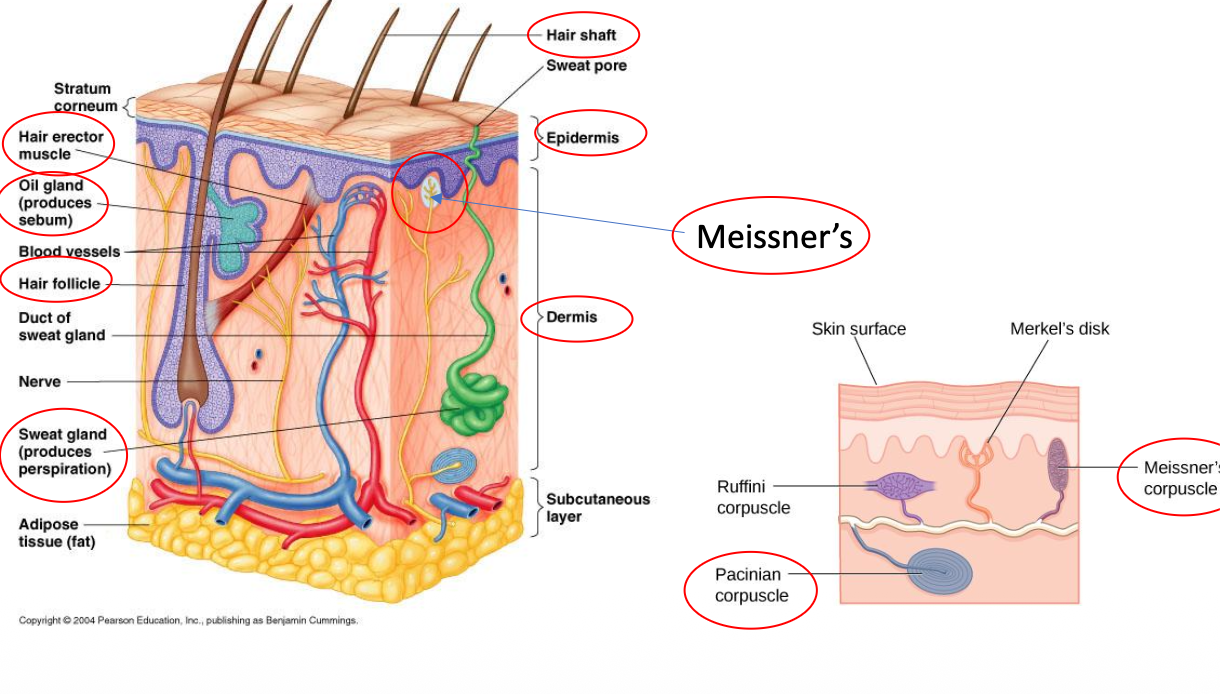

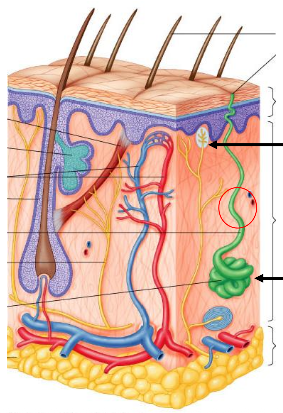
Meissner’s Corpuscle


Hair Follicle


Nerve

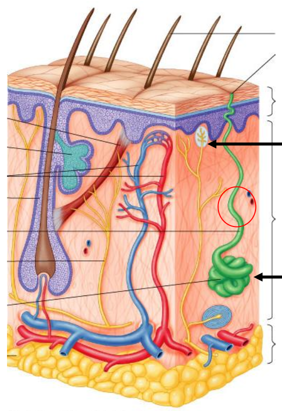
Sweat Gland

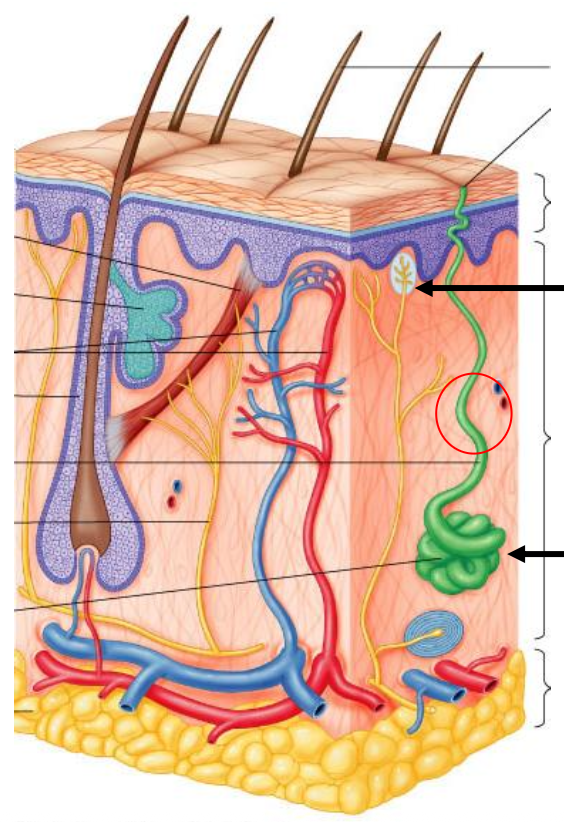
Adipose tissue
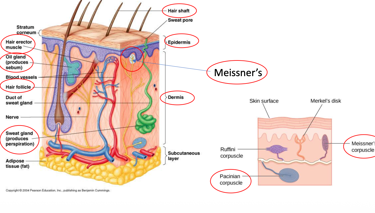

Sweat duct
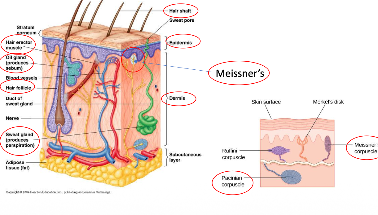

Epidermis
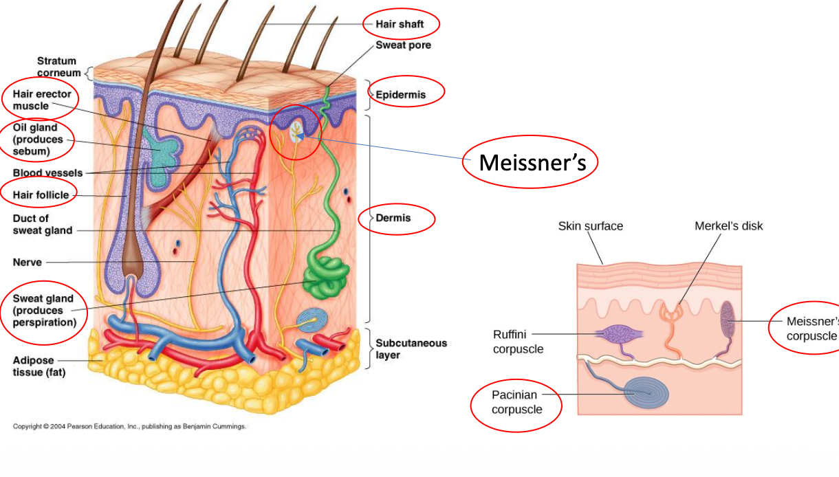

Dermis


Hypodermis (Subcutaneous Layer)

Pacinian Corpuscle
Name the layers of the Epidermis
Stratum basale, stratum spinosum, stratum granulosum, stratum lucidum, stratum corneum


What part of the body do you see? What special structures are pointed to?
Lip
Hair follicles

What is A? What is B?
A) Spongy Bone
B) Compact Bone

ID 1.
Skull
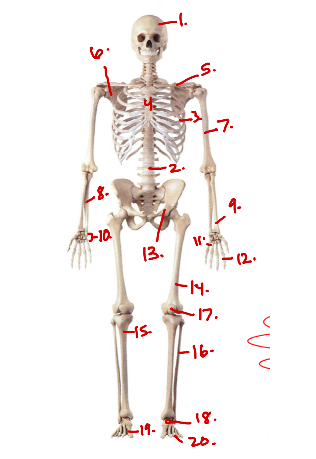
ID 2.
Vertebrae

ID 3.
Ribs

ID 4.
Sternum

ID 5.
CLAVICLE

ID. 6
SCAPULA

ID 7.
HUMERUS

ID 8.
ULNA

ID. 9
RADIUS

ID 10.
CARPAL BONES

ID 11.
METACARPAL BONES

ID 12.
PHALANGES

ID 13.
OS COXA OR COXAL BONES

ID. 14.
FEMUR

ID 15.
TIBIA

ID 16.
FIBULA

ID. 17.
PATELLA

ID 18
TARSAL BONES

ID. 19
METATARSAL BONES

ID 20.
PHALANGES OF FOOT
What does the ACL of the KNEE stand for?
Anterior Cruciate Ligament
WHAT DOES PCL of the KNEE stand for?
Posterior Cruciate Ligament
What is the MCL of the KNEE?
Medial collateral ligament
What is the LCL of the KNEE?
Lateral collateral ligament
What are the 3 parts of the Sternum from Superior to Inferior?
Manubrium, Body, Ziphoid Process

What are the ‘true ribs” ?
Ribs 1-7
What are the ‘false ribs’ ?
Ribs 8-12
What are the ‘floating ribs’?
Ribs 11-12

Identify A.
Lateral Femoral Condyle

Identify B.
Medial Femoral Condyle