Bacterial, fungal & yeast pathogens of the skin
 Call Kai
 Learn
 Practice Test
 Spaced Repetition
 Match
 Flashcards
 Knowt Play
1/35

There's no tags or description
Looks like no tags are added yet.
36 Terms

skin layout
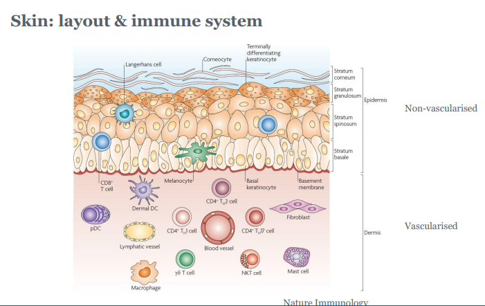

skin: layout and immune system
• Mechanical defense: keratinocytes, keratin layer, continuous renewal
• Chemical defence: antimicrobial compounds
• Relatively low pH
• Immune defense: innate and adaptive responses
• Toll-like receptors (TLRs) recognising specific compounds
• Activation of TLRs > production of chemokines > attracts immune cells
• Immature dendritic cells (Langerhans): presents antigens in MHC
innate immune system
The innate immune system recognises
• PAMPs: pathogen associated molecular patterns
• Peptidoglycan fragments (bacteria)
• proteins (bacteria)
• nucleic acids (viruses and bacteria)
• cell wall lipids (gram-negative bacteria)
• carbohydrates (fungi, bacteria)
• DAMPs: damage-associated molecular patterns
• Stress-induced proteins (Heat shock proteins)
• Nuclear proteins
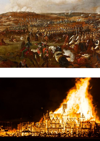

skin microflora
• Skin is not sterile


Types of diseases associated with skin infections
• pruritis (itchiness)
• alopecia (hair loss)
• scaling +/ crusts
• pyoderma (pustules, superficial or deep)
• other: Erosions, ulcers, pigment disorders, nodules and tumours, sinus tracts
second pic- staphs is staphilococcus cocci check name

alopecia
bacteria colonise/infect hair follicle
infection triggers innate and adaptive immune responses
• folliculitis
• damage to hair shafts
• atrophy

Folliculitis / sinus formation

pyoderma
Commonly caused by Staphylococci / Streptococci
• cats, dogs: S. pseudintermedius
• horses, cattle, sheep: S. aureus
• Other organisms involved
— Group A streptococci
— Pasteurella
Types of pyoderma
superficial:
• coagulase+ staphylococci
— cats, dogs: S. pseudintermedius
— horses, cattle, sheep: S. aureus
• Pasteurella, streptococci
• exudative dermatitis: Staphylococcus hyicus, Dermatophilus congolensis
deep
• subcutaneous abscess
• cellulitis
• necrotising fasciitis (rare) > Streptococcus canis
pustule
neutrophils starts opsonizing the bacteria
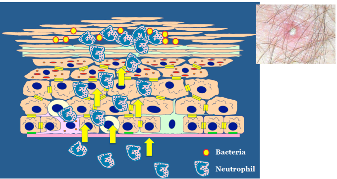

Pyoderma example
the PAMPs proliferate and cause damage → DAMPs - triggers immune response

Staphylococci
• aerobic / facultative anaerobic
• Gram-positive, catalase positive
Two major subtypes
• Coagulase-negative (CoNS, CNS): S. epidermidis
Commensal on skin, opportunistic pathogen
• Coagulase-positive: S. pseudintermedius, S. intermedius, S. aureus
Commensal (?) (S. pseudintermedius), trauma of skin or underlying
conditions
Virulence mechanisms
either trauma of skin or underlying conditions
Large array of virulence factors/colonisation factors
• Cell wall polymers: peptidoglycan, techoic acid
• Adhesins: Ig-binding, Fibronectin-binding proteins
• Toxins (enterotoxin, epidermolytic toxins)
• Lysins
• coagulase
• DNase
Group A streptococci
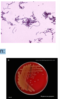
Group A streptococci (GAS) primarily Streptococcus pyogenes
— Aerobic/facultative anaerobic
—Gram+, catalase-, oxidase+, Beta-haemolytic
Pathology is the result of toxin and aggressin production:
• Streptolysin - two types (S and O).
• Streptokinase (fibrinolysin)
• Hyaluronidase (disrupts tissue connectivity)
• Erythrogenic toxin (rash of scarlet fever).
• DNase
• Exotoxins
Conclusion bacterial infections
• Skin defence/immune system controls microflora and pathogens
• Major bacterial causes of skin disease are Staphylococci and Group A
streptococci
• Bacteria have array of virulence factors for causing disease
• Often opportunistic pathogens
bacterial diseases of production animals
Examples:
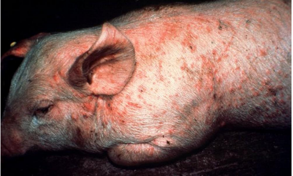
• Erysipelothrix in pigs
• Foot rot in sheep, goats, cattle, horses

Erysipelothrix in pigs
Erysipelothrix rhusiopathiae
• Gram-positive, catalase-negative, rod-shaped, non-spore-forming
• Produces H2S
• hon-haemolytic
Erysipelothrix rhusiopathiae
• Present in tonsillar tissues, shed in faeces
• Symptoms: febrile episodes, painful joints, lethargy, distress
• "Diamond skin"
• Well detected in acute phase, more difficult in the chronic phase
• Youngs: passive immunity maternal antibodies, vaccination
• Can be treated with penicillin, tetracycline

Foot rot in sheep, goats, cattle, horses
Two step infection
• Starts with Fusobacterium necrophorum
• Gram-negative rod, strict anaerobe
• found in environment, alimentary tract
• Associated with sore throat in humans
• Can enter foot through breakage or weakness of the skin surrounding the hoof

second stage of foot rot
• Second stage: Dichelobacter nodosus
• Gram-negative anaerobe
• Fimbrial adhesion structures
• Treated with antibiotics
• Only bacterium initiating invasion of the hoof matrix

Development of footrot lesions depends on:
• the presence of D. nodosus and the particular strain involved
• host susceptibility (younger sheep are generally more susceptible than older
sheep, merinos are generally more susceptible than crossbreds)
• environmental factors (temperature of >10°C, adequate moisture, pasture
length/density
• predisposing infection with other bacteria between the claws (i.e. F.
necrophorum)
Fungi: stages of development
remember not all fungi are pathogens

Signs/gross pathology seen in fungal diseases
• Pruritus
• Alopecia
• Scaling (+/- crusts)
• Pustules (+/- crusts)
• Nodules and tumours
Types of fungal disease
Superficial infections
• located in the epidermis
• often endogenous, already present on the skin, contact-spread or spores
• dermatophytes & yeasts
Candida albicans
Malassezia yeast
ringworm
Subcutaneous infections
• mostly exogenous (environmental, soil, vegetation)
• located in dermis and subcutaneous tissues
• inserted by trauma.
mycetomas
sporotrichosis
Dermatophytes
ringworm, "skin plants".
• Three important anamorphic genera: Microsporum, Trichophyton, and Epidermophyton
• Dermatophytes are keratinophilic - "keratin loving". Keratin is a major
protein found in horns, hooves, nails, hair, and skin (non-living tissue).
• Ringworm - disease called ‘herpes' by the Greeks, and by the Romans
‘tinea' (which means small insect larvae).
• General signs: scaling, alopecia
Ringworm
Subdivided on source preference:
• anthropophilic (humans)
• zoophilic (animals)
• geophilic (soil)

Trychopython spp
Common soil dermatophilic fungus.
Rarely causes infection in man or animals (cattle, dogs, horses, squirrels).
Readily isolated from soil by hair baiting.
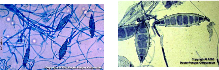
Cigar-shaped macroconidia with smooth ends.

Microsporum canis
Macroconidia are abundant, thick-walled with many septa, up to 15 septa
Macroconidia are often hooked or curved at ends.
Microconidia are small and clavate (club-shaped).
Dermatophytes of cattle
• Most common: Trichophyton verrucosum, but others possible
• Dermatophytosis most commonly in calves
nonpruritic lesions
generalized skin disease may develop
Lesions are characteristically discrete, scaling patches of hair loss with gray-white crust formation
• herd health problem more common winter
• more common in English rather than Zebu breeds
• spontaneous recovery common
• topical treatment + vaccination

Dermatophytes of dogs and cats
• in dogs: Microsporum canis (70%), M. gypseum (20%) and Trichophyton
mentagrophytes (10%)
• in cats, almost always Microsporum canis
• clinical signs
— dogs: alopecia, scaly patches, broken hairs; sometimes regional or
generalized folliculitis and furunculosis with papules and pustules.
— cats: focal alopecia, scaling, and crusting around ears, face or
extremities.
• Often self-limiting in shorthaired pets
• Treatment + hair clipping in longhaired pets

Dermatophytes of horses
Primary causes: Trichophyton equinum and T. mentagrophytes
Clinical signs: one or more patches of alopecia and erythema, scaling, and
crusting, especially in saddle and girth areas
Transmission by direct contact & grooming equipment
Topical treatment
• whole-body rinses
• treatment of individual lesions
Isolation of affected horses

Dermatophytes of pigs, sheep and goats
• in pigs: usually Microsporum nanum, often asymptomatic, not zoonotic
• in sheep/goats: M. canis, M. gypseum, T. verrucosum
• in show lambs problematic, but not commonly in production flocks of sheep
and goats
• Often self-limiting, but may not be apparent before shows.

Dermatophytes
Severity of ringworm disease depends on (1) strains or species of fungus
involved and (2) sensitivity of the host to a particular pathogenic fungus.
More severe reactions occur when a dermatophyte crosses non-host lines
(e.g., from an animal species to man).
• Geophilic species - keratin-utilizing soil saprophytes (e.g., M.
gypseum, T. ajelloi).
• Zoophilic species - keratin-utilizing on hosts - living animals (e.g., M.
canis, T. verrucosum).
• Anthropophilic species - keratin-utilizing on hosts - humans (e.g., M.
audounii, T. tonsurans)
Dermatophytes: diagnosis & treatment
• hair plucks / coat brushings, UV light (Wood's lamp)
• Look for fungal hyphae
• Growth > very slow, start treatment straight away
• Antifungals (examples: griseofulvin, ketoconazole or itraconazole)
• Dip or shampoo
• Clipping of hairs may reduce spreading
• Keep pets/animals in isolation
• Can be zoonotic / spread > PPE

Malassezia
• Found on skin and in ears of healthy dogs and cats
• Can cause dermatitis
• Most common cause: Malassezia pachydermatis
• grows in the stratum corneum
• Clinical signs:
— Erythema and pruritus
— Alopecia
— Usually secondary to underlying disease
• Diagnosis: Acetate strip cytology

Sporotrichosis
• reported in dogs, cats, horses, cows, camels, dolphins, goats etc.
• Zoonotic, especially via cats
• caused by Sporothrix schenckii
• dimorphic and forms mycelia on vegetation
• yeast-like in tissue and media at 37°C
• difficult to treat, very contagious

Environmental fungal organisms
• rare causes of skin disease
• usually present as nodules with/without discharging sinus (e.g. mycetoma)
— Madurella mycetomatis
• Opportunistic subcutaneous fungal infections
• Cutaneous and systemic mycoses (rare in the UK)
Portal of entry
• primary mycoses – respiratory portal; inhaled spores
• subcutaneous - inoculated skin; trauma
• cutaneous and superficial – contamination of skin surface