ADH3 Exam 2 Combined
 Call Kai
 Learn
Learn Practice Test
 Spaced Repetition
 Match
Match Flashcards
 Knowt Play
1/61

There's no tags or description
Looks like no tags are added yet.
Name | Mastery | Learn | Test | Matching | Spaced | Call with Kai | Chat |
|---|
No analytics yet
Send a link to your students to track their progress
62 Terms
CAD clinical manifestations: (3)
stable angina
unstable angina
prinzmetal’s angina

what distinguishes stable (3) from unstable angina (4)?
stable: (3)
CP with activity
linked to fixed plaque
relieved by meds and rest
unstable: (4)
CP at rest (and activity)
most concerning
marks the start of ACS
treated as emergency
what causes Prinsmetal’s angina?
when does it typically occur?
a variant of unstable angina caused by coronary spasms rather than plaque (but atherosclerotic changes are present)
→ occurs @ rest and in clusters b/w midnight and 8AM

What labs would you want to draw for CAD? (7)
total cholesterol
triglycerides
LDL
HDL
CK (creatinine kinase)
⬆= muscle damage
CK-MB (mostly in heart muscle)
⬆= HEART muscle damage
troponin
Whats the gold standard diagnostic test for CAD?
what does it help determine?
Coronary Angiography (left sided heart cath)
→ determines if PTCA/Stent placement is an option
(Percutaneous Transluminal Coronary Angioplasty aka Percutaneous Coronary Intervention)
What defines Coronary Artery Bypass Grafting (CABG):
arteries (chest/arms) or veins (legs) are grafted to bypass blocked coronary arteries while cardiopulmonary bypass (CBP) maintains perfusion
heart is stopped during this to provide bloodless field to work in

whats a complication of CAD?
Acute coronary syndrome (ACS)
aka MI
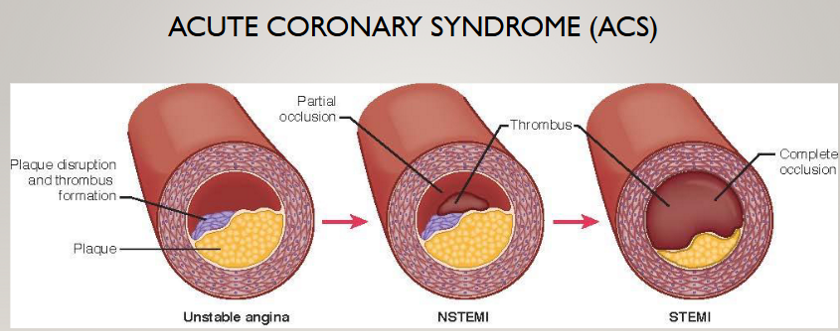
STEMI vs NSTEMI
Key differences b/w STEMI and NSTEMI:
STEMI: (BAAAD)
infarction w/ complete obstruction of blood flow
NSTEMI:
ischemia w/ partial obstruction
Aside from classic chest pain, what other symptoms should nurse watch for in MI? (5)
epigastric discomfort
pain b/w shoulders and jawline
diaphoresis
syncope
SOB
What cardiac biomarkers specific to myocardium and stays elevated the longest? (2)
Troponin I: Normal: < 0.04 ng/mL (MI: > 0.04)
Troponin T: Normal: <0.01 ng/mL
NSTEMI vs STEMI ECG changes:
NSTEMI:
ST depression
STEMI:
ST elevation

emergent treatment bundle for STEMI at presentation: (4)
aspirin
nitroglycerin
morphine
O2 if < 90%
Whys aspirin given 1st in ACS?
Whats the 2 doses?
Key note when taking aspirin?
Given first to ⬇️platelet aggregation (stops platelets from sticking together= ⬇clot formation)
initial dose: 325 mg CHEWED (quick onset)
2nd: 81 mg indefinitely
NOTE: dont take with other NSAIDS—prothrombic!!
What does nitro do?
What to monitor for?
Contraindications: (2)
Nitro is a vasoDILATOR
Monitor: hypotension, HA
Contraindicated: (2)
right ventricular MI
use of PDE-5 inhibitors aka Viagra
*life-threatening hypotension
vasodilation → ↓ preload/BP
How is Nitro given?
Sublingual 0.4 mg q5mins x3

How is morphine used in ACS? (2)
avoid in what and why?
used as adjunct to nitro (also vasodilator)
small doses!!!
1-2 mg IV q5-15 mins if CP not relived by Nitro
AVOID in right ventricular MI
vasodilation → ↓ preload/BP
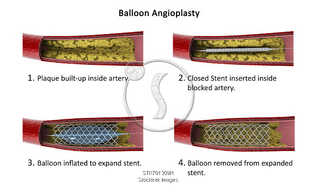
whats the door-to balloon time goal for STEMI treated w/ PCI?
90 mins!!!
Door = hospital arrival
not 90 minutes from when symptoms started…
Balloon = PCI opens the blocked artery
What are signs of a retroperitoneal bleed/Hematoma??? (RBH) (5)
catheter injures femoral artery → blood leaks into retroperitoneal space (behind abdominal organs)
hypotension
tachycardia (may be masked by beta blockers)
abdominal distention
back/groin pain (LATE)
flank ecchymosis (LATE)
how would u treat a retroperitoneal hematoma? (3)
hold pressure
percutaneous intervention w/ balloon tamponade
blood transfusion

Indications (2) for fibrinolytic therapy and whats the biggest risk?
indications: (2)
pain < 6 hrs with STEMI
ST elevation > 1 mm in 2+ leads
risk: BLEEDING!!!
stop anticoags
monitor PT/INR
monitor fibrinogen, urine output, BUN/Cr
give cryoprecipitate and platelets
neuro assessments (brain bleed)
NO punctures, invasive devices
If unable to get to cath lab in time, what would u give for a STEMI? (2)
TNKase (tenecteplase)
rtPA (Activase)
rural areas
Big 5 discharge meds after ACS? (5)
ASA—indefinitely
P2Y12 receptor blockers (Plavix/clopidogrel)
at least 1 yr!!
BB—indefinitely
Statin—indefinitely
ACE-I (or ARB)
EF < 40% or anterior MI
How should P2Y12 inhibitor typically be continued after ACS, and why does this matter for teaching?
At least 1 year
Plavix
continuing dual antiplatelet therapy for full yr is critical
if stopped early = stent thrombosis
preload measured on the Right vs Left side (and normals)
→ Right: RAP (right atrial pressure)
CVP
normal 2-6 mmHg (nickel)
→ Left: PAOP/PAWP
normal: 8-12 mmHg (dime)
Explain central line vs swan-ganz catheter:
Central line: Give meds/fluids through a large vein.
Location: internal jugular, subclavian, or femoral vein.
Measures: CVP (right atrial pressure)
Used for: Vasopressors (NE), long-term IV access (antibiotics)
Swan-Ganz/ Pulmonary Artery Catheter (PAC): measures pressures inside the heart and lungs
Location: passed through the R.A and R.V, and advanced into the pulmonary artery.
Measures: Pulmonary artery pressure (PAP), pulmonary artery wedge pressure (PAWP), and CO.
Used for: determining how well heart pumps and understand pressure and blood-flow problems.
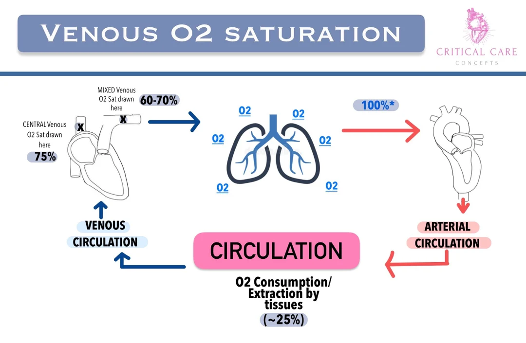
whats SVO2 (3)
hows it drawn?
relationship b/w O2 delivery and O2 extraction at the tissue level
tells u how pt Utilizes O2
% of O2 bound to hgb in blood left over, returning to right side of heart
→ drawn from central line or Swan-Ganz
SVO2 normal value?
High vs low:
normal: > 70% (3 quarters)
High: 70-100 % (aka normal)
Low:
tissues extracting MORE O2 than normal
⬇O2 delivery
SVO2 < 70% means?
tissue extracting more O2 than normal
septic
Cause of ⬇DO2 delivery? (3) and Increased O2 consumption? (6)
DO2:
⬇hemoglobin
⬇CO
⬇SpO2/SaO2
VO2:
shiver
increased work of breathing
fever
infection
agitation
nursing care
giving a full bed bath tires pt out
A- Line measures what? (2)
MAP
< 65 bad = Shock
> 65 good perfusion
BP
How is pressure bag and transducer for A-line set up?
inflate pressure bag to 300mmHg to prevent arterial blood from escaping!!
0.9 NS
transducer at Phlebostatic axis!!

Phlebostatic axis location?
Left atrium @ 4th ICS, MAL

What to NEVER do with an A-Line?
Infuse/flush anything through it!!
- its arterial, not venous
What is Pulses Paradoxus?
causes (3)
exaggerated BP variation w/ respiratory cycle
large drop (> 10 mmHg) in systolic BP when the patient breathes IN.
causes:
cardiac tamponade
COPD
asthma
what 3 meds need A-Lines?
whats their MOA?
NE
Vasopressin
Phenylephrine
all cause ⬆BP, CO
shunts blood back to heart
What meds need/ doesnt need to be tritiated?
Needs:
NE
Phenylephrine
Doesnt:
Vasopressin
NE (6)
monitor
first line med to raise BP
monitor for extravasation
go into tissue instead of blood stream → necrosis
digit/gut ischemia
bradycardia
HTN
renal artery vasoconstriction
⬇urine output < 30mL/hr x 2 hrs
titrate!!!
Vasopressin (3)
2nd line—additive
also for GI bleeding and Diabetes Insipidus
no need to titrate—its a set amount
Phenylephrine
monitor for: (5)
extravasation
reflex bradycardia d/t vasoconstriction and ⬆BP
arrythmias
HTN
CP
titrate!!
Central venous pressure shows what?
placement?
normal pressure?
measures RIGHT side of heart preload
jugular, subclavian, femoral vein
normal CVP: 2-6 mmHg (nickel)
Low vs High CVP number:
Low: “dry
fluid volume deficit
High: fluid volume overload
Pulmonary Artery Catheter (Swan-Ganz) measures what?
right side of heart pressure (PAP)
high vs low
normal BP in pulmonary artery: 25/10 mmHg
high: pulmonary HTN
low: impaired right side HF

Left side: PAOP/PAWP measures what?
Left Atrial pressure
normal: 8-12 mmHg (dime)
PA catheter key balloon rule?
balloon inflated only when moving FORWARD!! never backwards!!
PAWP/PAOP if high means? (6)
fluid volume overload
LV failure
cardiac tamponade
constrictive pericarditis
high PEEP
mitral stenosis
infective endocarditis key assessment findings: (4)
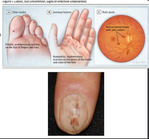
oslers nodes
red, painful nodes in pads of fingers and toes
janeway lesions
red, painless spots on palms and soles
splinter hemorrhages
tiny blood clots that run vertically under nails
murmur
turbulent blood flow across a heart valve
dx test for infective endocarditis: (4)
TTE (transthoracic echo)
regular echo
TEE (transesophageal echo)
more invasive; goes through esophagus to see backside of heart
blood cultures
⬆WBCs

infective endocarditis main tx:
for how long?
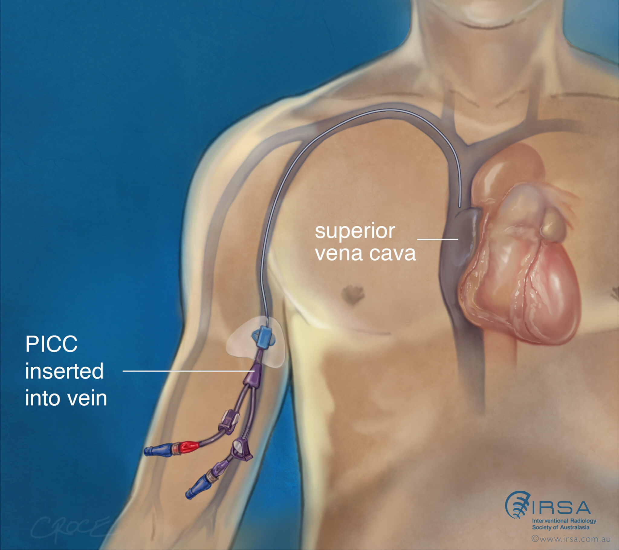
IV abx therapy (PICC line)
standard tx 4-6 weeks but may be longer for prosthetic valves
major complications of endocarditis:
embolic events!!
left side: TIA, stroke, kidney, spleen, liver
right (lungs): pulmonary embolism

nursing actions endocarditis (4)
PICC line for long term abx
standard 4-6 weeks
good oral hygiene
bleeding gums = bacteria entry into blood
inform of previous dental/invasive procedures
prophylactic abx may be prescribed
myocarditis assessment findings:
cardiogenic shock
CP
SOB
palpitations
myocarditis dx tests: (2)
whats definitive?
labs:
C-reactive proteins (inflammatory marker)
troponins
BNP (fluid volume overload)
myocardial biopsy: definitive test—reserved for actue cases!

main nursing education for myocarditis:
avoid strenuous activities!!
athletes no competitive sports while inflammation present
re-evaluate 3-6 months before continuing sport
complications for myocarditis: (2)
dilated cardiomyopathy
heart chamber enlarges and lose contractility
sudden cardiac death in young athletes

pericarditis key findings: (6)
pleuritic CP
pericardial friction rubs: scratchy sounds that occur w/ each heart beat
muffled heart sounds
pericardial effusion
EKG changes: widespread ST Elevation / PR Depression
pulses paradoxus
cardiac tamponade

how is pleuritic CP different from MI?
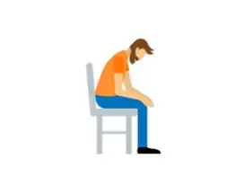
Pleuritic CP relieved by sitting up and leaning forward compared to MI
Dx testing in Pericarditis? (2)
EKG: widespread ST elevation / PR depression
CXR:
cardiomegaly + clear lung fields = pericardial effusion (fluid build-up in pericardial sac)

Complications of pericarditis? (2)
pericardial effusion
fluid build up >20-60 mL in pericardial space
tx: pericardiocentesis
pericardial tamponade
Becks Triad
JVD, muffled heart sounds, hypotension

Nursing interventions for Pericarditis: (3)
sit up and lean forward
ASA, NSAIDs
raise HOB relieves SOB

Stenosis definition
result
narrowed valve opening = ⬇blood flow
ventricular hypertrophy: occurs to generate adequate force to open valve
over time the ventricle will fail and progress to HF

Regurgitation definition
valve leaflets dont properly close = backflow of blood into atrium
signs of L/R sided HF develop

Prolapse definition
valve flaps dont close smoothly (they balloon upward as ventricles contract)
progress to regurgitation
