OBGYN: CTL 2nd/3rd Trimester Pathology 37% (Pt 1)
 Call Kai
 Learn
 Practice Test
 Spaced Repetition
 Match
 Flashcards
 Knowt Play
1/134

There's no tags or description
Looks like no tags are added yet.
135 Terms
Severe chest and lung hypoplasia are suspected if the thoracic circumference is _____.
a. <5th percentile
b. <15th percentile
c. <18th percentile
d. <25th percentile
a
Which of the following is the most common type of non-lethal fetal skeletal dysplasia?
a. heterozygous achondroplasia
b. osteogenesis imperfecta type II
c. thanatophoric skeletal dysplasia
d. achondrogenesis
a
Common complications of a velamentous cord insertion includes vasa previa, IUGR, and _____.
a. placental abruption
b. complete molar pregnancy
c. cord entanglement
d. choriocarcinoma
a
The most common cardiac defect seen with trisomy 21 is?
a. ventricular septal defect
b. patent ductus arteriosus
c. atrial septal defect
d. AV canal defect or AVSD
d
A patient presents for her first ultrasound at 22 weeks. A twin pregnancy with a single placenta is identified. Fetus A demonstrates an increased C/T ratio, mild pleural effusion, and anasarca. No cardiac activity is identified in fetus B. There are also no obvious cranial or cerebral structures. These findings are most suggestive of:
a. twin reversed arterial perfusion sequence
b. vanishing twin syndrome
c. fetus A - hydrops; fetus B - triploidy
d. dichorionic/diamniotic twins
a
Which of the following conditions is the most common cause of hydronephrosis in a fetus?
a. ureteropelvic junction obstruction
b. bladder exstrophy
c. aqueductal stenosis
d. posterior urethral valves
a
Which of the following statements is true regarding the sonographic appearance of fetal bowel?
a. dilated bowel can be differentiated from a dilated ureter by identifying peristalsis in the bowel segment
b. as the fetus ages, the bowel will become more hypoechoic
c. anal atresia will lead to the inability to visualize the fetal bowel due to collapse and atrophy of the digestive tract
d. fetal bowel is not normally visible until after 22 weeks
a
A patient presents for a fetal anatomy scan at 26 weeks. The HC, BPD, and FL measure 25.5 weeks. The AC measures 28.5 weeks. These findings are most suggestive of:
a. hepatic mesenchymal hamartoma
b. symmetric IUGR
c. asymmetric IUGR
d. hydrops
a
A monochorionic diamniotic twin pregnancy demonstrates 2 female fetuses with normal fetal heart rates. Fetus A is 25% larger than fetus B. Polyhydramnios is present with fetus A and oligohydramnios is demonstrated with fetus B. These findings are most suggestive of:
a. twin reversed arterial perfusion sequence
b. twin to twin transfusion
c. vanishing twin syndrome
d. molar twin pregnancy
b

What is demonstrated on the image?
a. club foot and syndactyly
b. arthrogryposis and polydactyly
c. club foot and polydactyly
d. rocker bottom foot and polydactyly
c
What are the expected US findings in vasa previa?
a. placenta percreta where the endometrium has invaded the myometrium and serosa causing hemorrhage
b. the internal os is covered by the umbilical vessel attachment to a low lying or marginal placenta
c. the internal os is covered by a large blood clot caused by placental abruption
d. the internal os is completely covered by the base of the placenta
b
In TTTS, the donor twin demonstrates _________ and in TRAP, the donor twin demonstrates ______.
a. growth restriction, hydrops
b. polyhydramnios, oligohydramnios
c. pleural effusion, ascites
d. CHF, no cardiac structure
a
You are performing a 4 week follow up on twins that should be 14 weeks. On today’s exam, there is only a single normal fetus present and an irregular anechoic area adjacent to the normal sac. The patient has not experienced any bleeding. These findings are most suggestive of:
a. stuck twin syndrome
b. vanishing twin syndrome
c. twin to twin transfusion
d. parabiotic twin
b
Which of the following statements regarding Dandy Walker Malformation (DWM) is true?
a. DWM is more commonly an isolated finding, without other defects
b. a neonate with DWM will usually develop normal intelligence and no long term effects on brain function
c. Dandy-Walker syndrome is frequently associated with agenesis of the corpus callosum, malformations of the heart, face, limbs, and digits
d. DWM is caused by cystic dilatation of the 3rd ventrile
c
What is the most common congenital cardiac anomaly?
a. VSD
b. patent ductus arteriosus
c. Tetralogy of Fallot
d. ASD
a
While performing a scan on a 21 week fetus you identify ectopia cordis, club foot, and an asymmetric cleft lip/palate. Which of the following is the most likely cause of this finding?
a. Patau syndrome
b. Pentalogy of Cantrell
c. amniotic band syndrome
d. Tetralogy of Fallot
c
Premature delivery is birth of the fetus that occurs before ____ weeks gestation.
a. 33
b. 35
c. 38
d. 37
d
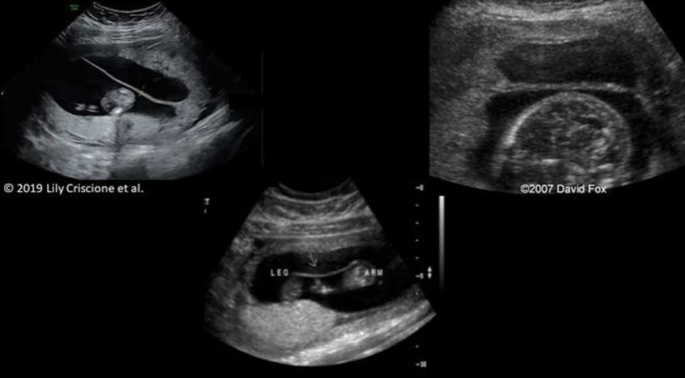
Which image is most suggestive of uterine synechia?

Which of the following is NOT considered a critical finding on a fetal US exam?
a. circummarginate placenta
b. cervical pregnancy
c. placental abruption
d. umbilical cord knot
a
Which of the following skeletal anomalies usually occurs as an isolated finding?
a. arthrogryposis
b. phocomelia
c. rhizomelia
d. talipes equinovarus
d
You are performing a follow up exam for limited fetal anatomy due to non-visualization of the stomach and kidneys at 15 weeks. The fetus should be 21 weeks today. You identify two normal kidneys, but the stomach remains barely visible. Borderline polyhydramnios is also present. These findings are most suggestive of:
a. anal atresia
b. trisomy 21
c. esophageal atresia
d. situs inversus
c

What cardiac defect is seen on the image?
a. muscular VSD
b. patent foramen ovale
c. aortopulmonary window
d. sinus venosus ASD
a
Tetralogy of Fallot consists of a group of 4 primary cardiac defects that include right ventricular hypertrophy, VSD, pulmonary stenosis, and:
a. overriding aorta
b. interrupted IVC
c. aortic coarctation
d. small left ventricle
a
If an isolated cystic hygroma is present, AFP levels will be ________. If a cystic hygroma is present in a fetus with Turner syndrome, AFP levels will be ________.
a. decreased, increased
b. normal, decreased
c. normal, increased
d. increased, decreased
b
What is the most common cause of death in lethal skeletal dysplasia?
a. hypoplastic heart
b. umbilical cord knot
c. pulmonary hypoplasia
d. renal failure
c
While performing a scan on a 27 week fetus, you document echogenic foci in the liver and a thick walled cyst in the fetal abdomen. Several small bowel loops appear dilated with limited peristalsis. Fetal ascites and polyhydramnios are also present. These findings are most consistent with:
a. umbilical cord knot
b. trisomy 21
c. duodenal atresia
d. meconium peritonitis
d

Which of the following does NOT cause the abnormality seen on the image?
a. teratoma
b. Chiari malformation
c. anencephaly
d. aqueductal stenosis
c

_________ is demonstrated on the image and is associated with ________.
a. Vein of Galen aneurysm, CHF
b. agenesis of the corpus callosum, triploidy
c. middle cerebral artery aneurysm, fetal demise
d. dilated 3rd ventricle, spina bifida
a

What is demonstrated on the image?
a. cardiomegaly
b. congenital cystic adenomatoid malformation
c. diaphragmatic hernia
d. normal thoracic cavity
c
Asymmetric IUGR:
a. involves shunting of arterial blood flow due to head sparing
b. usually presents early in the 2nd trimester
c. demonstrates a head circumference more than 2 SD smaller than the abdominal circumference
d. is typically related to aneuploidy
a
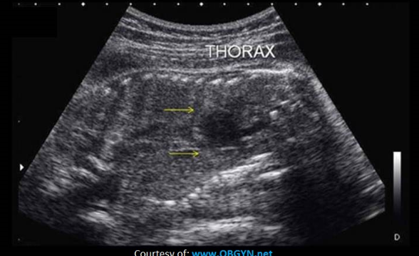
Which of the following are associated with the findings on the image?
a. congenital cystic adenomatoid malformation and pulmonary sequestration
b. Potter syndrome and diaphragmatic hernia
c. Vein of Galen malformation and cardiomegaly
d. thanatophoric skeletal dysplasia and osteogenesis imperfecta
d

What fetal skeletal anomaly is displayed on the images?
a. syndactyly
b. club foot
c. rocker bottom foot
d. clinodactyly
b
Which of the following correctly describes the sonographic appearance of the pump twin in a TRAP sequence?
a. skin edema, ascites, CHF
b. asymmetric IUGR with aged placenta
c. donor twin usually appears normal
d. symmetric IUGR with normal placenta
a
What maternal condition has been linked to umbilical vein thrombosis?
a. non-immune hydrops
b. trisomy 13
c. diabetes
d. anemia
c
__________ occurs when AV malformations in the placenta shunt blood from one twin to the other twin and the donor twin develops growth restriction.
a. TTTS
b. acardiac twin
c. umbilical cord knot
d. hydrops of the donor twin
a
Which of the following is the most common facial anomaly?
a. hypotelorism
b. micrognathia
c. hypertelorism
d. cleft lip/palate
d
Symmetric IUGR occurs when:
a. all parts of the fetus is reduced in size by >2 standard deviations for the current gestational age
b. the HC measures more than 2 standard deviations smaller than AC for the current gestational age
c. the AC measures more than 2 standard deviations smaller than HC for the current gestational age
d. the limbs are abnormally shortened with a relatively normal HC and AC
a
What is the most common abnormality of the umbilical cord?
a. single artery
b. cord varix
c. cord knot
d. cord cyst
a
Ebstein anomaly is defined as:
a. abnormal placement of the tricuspid valve more inferiorly into the apex of the left ventricle
b. abnormal placement of the tricuspid valve more inferiorly into the apex of the right ventricle
c. abnormal placement of the mitral valve more inferiorly into the apex of the left ventricle
d. abnormal placement of the mitral and tricuspid valves more inferiorly into the apex of the ventricles
b
You are evaluating the aortic arch and descending aorta on a 25 week fetus. There is a narrowing of the aorta just below the origin of the left subclavian artery. Color doppler demonstrates aliasing at the narrowed area with some flow moving distally in the descending aorta. Which of the following best describes these findings?
a. stenosis of the ductus arteriosus
b. interrupted aortic arch
c. coarctation
d. truncus arteriosus
c

The image demonstrates ________, which is a common finding in cases of ______.
a. hypertelorism, trisomy 21
b. cystic hygroma, monosomy x
c. hypotelorism, trisomy 13
d. cerebellar agenesis, Dandy Walker syndrome
c
An encephalocele can easily be mistaken for ______.
a. cystic hygroma
b. gastroschisis
c. epignathus
d. porencephaly
a
Monochorionic twins over 6 weeks GA should be evaluated every 2 weeks for possible complications. Doppler evaluation of the ______ for discordant flow resistance and evaluation of the _________ for discordant flow velocities must be included in the exams.
a. umbilical artery, MCA
b. MCA, umbilical artery
c. ductus venosus, uterine artery
d. uterine artery, umbilical artery
a
__________ twins have the highest incidence of umbilical cord knotting.
a. monochorionic/diamniotic
b. monochorionic/monoamniotic
c. conjoined
d. dichorionic/diamniotic
b

Which of the following is the correct description of the placenta demonstrated on the images?
a. placenta previa
b. circumvallate placenta
c. normal grade II placenta
d. placental abruption
b
What urinary anomaly is caused by a urinary obstruction in the first trimester and leads to renal cyst formation and ureteral atresia?
a. adult PCKD
b. multicystic kidney disease
c. infantile PCKD
d. Potter syndrome
b

Which of the following best describes the image displayed?
a. there are normal kidneys seen on either side of normal lumbar spine
b. there are two normal kidneys but spina bifida is present
c. there are two normal kidneys seen but there is a cystic hygroma present
d. there are no kidneys identified on either side of the normal lumbar spine
b
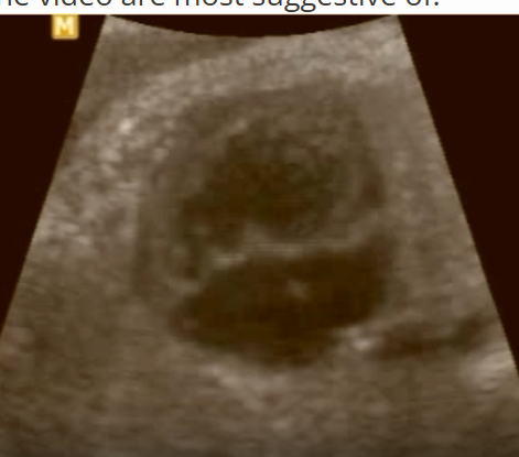
The findings on the video are most suggestive of:
a. normal 4 heart chamber
b. AVSD
c. Tetralogy of Fallot
d. hypoplastic left heart
b
In a 32 week pregnancy, a cervix that is less than ______ in length and dilated more than _____ is considered incompetent.
a. 3 cm, 2 cm
b. 2 cm, 2 cm
c. 3 cm, 3 cm
d. 4 cm, 2 cm
a
While scanning a 22 week fetus, you identify normal long bones in the lower extremities but the feet are very small with shortened tarsals and metatarsals. Which of the terms describes this isolated finding?
a. clinodactyly
b. rhizomelia
c. acromelia
d. phocomelia
c

What intracranial anomaly is commonly associated with the abnormality on the image?
a. holoprosencephaly
b. Vein of Galen aneurysm
c. lissencephaly
d. Dandy Walker malformation
a
A patient presents at 20 weeks with their size measuring 12 days behind where they should be according to their first trimester US. The cranial bones appear less echogenic and the humerus and femur appear slightly bowed. The small bones in their extremities are also somewhat difficult to identify due to decreased echogenicity. These findings are most suggestive of:
a. thanatophoric dwarfism
b. osteogenesis imperfecta
c. IUGR
d. achondroplasia
b
What is the most commonly identified fetal chest mass?
a. diaphragmatic hernia (digestive tract segment above the diaphragm)
b. congenital cystic adenomatoid malformation (CCAM)
c. pulmonary sequestration
d. cardiac sarcoma
b
You are performing a follow up exam for limited fetal anatomy due to nonvisualization of the stomach and kidneys at 15 weeks. The fetus should be 21 weeks today. On today’s exam, you identify a single normal kidney, but the stomach remains barely visible. Borderline polyhydramnios is also present. The head measures 3 standard deviations lower than the abdomen and femoral measurements. Rocker bottom feet are also present. These findings are most consistent with:
a. Noonan
b. Ellis Van Creveld
c. Trisomy 13
d. Trisomy 18
d

If the fetus is in breech position, what is demonstrated on the image?
a. encephalocele
b. spina bifida
c. teratoma
d. chorioangioma
c
When evaluating the abdominal circumference of a 30 week fetus, you note that the umbilical vein curves to the left toward the stomach and the GB is medial to the vein. The AC measurement and the skin thickness are normal. These findings are most suggestive of:
a. normal anatomy
b. absent ductus venosus
c. persistent right umbilical vein
d. situs inversus
c
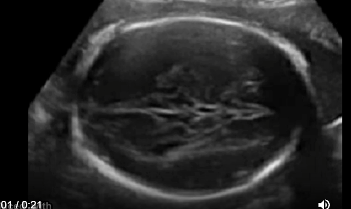
The findings on the video are most suggestive of:
a. normal fetal anatomy
b. ACC
c. Chiari malformation
d. schizencephaly
b

Most forms of skeletal dysplasia demonstrate a foreshortened femur throughout pregnancy, except ________ that typically has a normal femur length until later in the 2nd trimester.
a. heterozygous achondroplasia
b. thanatophoric skeletal dysplasia
c. osteogenesis imperfecta
d. campomelic dysplasia
a

What is demonstrated on the coronal view of the thoracic and lumbar spine?
a. an enlarged echogenic kidney
b. renal agenesis
c. normal lung tissue
d. abnormal splaying of the vertebral bodies
b
A 20 week fetus demonstrates an echogenic mass below the liver and a dilated duodenum. Color doppler demonstrates the “whirlpool” sign adjacent to the mass. The AFI measures 28 cm. These findings are most suggestive of:
a. midgut volvulus
b. gastroschisis
c. duodenal atresia
d. allantoic cyst
a

Which of the following abnormalities is displayed on the image provided?
a. ascites
b. pericardial effusion
c. pleural effusion
d. hydronephrosis
c
In the 2nd trimester, documenting a normal lens in each eye on the fetal US can rule out which of the following abnormalities?
a. holoprosencephaly
b. arhinia
c. stigmatism
d. cataracts
d
IUGR is suspected when the EFW% is less than _____ for the GA.
a. 3rd percentile
b. 15th percentile
c. 10th percentile
d. 5th percentile
c
Which of the following characteristics indicates multicystic kidney disease is present?
a. multiple cysts that replace the renal sinus with a normal renal cortex
b. multiple noncommunicating cysts that do not connect to a dilated ureter
c. multiple communicating cysts that connect to a dilated ureter
d. multiple microcysts that cause increased echogenicity of the kidney
b

A patient presents for a fetal ultrasound due to higher than expected bhCG levels. Her LMP was 8 weeks ago. These findings are most suggestive of:
a. normal twin pregnancy
b. twin pregnancy in bicornuate uterus
c. twin pregnancy in septate uterus
d. heterotropic pregnancy
d
Which anomaly is almost always associated with a tracheoesophageal fistula?
a. megacystis
b. Bochdalek hernia
c. duodenal atresia
d. esophageal atresia
d
What delivery complication is most commonly associated with velamentous cord insertion?
a. premature rupture of membranes
b. fetal respiratory distress
c. meconium peritonitis
d. vasa previa
d
You are evaluating a 20 week fetus for size and anatomy evaluation. A mildly hyperechoic, homogeneous, triangular shaped mass is identified inferior to the level of the heart in the RT chest cavity. The remainder of the chest cavity and the heart are normal. These findings are most suggestive of:
a. pulmonary sequestration
b. rhabdomyoma
c. type I CCAM
d. normal thymus gland
a

A pregnant patient presents with a recent acute onset of hypertension, nausea, and vomiting. She has no pain, cramping, or vaginal bleeding. A prior ultrasound exam at 6 weeks indicates the pregnancy should now be 17 weeks 6 days in GA. The findings demonstrated on the video should cause significant suspicion of:
a. placental abruption
b. gestational trophoblastic disease
c. omphalocele
d. subchorionic hemorrhage
b
What is anasarca commonly seen with?
a. fetal hydrops
b. IUGR
c. achondrogenesis
d. thanatophoric dwarfism
a
Which of the following is the most common type of lethal fetal skeletal dysplasia?
a. osteogenesis imperfecta type ii
b. homozygous achondroplasia
c. achondrogenesis
d. thanatophoric skeletal dysplasia
d
Which of the following describes the sonographic appearance of possible intestinal atresia in a 29 week fetus?
a. small bowel loops dilated at 3 mm luminal diameter
b. pleural effusion, megacystis, and ascites
c. decreased or absent peristalsis of bowel
d. hyperechoic bowel with increased peristalsis
c
Which of the following statements regarding micrognathia is true?
a. micrognathia is seen with polyhydramnios in 70% of cases due to the presence of dysphagia
b. micrognathia is seen with oligohydramnios in 70% of cases due to the presence of dysphagia
c. micrognathia is seen with oligohydramnios in 70% of cases due to the presence of associated renal anomalies
d. micrognathia is seen with polyhydramnios in 70% of cases due to the presence of associated renal anomalies
a
A circumvallate placenta:
a. is commonly associated with chromosomal anomalies
b. is caused by back folding of the placenta and the fetal membranes toward the chorionic surface
c. causes placental thickening associated with fetal hydrops
d. causes placental insufficiency and IUGR
b
Gestational diabetes:
a. is associated with macrosomia
b. typically causes IUGR in the 3rd trimester
c. causes placental thinning and calcification formation
d. results in sirenomelia in most pregnancies
a
Which of the following is NOT an expected post-partum complication?
a. thrombosis/embolism
b. gestational diabetes
c. metritis
d. hemorrhage
b
If posterior shadowing from the skull, ribs, and long bones are demonstrated, which of the following anomalies can be ruled out?
a. hypophophatasia
b. Ellis van Creveld syndrome
c. arthrogryposis
d. craniosynostosis
a
A small, underdeveloped right ventricle, right atrium, and pulmonary artery are collectively referred to as:
a. double outlet right ventricle
b. Noonan syndrome
c. Ebstein anomaly
d. hypoplastic right heart syndrome
d

A solid mass is identified attached to the ventral wall of a 22 week fetus. There appears to be no abdominal wall or fetal skin covering the mass, but the abdominal vasculature appears to be shared between the fetus and the mass. Which of the following is the most likely diagnosis?
a. parasitic conjoined twins
b. prune belly syndrome
c. gastroschisis
d. omphalocele
c
You are evaluating the heart on a 28 week fetus. A single large trunk vessel exits the heart and the pulmonary artery appears to branch from it instead of from the right ventricle. These findings describe:
a. total anomalous pulmonary venous return
b. partial anomalous pulmonary venous return
c. double outlet left ventricle
d. truncus arteriosus
d
All the following can cause a false positive diagnosis of club foot, except:
a. 8 cm intramural fibroid
b. footling breech position
c. uncooperative fetus
d. oligohydramnios
c
What urinary anomaly usually affects one kidney, but can lead to bilateral renal enlargement due to contralateral renal compensation?
a. adult PCKD
b. infantile PCKD
c. Potter syndrome
d. multicystic kidney disease
d
Which of the following is true regarding autosomal dominant polycystic kidney disease?
a. usually occurs unilaterally
b. requires 2 abnormal genes to occur in the fetus
c. usually associated with early fetal demise
d. if it occurs in a fetus, at least one parents has ADPKD
d
A mother presents with a history of heterozygous achondroplasia. The father does not share this characteristic. What is the chance that their fetus will have the same skeletal dysplasia?
a. 50%
b. <25%
c. 100%
d. 25%
a

A patient presents for a fetal ultrasound for a routine anatomy evaluation. In addition to the findings on the image, an encephalocele and polydactyly are also noted. These findings are most suggestive of:
a. Turner syndrome
b. trisomy 18
c. Meckel Gruber syndrome
d. trisomy 13
c
Which of the following statements regarding fetal hydrops is correct?
a. non-immune and immune fetal hydrops can be distinguished from one another by their very different sonographic appearance/characteristics
b. if the fetal hydrops is caused by Rh incompatibility, it is labeled as non-immune
c. immune hydrops can be caused by over 120 different diseases/abnormalities/congenital defects
d. fetal ascites, scalp edema, and skin edema are common sonographic features of fetal hydrops
d

What facial anomaly on the image is commonly associated with Beckwith-Wiedemann syndrome?
a. frontal bossing
b. proboscis
c. macroglossia
d. midface hypoplasia
c

What are the arrows pointing to on the 4 chamber heart image?
a. multiple echogenic intracardiac foci
b. hypoplastic left heart
c. rhabdomyoma
d. pulmonary sequestration
c

Which of the following is correct regarding the abnormality demonstrated on the image?
a. the fetus most likely has Potter syndrome
b. the fetus is most likely female
c. the fetus is most likely male
d. the fetus most likely has Turner syndrome
c
While scanning a 21 week fetus, you identify splaying of the cerebellar lobes with absence of the cerebellar vermis. The 13 mm cisterna magna is anechoic and the 4th ventricle is dilated. These findings are most consistent with:
a. trisomy 13
b. schizencephaly
c. hydranencephaly
d. Dandy Walker malformation
d
If the LVOT and RVOT are parallel as they exit the fetal heart,
a. complete transposition of the great vessels is demonstrated
b. tetralogy of Fallot is demonstrated
c. the fetus has normal cardiac outflow anatomy
d. truncus arteriosus is demonstrated
a

Trisomy 21 is suspected. Which of the above images is most suggestive of that diagnosis?
D

Which of the following best describes the findings on the image?
a. there is a large amount of meconium funneled toward the cervix
b. complete placenta previa is present
c. the maternal bladder is too full to accurately evaluate the cervix
d. a chorioangioma has formed at the internal os
b
Microtia is a strong indicator of:
a. trisomy 18
b. trisomy 21
c. triploidy
d. Turner syndrome
b

Potter syndrome is suspected. Which of the above images is most suggestive of that diagnosis?
D
A trident hand is commonly seen with which skeletal abnormality?
a. spina bifida aperta
b. heterozygous achondroplasia
c. spina bifida occulta
d. osteogenesis imperfecta II
b
Which of the following correctly describes asymmetric IUGR?
a. the limbs are abnormally shortened with a relatively normal HC and AC
b. the HC measures more than 2 standard deviations smaller than AC for the current GA
c. the AC measures more than 2 standard deviations smaller than HC for the current GA
d. all parts of the fetus to be reduced in size by more than 2 standard deviations for the current GA
c

_________ is demonstrated on the image and is commonly associated with _________.
a. frontal bossing, trisomy 21
b. macroglossia, Beckwith-Weidemann syndrome
c. micrognathia, trisomy 18
d. midface hypoplasia, trisomy 13
c

The findings in the video are most suggestive of:
a. acrania
b. anencephaly
c. absence of corpus callosum
d. encephalocele
b
Malformation of the cloaca:
a. is the most common cause of fetal urethral obstruction in females
b. leads to significant polyhydramnios
c. most commonly occurs as an isolated anomaly
d. is commonly associated with a cleft lip/palate
a