OMFPR 20 - Reactive Lesions (Dr. Lerman)
 Call Kai
 Learn
 Practice Test
 Spaced Repetition
 Match
 Flashcards
 Knowt Play
1/133

There's no tags or description
Looks like no tags are added yet.
134 Terms
Epulis fissuratum
What condition?
- Inflammatory fibrous hyperplasia, denture epulis
- Tumor-like hyperplasia associated with flange of poorly-fitting denture
Epulis
Define the following:
Tumor of gingiva or alveolar mucosa
Epulis fissuratum
What condition has the following characteristics?
- Single or multiple folds of hyperplastic tissue in alveolar vestibule
- Fibroepithelial polyp/leaf-like denture fibroma may be seen along palatal mucosa
Epulis fissuratum
ID the pathology:

Epulis fissuratum
ID the pathology:

Epulis fissuratum
70 y/o edentulous patient presents with multiple folds of hyperplastic tissue in maxillary alveolar vestibule. Patient has had same denture for 15 years. Histopathology shows papillary hyperplasia and/or pseudoepitheliomatous hyperplasia with hyperplasia of connective tissue. What is the diagnosis?

Epulis fissuratum
80 year old patient who has worn a denture for the last 10 years. What condition do you suspect?

Epulis fissuratum
ID the pathology:

Epulis fissuratum
ID the pathology:

vestibules
What is most common location for Epulis Fissuratum in oral cavity?

Epulis fissuratum
What condition has the following histopathologic features?
- Hyperparakeratosis or hyperorthokeratosis
- Papillary hyperplasia and/or pseudoepitheliomatous hyperplasia
- Hyperplasia of connective tissue
- Surgical removal
- Remake denture to prevent recurrence
What are two treatments for Epulis Fissuratum?
Inflammatory Papillary Hyperplasia
What condition has the following characteristics?
- Denture papillomatosis
- Reactive tissue growth
- Associated with poorly-fitting dentures, poor denture hygiene, and/or continuous denture wear
Inflammatory Papillary Hyperplasia
ID the pathology:
- Hard palate beneath denture base
- Pink or red pebbly mucosa
- Erythema suggests secondary candidal infection
Inflammatory Papillary Hyperplasia with secondary candidal infection
75 y/o patient presents with a sharply demarcated red pebbly erythematous palatal mucosa when the denture is removed. Patient is known to have less than acceptable oral hygiene. Histo path reveals pseudoepitheliomatous hyperplasia. What condition do you suspect?

Inflammatory Papillary Hyperplasia
ID the pathology:

Inflammatory Papillary Hyperplasia with secondary candidal infection
ID the pathology:
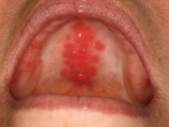
secondary candidal infection
if a patient with Inflammatory Papillary Hyperplasia notices that the palatal mucosa appears to display erythema, what is that indicative of?

Inflammatory Papillary Hyperplasia
ID the pathology:
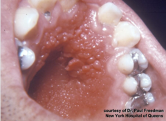
Inflammatory Papillary Hyperplasia
What condition has the following histopathologic features?
- Papillary growths on surface
- May show pseudoepitheliomatous hyperplasia
- Sialadenitis

true
t/f: mild cases of Inflammatory Papillary Hyperplasia may resolve with removal of dentures
relining/refabricating denture following excision of hyperplastic tissue
What is the course of treatment for established cases of Inflammatory Papillary Hyperplasia?
true
t/f: antifungal therapy can be used for treatment of inflammatory papillary hyperplasia if necessary
True
T/F: Traumatic ulcer is one of the MOST COMMON oral pathologies
Traumatic ulcer
ID the pathology:

Traumatic ulcer
patent presents with ulceration on the ventrolateral border of the tongue. Patient has a broken restoration on the same side and patient complains that the tongue keeps "getting snagged" on it. Histo report shows granulation tissue. You follow up with patient in two weeks and the ulceration is gone. What is the diagnosis?

Traumatic ulcer
What condition has the following histopathologic features?
- Ulcerated surface with fibrinopurulent membrane
- Granulation tissue
- Variable amounts of inflammation
Traumatic ulcer
ID the pathology:

Anesthetic-associated lip bite
- History of recent dental treatment
- Often secondary to inferior alveolar nerve block
What is a common, dental-related cause of a traumatic ulcer?
Traumatic ulcerative granuloma
patient presents with a deep ulceration with a white hyperkeratotic rim and irregular borders on the dorsal tongue. You decide to watch it, and follow up with patient in 2 weeks, but patient didn't return to your office for 6 weeks, and the ulceration has not resolved yet. Histo report shows granulation tissue with numerous eosinophils. What is the diagnosis?

Traumatic ulcerative granuloma
ID the pathology:
- Not a true granuloma
- Slow-healing, penetrating ulcer takes weeks to months to resolve
- May be mistaken for maligancy
Traumatic ulcerative granuloma
ID the pathology:
- Male predilection
- Most common on tongue
- Often surrounded by white hyperkeratotic rim
- - May help distinguish from aphthous ulcers or SCCA
Traumatic ulcerative granuloma
ID the pathology:

Traumatic ulcerative granuloma
ID the pathology:

white hyperkeratotic rim
Although you STILL need to perform a biopsy, what feature of a Traumatic Ulcerative Granuloma might help distinguish it from aphthous ulcers or SCCA?
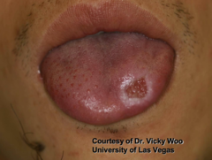
tongue, males
Traumatic Ulcerative Granuloma commonly occur in what location and in what gender?

Traumatic ulcerative granuloma
ID the pathology:
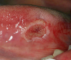
Traumatic ulcerative granuloma
ID the pathology:

- Aphthous ulcers
- SCCA
When detecting a Traumatic Ulcerative Granuloma, what other oral pathologies might be in the differential diagnosis?

Traumatic ulcerative granuloma
What condition has the following histopathologic features?
- Numerous eosinophils
- - May be called "traumatic ulcerative granuloma with stromal eosinophilia (TUGSE)"
- Inflammation of skeletal muscle
- Granulation tissue

- Remove irritant
- Excision of excess tissue may be necessary
- Topical and/or intra-lesional steroids
What are three treatments for a traumatic ulcerative granuloma?
false
t/f: Traumatic Ulcerative Granuloma are true granulomas
false, papillary but not associated with HPV
t/f: a Verruciform Xanthoma is papillary and associated with HPV
Verruciform Xanthoma
ID the pathology:
- Hyperplastic epithelium of mouth, skin, and genitalia
- Lipid-laden macrophages beneath epithelium
- Papillary, but not associated with HPV

Verruciform Xanthoma
ID the pathology:
- 50% of oral lesions on gingiva or alveolar mucosa
- May be pink, white, red, yellow, or orange
- Well-demarcated verrucous mass
- May resemble squamous papillomas or early carcinomas
Verruciform Xanthoma
ID the pathology:

Verruciform Xanthoma
ID the pathology:
- Lipid-laden macrophages beneath epithelium

Verruciform Xanthoma
ID the pathology:
- HPV negative

Verruciform Xanthoma
patient presents with a round flat lesion on the maxillary palatal gingiva that is well demarcated, verrucous in texture, and has pink, red, white and yellowish tones. Patient is HPV negative.
Histo report shows a papillary, acanthotic surface covered by parakeratin and large macrophages with foamy cytoplasm in the connective tissue. What is the diagnosis?

Verruciform Xanthoma
ID the pathology:
- Lipid-laden macrophages beneath epithelium
- HPV negative

gingiva or alveolar mucosa
What is the most common location for a Verruciform Xanthoma to present in oral cavity (50%)?

- Squamous papillomas
- Early carcinomas
When detecting a Verruciform Xanthoma, what other oral pathologies might be in the differential diagnosis?
Verruciform Xanthoma
What condition has the following histopathologic features?
- Papillary, acanthotic surface covered by parakeratin
- Large macrophages with foamy cytoplasm

Conservative excision
(Recurrences rare)
What is the treatment for Verruciform Xanthoma?
- Aspirin
- Mouthwashes / Hydrogen peroxide
- Tooth-whitening products
What are three type of factitial medicaments that cause oral chemical injuries?
Aspirin burn
A 42-year-old patient presents with a white, sloughing, painful lesion on the buccal mucosa adjacent to a maxillary molar. He reports placing an aspirin tablet against the tooth for pain relief the night before. What is the most likely diagnosis?
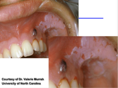
Aspirin burn
Patient presents to your office with a well demarcated white sloughing of the vestibular mucosa. Patient does not use tobacco products. Patient claims that their were experiencing tooth pain and "put a pill in the area to make it better". What most likely causes the reaction?

Aspirin burn
A 38-year-old patient presents with a white, leathery patch on the right buccal mucosa that is tender and partially sloughs off when gently scraped, revealing an erythematous surface. He reports holding an aspirin tablet against his cheek for a toothache. What is the most likely diagnosis?
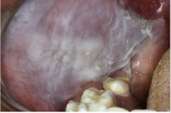
mouth wash
patient present to your office complaining that their "mouth skin is peeling". Patient has excellent oral health and is a self-proclaimed germaphobe. Patient says they brush 3 times a day, use mouth wash every 2 hours, and floss after every meal. What most likely causes the reaction?

- Silver nitrate
- Phenol
- Endodontic materials
- Cotton roll "burn"
What are four types of iatrogenic injuries that cause oral chemical injuries?
Rubber dam application
What can reduce incidence of iatrogenic injuries that cause oral chemical injuries?
Cotton roll "burn"
What is the condition?
- Medicament concentrated against the tissue
- - e.g., Acid etch solution
- Removal of roll without first moistening strips mucosa

all are complications of chemotherapy
Which of the following are complications of chemotherapy? Select all that apply.
a. mucositis occurs within days
b. bone marrow suppresion (thrombocytopenia, agranulocytsosi)
c. opportunistic infections (HSV, candiasis)
Mucositis
ID the pathology:

Mucositis
ID the pathology:

Mucositis
Patient presents with severe loss of mucosal epithelium, bleeding and ulceration. Patient complains of trouble with eating, talking and swallowing. Patient recently had chemotherapy. What is the diagnosis?

- Medication
- Radiation
- Infections
- Chemicals
- Trauma
- Idiopathic
What are six causes of osteonecrosis of the jaws?
- Bisphosphonates
- Denosumab (prolia, Xgeva)
What are two anti-resorptive agents that have been associated with causing Osteonecrosis of the jaws?
- Monoclonal antibodies (Bevacizumab)
- Tyrosine kinase inhibitors (Sunitinib, Sorafenib)
What are two anti-angiogenic agents that have been associated with causing Osteonecrosis of the jaws?
osteoclast inhibition
what is the mode of action for the drugs that can cause Osteonecrosis of the jaws?
Osteonecrosis of the jaws
patient presents with visual necrotic bone on the mandibular myohyoid ridge area. Patient denies having cancer or radiotherapy. Patient is currently being treated for osterporosis and is taking Bisphosphonates. What is the diagnosis?

- Osteoporosis/osteopenia
- Multiple myeloma
- Metastatic carcinomas to bone
What are the three most common indications for being prescribed Bisphosphonates?
osteonecrosis of the jaws (MRONJ)
This is the diagnostic criteria for what?
- Current or previous treatment with anti-resorptive OR anti-angiogenic agents
- Exposed bone OR bone that can be probed through a sinus tract persisting > 8 weeks
- No h/o radiation or obvious metastasis to the jaws

osteonecrosis of the jaws (MRONJ)
All of the following are conditions to consider and rule out with what?
- Alveolar osteitis
- Gingivitis/periodontitis
- Sinusitis
- Caries
- Periapical pathology
- Fibro-osseous diseases
- Cancer
- Condensing osteitis
- Temporomandibular disorders
osteonecrosis of the jaws (MRONJ)
The following are pathogenesis for what?
- Anti-osteoclastic
- Anti-angiogenic
- Inflammatory/infectious
- Immune dysfunction
- Soft tissue toxicity
C. Etidronate (equivalent to Clondronate too!)
Which of the following bisphosphonates has the lowest relative potency?
A. Tiludronate
B. Pamidronate
C. Etidronate
D. Alendronate
D. Zoledronic acid
A patient with metastatic breast cancer receives monthly IV bisphosphonate therapy. Which medication carries the highest risk for medication-related osteonecrosis of the jaw (MRONJ)?
A. Alendronate
B. Risedronate
C. Ibandronate
D. Zoledronic acid
True
T/F: Nitrogen-containing bisphosphonates have significantly higher antiresorptive potency.
Bisphosphonates
All of the following are indications for what medication?
- Osteoporosis/osteopenia
- Multiple myeloma
- Metastatic carcinomas to bone
- Paget disease
- Osteogenesis imperfecta
Bisphosphonates
A 67-year-old woman with a history of osteoporosis, recently diagnosed multiple myeloma, and new bone pain from metastatic breast carcinoma, is started on an IV medication to reduce skeletal-related events. This drug is also commonly used in patients with Paget disease and osteogenesis imperfecta. Which class of medications is she most likely receiving?

Bisphosphonates
A patient with osteoporosis, multiple myeloma, and bone metastases is prescribed a medication also used for Paget disease and osteogenesis imperfecta. Which drug class is this?

osteonecrosis of the jaws (MRONJ)
A 70-year-old woman taking alendronate for osteoporosis presents with exposed mandibular bone that has not healed for 8 weeks following a tooth extraction. She denies prior radiation therapy. What is the most likely diagnosis?

stage 0
a patient that has osteonecrosis of the jaws and the bone is not exposed would be in what AAOMS stage?
stage 1
a patient that has osteonecrosis of the jaws and the bone is exposed but the patient is asymptomatic would be in what AAOMS stage?
stage 2
a patient that has osteonecrosis of the jaws and the bone is exposed AND the patient is symptomatic would be in what AAOMS stage?
stage 3
a patient that has osteonecrosis of the jaws and the bone is exposed, the patient is symptomatic and the disease so extensive that it caused a mandibular fracture would be in what AAOMS stage?
True
T/F: Increased radiopacity may be noted before clinical evidence of necrosis in osteonecrosis of the jaws
mandible
which jaw region is most commonly affected by osteonecrosis of the jaws?
mandible
which jaw region is most commonly affected by Osteoradionecrosis?
Prevention
What is the best treatment for osteonecrosis of the jaw?
- Chlorhexidine rinse
- Smooth rough edges of exposed bone
- Soft splint
What are three treatments for a osteonecrosis of the jaws patient that is asymptomatic?
- Systemic antibiotic therapy
- Chlorhexidine
(Hyperbaric oxygen has not been beneficial)
What are two treatments for a osteonecrosis of the jaws patient that is symptomatic?
osteonecrosis of the jaws (MRONJ)
All of the following are dental considerations for what pathology?
- Prophylactic dental care
- Less invasive procedures recommended over more invasive
- If multiple extractions indicated, consider performing by quadrant
Radiotherapy
All of the following are complications of what?
- Saliva thickens at 1 week
- Salivary gland hypofunction
- Mucositis occurs 1-2 weeks following irradiation; desquamation and ulceration
- Osteoradionecrosis (radiation- induced osteonecrosis, ischemic necrosis, infection not necessarily present)
- Pain, fracture
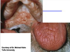
Radiotherapy
All of the following are complications of what?
- Hypofunction, with/without xerostomia, 1 month following therapeutic irradiation
- Irreversible damage
chemotherapy
Mucositis that occurs within days and spares keratinized tissues is a complication associated with what type of therapy?
radiotherapy
Mucositis that occurs within weeks and can appear on any mucosal surface leading to desquamation and ulceration is a complication associated with what type of therapy?
chemotherapy
Bone marrow suppression is a complication associated with what type of therapy?
chemotherapy
Opportunistic infections like candidiasis and herpetic infections are complications associated with what type of therapy?
radiotherapy
salivary gland hypofunction is a complication associated with what type of therapy?