Year 2 Chemistry (spectroscopy)
 Call Kai
 Learn
Learn Practice Test
 Spaced Repetition
 Match
Match Flashcards
 Knowt Play
1/34

There's no tags or description
Looks like no tags are added yet.
Name | Mastery | Learn | Test | Matching | Spaced | Call with Kai | Chat |
|---|
No analytics yet
Send a link to your students to track their progress
35 Terms
What is spectroscopy?
The analysis and classification of different molecules using their characteristic and structural differences
The interaction of electromagnetic radiation with matter
How does a spectrometer work?
The source of the radiation emits radiation that enters into the entrance slit of a monochromator. The diversion device then splits the radiation into its constituent wavelengths which exits through the exit slit as one wavelength of light. The light then hits the sample and the resultant penetrating light hits the detector
What is the purpose of spectroscopy?
Determining structure or changes in structure. This allows us to monitor product formation in real time (determining rate of reaction etc), determine unexpected results (impurities) and rapidly assess biomolecule structure
Practical usage of spectroscopy
In order to produce a cure for a disease, the interaction of the disease with biomolecules should be determined which relies on the chemical structure and composition of the biomolecules.
How the three different methods of imaging work
Cryo EM - electron microscopy that requires cryogenics. By plunging the sample in liquid ethane so the water forms a glassy solid rather than ice crystals that damage/change the sample. Using TEM then to shoot electron beams through teh sample making a 2d projection of the sample. A 3D image is created using a composite of numerous 2D images.
X-ray Crystallography - The technique used by the Big RF to determine the structure of DNA. - Purify the sample and form a highly ordered repeating 3D crystal lattice. A high intenisty x-ray is then used anf the electrons diffract the X rays into a pattern and computers are then used to measure the position and intensity of the difracted spots calculating the electron density, Which can then be modelled to the atom and showing every chemical bond.
NMR - Isotopes with a magnetic spin act as magnetics within a powerful magnetic field. A pulse of radiowaves knock the the nuclei out of alignment and the electromagnetic signals released from the return to the original states is measured. The frequency the nucleus emits is dependent of the local chemical environment and will produce distinct peaks.
Comparing and contradting the different imaging methods and their limitations
Cryo EM
is best at large complexes >50 kDa
Is good at capturing motion in snapshots
Very little sample is required
X-ray Crystallography
Can do small and medium sized samples
Can’t track motion
Requires high concentration samples
NMR
Limited to smaller molecules within the 30-50 kDa range
Great at tracking movement of atoms
Requires highly concentrated isotopic labelling
𝐸 = ℎ𝜈 = ℎ𝑐 𝜆 = ℎ𝑐�
? = wavenumber (v with a wave above it)
The wavenumber is directly proportoinal to energy
IR gives the range where the bonds stretch and bend
Plotted as transmission against wavenumber (1/wavelength) but are arranged in order of wavelength
The different motions of bonds in an IR spectrum
Bond vibration related to IR spectroscopy
When a bond vibrates, the behaviour is similar to a spring and can be modelled as such according to Hookes law.
F = -kx where:
F is the restoring force
k is the spring constant that represents bond stiffness
x is the stretch or compression distance from the bond length at equilibrium
The more bonds present provides a higher value of k vibrating at a higher frequency.
Lighter atoms or combinations with smaller reduced mass vibrate at a higher frequency when displaced
The spring like behaviour explains why molecules absorb specific frequencies of infrared light causing bond stretching, bending or rocking at resonant frequencies.
What is reduced mass
The categorisation of a two body system down to a one body system.
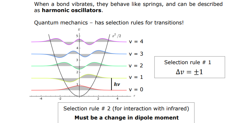
A diatomic molecule behaving like a quantum spring
The parabola representing the potential energy of the bond
The quantized energy levels (v) - a chemical bond can only exist at specific vibrational energy levels by quantum number (v)
Wavefunctions represented by the wavy lines and the shaded regions represent the probability density of finding atoms at a given stretch distance
The energy gap between each level is perfectly equal indicating that to jump up each level a molecule must absorb a photon with energy equal to hv
What are the selection rules for the bonds?
1st rule - stating how the quantum energy states can change
A molecule can only absorb or emit a photon by going up or down energy level one at a time.
Jumps greater than 1 are highly improbable according to the ideal harmonic oscillator model
Anharmonic oscillators
Bonds don’t obey Hookes law fully as the force required to compress a bind is larger than the force required to stretch it and bonds if stretched enough will dissociate. At low energies the vibrations are harmonic but become less harmonic as energy increases
Molecular orbitals
Bonds are formed by a combination of atomic orbitals giving firm to molecular orbitals
Atomic orbitals in phase are examples of constructive overlap
Atomic orbitals out of phase cause destructive overlap
These destructive bonds are sigma* anti-bonds and pi* anti-bonds where the orbitals are as far away from the bonds
Non-bonding orbitals (lone pairs) are comprised of orbitals that remain with their parent atom
The more orbitals out of phase, the more energy contained within the bonds.
HOMO and LUMO
Highest occupied molecular orbital
Lowest unoccupied molecular orbital
When you put an electron into an antibonding orbital, the bond is weakened
Electron transitions
From ground state to its excited state using the smallest jump in energy possible. Most likely from the HOMO to the LUMO
Etot + Eelec + Evib +Erot
When molecules absorb electromagnetic radiation, the excited state causes bonds to be longer. Therefore each electronic transsition is associated with change in vibrational and rotational states
Chromophores
All molecules will have electronic absorptions, however most sigma to sigma* transitions will be too high in energy to be observed on typical spectrometers <200 nm
Any molecule that will produce a homo lumo gap that is visible in typical spectrometers (UV visible spectra) is a CHROMOPHORE
Typically double and triple bonds or lone pairsTyheb
The Beer Lambert Law
Quantifying absorbance
A = ecl where:
c = concentration (M)
l = pathlength of the sample (cm)
e = extinction coefficient (permoleper cm)
If the extinction coefficient is unknown then build a calibration curve of known concentrations
The gradient of slope of Absorbance vs concentration gives the extinction coefficient
e = nW(etryp) + nY(etyr)
Where:
e = the extinction coefficient
nW means the number of tryptophan residues
nY means the number of tyrosine residues.
moldm-3
gmol-1
Multiplied together to gdm-3
= gl-1 = mg.mL-1
As the nucleobases all absorb, there are generic extinction coefficients for dsDNA, ssDNA, and ssRNA
When a molecule has spaces between double bomds is called conjugation
Highly conjugated molecules have many
The energy between HOMO and LUMO decreases with increased conjugation leading to redshift
Retinal vs Retinol
Retinal in the aldehyde form is active as there is increased conjugation causing a redshift as the absorbance is shifted to the right
Solvatochroism
The absorbance depends on the polarity of the solvent as a solvent stabilises the ground or excited state of the molecule changing the energy gap between them
Why bees see in UV electron environment
Slight change in the polarity of the binding pocket (electron environment)
Changing the absorbance of a molecule by chemical reactions such as in indicators can lead to characteristic shifts in the colour
Para nitrophenol
Removal of the H in basic conditions causes p nitrophenolate to form with a yellow hue
This can be used to observe the cleavage of bonds in model compounds
MTT assay
MTT - a molecule with 2 very different colours dependent on oxidation state.
Viable cells reduce MTT to formazan and can be used inhigh throughput assays
Lipase
Breaksdown fatty acid esters and catalyses the catalytic triad
Heme proteins
Iron containing protoporphyrin IX
Measuring IR spectra
Vibrational frequencies aren’t measured at every wavelength
Instead an interferogram is collected that is converted to a spectrum using a fourier transformation.
FTIR - Fourier transform InfraRed
Raman spectroscopy
Also measures vibrational states using a form of rayleigh scattering to provide info on vibrational states
Complementary to IR and shows vibrations that do not break a molecules symmetry