Eye diseases
 Call Kai
 Learn
 Practice Test
 Spaced Repetition
 Match
 Flashcards
 Knowt Play
1/56

There's no tags or description
Looks like no tags are added yet.
57 Terms
Types of benign lesions
Xanthelasma
Squamous cell papilloma
Skin tags
Basal cell papilloma
Dermatitis papulosa nigra
Retention cysts
Milia
Naevi
Port-wine stain
Haemangioma
Cutaneous horn (not always benign)
Xanthelasma
Soft yellowish plaques, variable size
Usually medial upper and lower eyelids, often bilateral
Lipid and cholesterol deposits/elevated serum lipid levels

Squamous cell papilloma
human papilloma virus (HPV)
Sessile or pedunculated
Excessive convoluted epithelium with central fibrovascular core

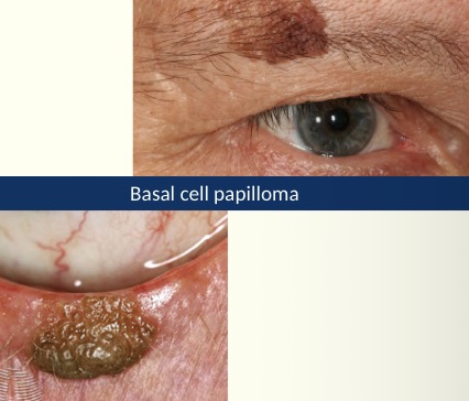
Basal cell papilloma
seborrheic keratosis or seborrheic wart
Benign, harmless skin growth- build up of skin cells
Smooth, waxy or warty surface
Slow growing
Flat or raised plaque
Skin coloured, yellow, grey, light brown, dark brown or mixed colours
Cosmetic removal
Dermatitis papulosa nigra (DPN)
multiple small diameter black to dark brown papules- face and neck
Dark skin colour
Incidence and number increase with age
Identical to small seborrheic keratoses

Skin tags
cutaneous papilloma or acrochordons
Small soft skin coloured growth
Variable size, shape colour and number
Clusters of collagen and blood vessels surrounded by skin
Associated with friction when skin rubs together

Retention cysts
Small, round, non-tender cysts
3 types: cyst of zeis, sebaceous cyst, cyst of moll
Retention cysts: cyst of zeis
white, cheesy sebaceous material
Sebaceous cysts are similar to this

Retention cysts
clear, fluid filled

Milia
tiny, superficial white/yellow dome-shaped cysts
Usually multiple- nose, chin and cheeks
Can happen at any age
Trapped keratin

Naevi

Naevus flammeus
port-wine stain
Vascular malformation (capillaries under the skin remain dilated)
Red/purple mark on the skin
Present at birth- may become more prominent with age

Capillary haemangioma
strawberry naevus- evident in neonatal period
Grows in first year then usually regressed by the age of 5
May be cutaneous, orbital or mixed
Systemic associations

Types of pre malignant lesions
actinic keratosis
Cutaneous horn
Keratoacanthoma
Actinic (Solar) keratosis
flat, scaly lesions, rough skin
Red, pink, brown or skin coloured
Happens with old age or history of sun exposure
May give rise to squamous cell carcinoma
Occasionally cutaneous horn

Cutaneous horn
keratin projection
Arise from benign, premalignant and malignant lesions
10% associated with squamous cell carcinoma

Keratoacanthoma
rapidly enlarges
Regresses/evolves into squamous cell carcinoma
Volcano shaped with keratin plug
Often difficult to distinguish from BBC or SCC
Arises from hair follicle skin cells

Types of malignant lesions
basal cell carcinoma
Squamous cell carcinoma
Sebaceous gland carcinoma
Malignant melanoma
Kaposis sacroma
Merkel cell carcinoma
Basal cell carcinoma (BCC)
periocular malignancy
Slow growing, painless
Often ulcerated
Do not metastasise but invade locally
Change in lid contour/lash direction
3 types: nodular, ulcerative, sclerosing

Squamous cell carcinoma (SCC)
May evoke inflammatory response
Symptomatic: irritation, itchy, may bleed
More likely to metastasise than BCC

Malignant melanoma
can arise as de nova (out of nowhere) or as a malignant transformation of a naevus
Itching, bleeding
Pigmentation changes (50% are non-pigmented)
Increase in size

Sebaceous gland carcinoma

Merkel cell carcinoma
rare
Highly malignant
Grows rapidly
25-30% fatal
Painless, red, hard nodule

Kaposis sarcoma

Ectropion
outward rotation (eversion) of the eyelid margin (usually lower)
Usually bilateral
Age related
Weakness of the orbicularis oculi and/or canthal tendons
Cicatricial: scarring contracture of skin and underlying tissue
Paralytic (VII nerve palsy)
Mechanical/inflammatory
Congenital
Exposed palpebral conjunctiva hyperaemia
Mucous discharge
Epiphora
Soreness/redness
Lower punctum is spontaneous visible

Entropion
inward turning (inversion) of the Tarsus and lid margin
Causes lashes to come into contact with the ocular surface
Degenerative changes result in horizontal lid laxity
Foreign body sensation
Watering
Vertical corneal/conjuctival staining
Localised conjunctival hyperaemia
Risk of corneal scarring
Cicatricial (scarring that forms when wound is healing)- pulls the lids inwards
Muscle spam
Congenital

Types of acquired ptosis
age-related (aponeurotic or involutional)
Myogenic
Neurogenic
Mechanical
Traumatic
Pseudoptosis
Age-related acquired ptosis
muscle is stretched out or disinsertion of the levator palpebrae superiosis
Increases with age
High tear upper crease
Compensatory brow lift
Low relative position in down gaze

Myogenic acquired ptosis
Weak muscle/muscle dystrophy

Neurogenic acquired ptosis
Problem with the nerve controlling the lid
third nerve palsy
Horners syndrome
Marcus gun jaw-winking
Myasthenia gravis

Mechanical acquired ptosis
Physical pushing to weighing the lid down
blepharochalasis
Eyelid tumours
Oedema

Traumatic acquired ptosis
The result of injury
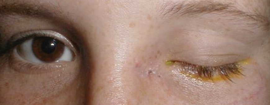
Pseudoptosis acquired ptosis
Normal lid, another anatomical anomaly
microphthalmia: one eye is smaller so eyelid comes down lower
Hypertropia (one eye is slightly higher than other)

Congenital ptosis
dysgenesis of the levator
Idiopathic
Can indicate other pathology
Risk of amblyopia/astigmatism

Lid retraction
suspected when eyelid margin is above or level with the superior limbus
Number of causes:
TED- thyroid eye disease: autoimmune condition where the eyes appear to bulge
Neurogenic- Marcus Gunn jaw winking
Mechanical: surgical overcorrection of a ptosis
Congenital

Lagophthalmos
inadequate eyelid closure: tear film disturbance, corneal desiccation
Grittiness, burning
Increased lacrimation

Floppy eyelid syndrome
generalised laxity of eyelid tissues
Unilateral/bilateral
Lid spontaneously evert during sleep
Non specific ocular irritation, redness
Easy distraction of lid from glow
Easy upper lid eversion
Lower lid Ectropion
Ptosis
Chronic papillary conjunctivitis
Whitish mucous discharge
Facial nerve palsy
Partial or complete paralysis of the facial nerve (VII CN)
Bell’s palsy: idiopathic lower motor neurone facial nerve dysfunction
Sudden onset
Unilateral
Weakness or inability to move one side of the face
Eyebrow droop/cant raise
Incomplete blink- corneal drying
Lagophthalmos
Ectropion
Epiphora
Possible changes in taste and salivation
Types of positional disorder muscle spasms
blepharospasm
Orbicularis myokymia
Blepharospasm

Orbicularis myokymia

Types of eyelash disorders
Trichiasis
Madorosis
Poliosis
Lash infestations
Trichiasis
inward misdirection of eyelashes towards the cornea
Ocualr discomfort, irritation and foreign body sensation
Watering and red eye
Corneal erosion
Corneal ulcer/infective keratitis

Madarosis
Loss of eyelashes
Poliosis
Whitening of lashes
Local infestations
pediculosis: headlice
Phthiriasis: pubic lice

Blepharitis
inflammation of eyelid margins
Chronic/relapsing
Typically bilateral
Ocular discomfort, grittiness, soreness, photophobia, itching, CL intolerance, burning

Posterior Blepharitis
thickened meibomian secretions
Microliths
Meibomianitis: passive retention of secretions (Chalazion,styes)
Foam in tear meniscus
Unstable tear firm

Anterior Blepharitis
Bacterial (staphylococcal)
crusting.collarettes/scales at base of lashes
Telangiectasia (dilated blood vessels)
Lash misdirection/loss
Seborrheic (gland of zeis)
greasy deposits at base of lashes
Demodex
cylindrical deposits extending up lashes
Lash misdirection/loss
General redness
Itching

External hordeolum
Acute bacterial infection of an eyelid gland, tender eyelid bump
lash follicle and associated gland of zeis or moll- base of eyelash

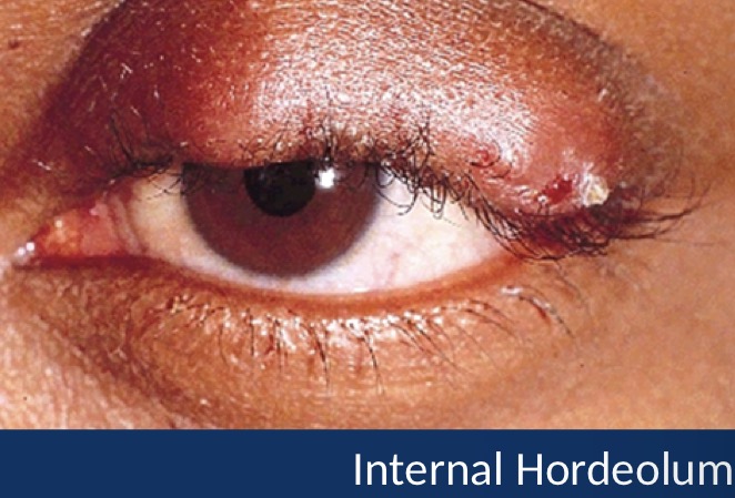
Internal hordeolum
Meibomian gland- tarsal plate treat as per plepharitis
Chalazion
common chronic lid lump
Well-define 2-8mm subcutaneous nodule in tarsal plate
May be associated with Blepharitis and astigmatism
Painless lid lump

Herpes simplex virus
Swollen lids and conjunctivitis

Herpes zoster virus (shingles)

Molluscum contagiosum
Umbilicated skin nodule

Impetigo
staph infection
Rash
Occasionally blisters on skin
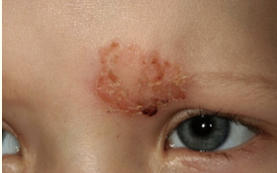
Fungal infections: Tinea faciei
