HDE 101: Muscle Physiology 1- Anatomy and Contraction
 Call Kai
 Learn
 Practice Test
 Spaced Repetition
 Match
 Flashcards
 Knowt Play
1/31

There's no tags or description
Looks like no tags are added yet.
32 Terms
Muscle Function
Move us, stuff, stuff through us, prevent movement
Muscle Body Composition
Skeletal muscle - 30-40% of body weight
Cardiac & Smooth muscle - 10% of body weight
Muscle Types
Skeletal Muscle
Multi-nucleated, voluntarily contracted , striated
Cardiac Muscle
Lots of lymphocytes , Intercalated Disk’s, Striated, Nucleus, Automatically controlled
Smooth Muscle
No striations, nuclei and muscle cells, Automatically controlled
Skeletal Muscle Structure
Whole muscle (cm)
Fascicle (mm)
Muscle Fiber (μm x 100
Myofibril (μm)
Sarcomere (μm)
Thick filament (nm)
Thin filament (nm)
Z-line
Thin filaments are joined at the Z-line
Sarcomere
Functional unit of sliding shortening
Sliding filaments
Composed of Thick filaments (μm) and Thin filaments (μm)
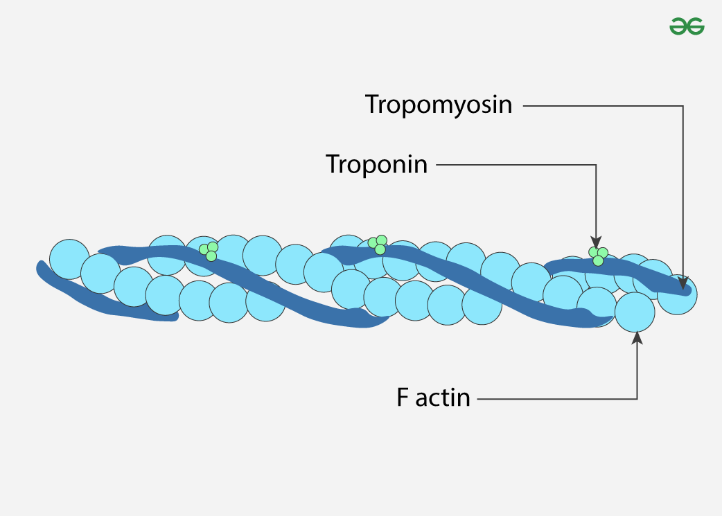
Thin Filaments (Regulatory)
Helical actin molecules
Myosin binding sites
Tropomyosin — thread like protein
Covers myosin binding sites
Troponin
Releases tropomyosin when Ca2+ is bound
Thick Filaments (Motor)
Myosin molecules - 2 golf club-shaped subunits
Tails aligned toward middle
Globular heads protrude out at regular intervals
Actin binding sites
ATP binding sites

Sarcomeric Structure
Z-line
Defines the border of the sarcomere
Thin filaments joined
I-Band
Only thin (actin) filaments , light-colored region
A-Band
Entire length of thick (myosin) filaments, including where they overlap with thin filaments
H-Zone
Containing only thick (myosin) filaments w/ no overlapping actin
M-Line
Dark line running down exact center of H-zone/sarcomere, where myomesin/other proteins anchor the thick filaments

Z-line
Defines the border of the sarcomere
Thin filaments joined
I-Band (Isotropic Band)
Only thin (actin) filaments , light-colored region
A-Band (Anisotropic Band)
Entire length of thick (myosin) filaments, including where they overlap with thin filaments
H-Zone
Containing only thick (myosin) filaments w/ no overlapping actin
M-Line
Dark line running down exact center of H-zone/sarcomere, where myomesin/other proteins anchor the thick filaments
What would cross-section look like at Z-Line?
Z-line included only thin filaments, shouldn’t include any thick filaments
Should have interconnections since thin filaments join at Z-line
Skeletal Muscle Fibers
Packed with myofibrils
Multinucleated → support many nuclei processes that req. protein
Many mitochondria → ATP production req. energy
Transverse Tubules
Extension of membrane through the cell → Allow ATP to propagate
Specialized organelles
Sarcolemma = Plasma membrane
Sarcoplasm = Cytoplasm
Sarcoplasmic Reticulum = Endoplasmic Reticulum
Skeletal Muscle Fibers- Specialized Organelles
Sarcolemma = Plasma membrane
Sarcoplasm = Cytoplasm
Sarcoplasmic Reticulum = Endoplasmic Reticulum
Motor Units
Motor unit
A single motor neuron and all of the muscle fibers it innervates
A muscle fiber is only innervated by ONE motor unit → “1 motor unit innervates 100 muscle fibers”
Motor units are intercalated within healthy muscle
Motor neurons determine muscle contraction
Motor Unit Size
Variable
Some <10 fibers
Some <1000 fibers
Amount of motor units activated are key to determining how much force the muscle will make
Neuromuscular Junction
Specialized synapse that transfers an AP in a nerve to a muscle fiber
Steps of AP in a nerve to a muscle fiber
Motor neuron action potential
Ca2+ enters voltage-gated channels
Acetylcholine release
Acetycholine binding opens ion channels
Na+ entry
Local current between depolarized end plate and adjacent muscle plasma membrane
Muscle fiber AP initiation
Propagated AP in muscle plasma membrane
Acetylcholine degradation
How does Botox stop muscle activity?
Prevents Acetylcholine (ACh) from being released into synaptic cleft
Myasthenia Gravis
Autoimmune disorder where antibodies mediate destruction of the acetylocholine receptor →There will be less Acetylcholine receptors
Which therapies could you imagine be used in myasthenia gravis?
Prevent re-uptake/degradation of Acetylcholine
Keep Acetylcholine in cleft longer by preventing acetylcholine esterase (enzyme) in using an inhibitor
Excitation-Contraction Coupling
AP propagated throughout muscle fibers by T-tubules
DHP receptors sense voltage change → Open ryanodine receptor coupled to it on the sarcoplasmic reticulum (SR) leading to Ca2+ release
SR has a really high Ca2+ conc. , once activated it releases them
Cytosolic Ca2+ binds to troponin → removes blocking action of tropomyosin→ exposes myosin binding site
Myosin cross-bridges cycle to generate force using ATP
Cystolic Ca2+ pumped back into Sarcoplasmic Reticulum (SR) through SERCA pumps using ATP
Low cystolic Ca2+ leads to Ca2+ removal from troponin → restores tropomyosin blocking action on myosin binding site
Simple Term for Excitation-Contraction Coupling
AP propagated along muscle fibers through T-tubules
DHP receptors sense voltage change → open ryanodine receptor →Ca2+ released from Sarcoplasmic Reticulum (SR)
Ca2+ binding to troponin removes blocking action of tropomyosin
Myosin cross-bridges bind and generate force using ATP
Ca2+ transported back into SR using ATP
Ca2+ removal from troponin restores tropomyosin blocking action
Excitation-Contraction Coupling
Ca2+ activates thin filament
Ca2+ binds to troponin
Troponin moves from inhibitor state
Tropomyosin slides off the actin binding site
Myosin cross-bridge can now bind
Excitation-Contraction Coupling - Low cytosolic Ca2+, relaxed muscle
Ca2+ doesn’t bind to troponin → doesn’t remove blocking action of tropomyosin → Energized cross-bridge cannot bind to actin
Excitation-Contraction Coupling - High cytosolic Ca2+, activated muscle
Ca2+ binds to troponin → troponin removes blocking action of tropomyosin → Myelin cross-bridge binding sites are exposed → Cross-bridge binds to actin and generates force
Which doesn’t change length during a shortening contraction?
A-band
A-band is where we have thick myosin filaments → Myosin filaments don’t shorten or change size
Areas defined by a lack of filament get smaller, but areas defined by the length of a filament won’t change
Which does change length during a shortening contraction?
I-Zone
Region containing only thin actin filaments shortens
H-Zone
Shorten and completely disappear during muscle contraction
Sarcomere length
Overall distance between Z-lines decreases
Latent Period
Latent Period (lag phase) in muscle contraction. The arrival of an electrical stimulus (AP) at the muscle fiber and start of a muscle contraction has about 1 to 10 millisecond delay bc of the excitation-contraction coupling at the NMJ.