Obstetrics Exam
 Call Kai
 Learn
 Practice Test
 Spaced Repetition
 Match
 Flashcards
 Knowt Play
1/133

There's no tags or description
Looks like no tags are added yet.
134 Terms

Identify the female reproductive anatomy

Explain a typical menstrual cycle
A typical menstrual cycle lasts 28 days, with menstruation only lasting 4-6 days and occurs when hormones stimulate the development of an egg and the thickening of the endometrium. If an egg is fertilised, the ovum implants itself into the endometrium and pregnancy will develop, whereas if the egg is not fertilised, the lining of the endometrium will shed.
What is abnormal vaginal bleeding and what are some common causes?
Bleeding that occurs for less than 21 days or greater than 36 days, more than 80mls or for longer than 9 days or that soaks at least one pad an hour for several hours.
Common causes include ectopic pregnancy, miscarriage, contraceptives, STIs, stress, medications, blood clotting disorders, hormonal imbalances, etc
What are common causes of post menopausal bleeding?
Cancer: cervical, uterine
Cervical Polyps
Prolapse
Atrophy due to lack of oestrogen
What is the prehospital management of abnormal menstrual bleeding?
Primary Survey - A-E
Specific obstetric/gynacological history
Onset
Duration (constant?)
Amount, size, colour and odour – clots, gushing, how many pads?
Menstrual History (Has this happened before)
Date of LMP
Contraception
Pregnancy?
Previous Medical History
Medication – Warfarin, aspirin
Allergies
Last blood test, Iron levels?
Symptoms: - Symptom Management
Pain
Dizziness
Syncope
Bruising / Petechiae
Nausea/Vomiting
Pallor
Clamminess
Diaphoresis
Altered conscious state
Assess the abdomen for tenderness or distention
NEVER perform an internal vaginal examination
Do not dam the flow
Manage any signs of hypovolaemia
What is Pelvic Inflammatory Disease?
PID is an infection or inflammation of the upper female genital tract, sometimes caused by a complication of an STD, IUD, or termination of a pregnancy in women, and can lead to scarring of the uterus, fallopian tubes and ovaries. PID can lead to ectopic pregnancy, infertility, chronic pelvic pain and abscesses. PID is most common in women under 25 and presents with fever, purulent discharge, irregular bleeding, malaise, nausea and vomiting, painful intercourse or painful urination. Mild cases are treated with antibiotics, whereas severe cases may require surgery and hospitalisation.
What is endometriosis?
Endometriosis is the presence of any endometrial tissue outside of the endometrial cavity. The endometrial tissue bleeds in cycle with the menstrual cycle, which results in inflammation and scarring of the peritoneum and adhesions. The cause is unknown and symptoms include pelvic pain, painful intercourse, and abnormal uterine bleeding. Treatment is NSAIDs and the oral contraceptive pill or in severe cases surgery or hysterectomy.
What is an ovarian cyst?
Ovarian cysts are follicular, luteal or corpus cysts that can develop on the ovaries. They can develop at any age and affect 30-50% of women. They can be asymptomatic or can present with aching, heaviness, pressure, sudden intermittent or sharp pain or with some vaginal spotting.
What is Mittelschmerz?
Pain typically experienced on only one side, in the lower abdominal region associated with normal ovulation. Mittelschmerz is German for “Middle pain” and is typically experienced around 14 days before menstrual cycle and marks the release of the egg from the ovary. Completely normal.
What is Dysmenorrhoea
Painful menstrual cramps, which can be primary, usually lasting the first two days of menstruation and reflecting prostaglandin-induced contractions of the myometrium or secondary, reflecting menstrual cramps from an identified pathology such as endometriosis, PID, pelvic adhesions, etc.
What are possible differential diagnoses for pelvic pain?
Gallbladder
Chronic Constipation
Diverticulitis
Urinary Tract Infections
Appendicitis
What is the prehospital management of pelvic pain?
Primary Survey - A-E
Specific obstetric/gynacological history
Onset
Duration (constant?)
Amount, size, colour and odour – clots, gushing, how many pads?
Menstrual History (Has this happened before)
Date of LMP
Contraception
Pregnancy?
Previous Medical History
Medication – Warfarin, aspirin
Allergies
Last blood test, Iron levels?
Assess the abdomen for discolouration, tenderness or distension
Pain Relief
What is Toxic Shock Syndrome?
Toxic Shock Syndrome is a multisystem diseases caused by an infection of Group A Strep bacteria what produce toxins in the body and has been linked with super absorbent tampons but can also result from infections such as pneumonia, or surgical wounds. Symptoms include a fever above 38.8 degrees, rash on trunk and flaky skin on palms, headache, hypotension, diaorrhaea and vomiting, and syncope.
Patients need to be monitored for sepsis red/amber flags.
What is Amenorrhoea?
The absence of menstruation and can be primary, where a woman has never experienced a period and which is associated eith genetic conditions that affect reproduction or seconday where menstruation stops for longer than 12 months.
What is the most common vaginal infection?
Vaginitis occurs when normal fauna of the vahina is distupted and replaced by gardenrella, mycoplasmas and anaerobe bacteria. While it is not considered an STI it is most common in sexually active women.
What is a bartholin abscess?
Bartholins glands lie at 5 and 7 oclock positions of the vagina and when infected they can cause large painful absesses to develop.
How long is the average pregnancy?
266 days (38 weeks) post ovulation or 280 days (40 weeks) post the last menstrual peroid.
Trimester 1 - 0-12 weeks
Trimester 2 - 12-28 weeks
Trimester 3 - 28-40 weeks
What symptoms categorise the first trimester of pregnancy?
Missed period - Cessation of menses
Nausea with or without vomiting
Breast tenderness and tingling
Increased breast size and vascularity
Fatigue
Increased frequency of urination
Maternal cardiovascular changes
What symptoms categorise the second trimester of pregnancy?
Nausea will usually have passed
Begin to be able to feel the foetus move
Skin discolouration on the face and abdomen
Stretch marks
Increased leucorrhoea - Thick vaginal discharge
Maternal blood volume increases and haemoglobin levels fall
Blood pressure decreases slightly while the heart rate increases 10-20bpm
UTIs are common but can cause preterm labour
Uterus is now considered an abdominal organ.
What symptoms categorise the third trimester of pregnancy?
Episodes of fatigue and dyspnoea
Increased urinary frequency
The enlarged uterus displaces the heart to the left, shifts the intestines and stomach and displaces the bladder up and out the pelvis
Blood pressure increases
Dependent Oedema is evident and venous return reduces
Foetus is easily palpatedf and movement is obvious
Centre of gravity shifts
Stress on the lower back and spine
What is the significance of the endocrine system in pregnancy?
Most of the physiologicla changes in pregnancy are cuased by hormon production and secretion managed by the endocrime system. Primarily oestrogen and progesterone are responsible for the maintenance of pregnancy, foetal growth and postpartum recovery.
What hormones are significant in pregnancy and why?
Progesterone - Relaxes smooth muscle in blood vessels, uterus and GIT, increases body temperature, causes nausea and vomiting and inhibits uterine contractility.
Oestrogen - Growth stimulator, increases the number of oxytocin receptors in the uterine wall at the onset of labour and triggers prostaglandins from the uterus and placenta to soften and thin the cervix.
Human Chorionic Gonadotropin (HCG) - Maintains steroid hormones in early pregnancy and maintains the pregnancy until the placenta takes over
Human Placental Lactogen (hPL) - Modifies maternal metabolism and stimulates growth of maternal and foetal tissues. Protects the foetus from rejection, reduces maternal glucose uptake so it’s available to the foetus and stimulates the growth and development of mammary glands for lactation.
Relaxin - Remodels connective tissue, softens elastic ligaments of pelvic bones, allows mobilisation and growth of the uterus into the abdomen, alters vascular compliance to help the body tolerate 30-50% surge in blood volume without spiking blood pressure.
Thyroid gland -Pregnancy mimics hyperthyroidism which increases body temperature, stimulates appetite and increases feelings of fatigue. In the first trimester the foetus relies on the maternal thyroid gland.
What does fundal height mean?
The height of the uterus equates to the number of weeks of gestation plus or minus 3cm. Fundal height is measured in cm from the pubic symphysis to the topmost portion of the uterus.
At 12 weeks, the fundus can be palpated at the pelvic brim
By 20 weeks, the fundus reaches the maternal umbilicus
By 36 weeks, the fundus can reach the lowest part of the sternum
In the last month, the foetus drops into the lower pole and the fundal height may reduce.

What are braxton hicks contractions?
Painless uterine tension felt through the abdominal wall that improve uterine blood flow and foetal oxygenation and help prepare the body for labour. Threy are generally centred in the lower groin and transient (not in the lower back), they do not increase in strength or intensity or dilate the cervix
What cervical changes occur during pregnancy?
the cervix increases in mass, length and width and the cervical glands secrete thickened mucus that forms a mucus plug and protects from ascending infections. The mucus plug is then released prior to or during labour and indicated that the cervix is preparing for labour.
What circulatory changes occur during pregnancy?
Increased metabolic demand
Expansion of vascular channels
Increase in steroid hormones
Cardiac output increases of 30% to 50%, which is essential for vascular supply to an enlarged uterus, hydration of foetal and maternal tissues, exchange of respiratory gases, nutrients and wastes and fluid reserve for loss after birth.
Plasma will increase by 50% but red cell mass will only increase by 30%, which can result in hypervolaemia, haemodilution, anaemia and a fall in Hb.
The heart increases in size by 12% and is displaced upwards and rotated forwards.
BP decreases in the second trimester due to peripheral vasodilation caused by hormonal changes but should return to pre-pregnancy levels by term.
A BP higher than 140/90 is a risk factor for preeclampsia and pregnancy-induced hypertension
What is the recommended position for a pregnancy woman and why?
Left lateral
Placing a pregnant woman supine will impede venous return from the inferior vena cava, decrease maternal and foetal oxygenation, increase hydrostatic pressure, increase oedema in the feet and ankles, decrease circulating volume by 30% and can cause supine hypotension with signs of shock.
What respiratory changes occur in pregnancy?
O2 requirements increase to meet accelerated metabolic rate and tissue growth, capillary dilation throughout the respiratory tract and engorgement of the nasopharynx, trachea and bronchi result in nasal and sinus stuffiness, epistaxis, voice changes and an increase in snoring and upper respiratory tract infections.
Ligaments of the ribcage relax and are displaced upward
The diaphragm is elevated by up to 4cm and breathing converts from abdominal to thoracic
Increases thoracic volume.
What is important when completing an antenatal assessment?
Gestational age
Presence of cramping, bleeding or ‘spotting’
Foetal heart rate
Determine position of foetus
Foetal movement
Membranes intact?
Gravida/Parity
Maternal BP
Pyrexia
Presence of disease
What is gravidity and parity?
Gravidity - Total number of times a woman has been pregnany regardless of the outcome (including the current pregnancy)
Parity - Number of times a woman has given birth to a goerus that reached vitability (including stillbirths that reached the viability threshold)
What is the placenta?
The placenta is formed by the fertalised ovum (egg) once it has implanted in the endometrium. The placenta begins to function by week 4, reaches full functionality from week 10 and is considered its own organ by week 14. The placenta from the maternal surface adheres to the uterine wall and is rough and dark red, the foetal surface is shiny, slightly grey and consists of membranes that cover the foetus and enclose it with amniotic fluid. The membranes equalise pressure and protect the foerus from injury. The umbilical cord goes from the foetus to the placenta and carried O2 and nutrients to the foetus and removes excreted waste via 2 arteries and 1 vein.
What is the amniotic sac?
Two fine membranes extending from the placenta around the baby called and amnion and chorion are collectively known as the amniotic sac and stabilise pressure and protect the foetus from injury.
What is uniquie about foetal circulation?
Foetal circulation varies significantly from adult circulation and is dependent on 3 circulstory shunts that deliver oxygenated blood to vital organs. Non-essential organs that are not fully functional in utero are bypassed.
The ductus venosus connects the umbilical bein to the inferior vena cava, the foramen ovale connects the right atrium to the left atrium and the ductus arteiosus shunts blood from the pulmonary artery to the aorta.
What are potential causes of vaginal bleeding in the first 20 weeks of pregnancy?
Infection
Polyps
Cancer
Ectopic Pregnancy
Miscarriage
Hydatidiform Mole
Trauma
What is a spontaneous miscarriage?
Unintentional termination of pregnancy before the foetus is viable and is classified as;
Threatened - Pregnancy loss is possible but may continue
Inevitable - Pregnancy cannot be saved
Incomplete - Not all products of conception have passed
Complete - All products of conception have been expelled
Silent or delayed miscarriage - Retention of deceased foetus for more than 8 weeks
Early miscarriage - Pregnancy loss before 12 weeks
Late miscarriage - Pregnancy loss between 12 and 20 weeks
Molar Pregnancy - AKA a hydatidiform mole or gestational trophoblastic disease occurs when tissue that normally becomes a foetus becomes an abnormal growth in the uterus. Even though it isn't an embryo, it triggers symptoms of pregnancy.
In many cases, the cause of a miscarriage is unknown or multifactorial; however, more than 70% are due to chromosomal abnormalities. Specific microorganisms, smoking or drug use, abnormalities of the uterus or an incompetent cervix are also known to increase the risk. Signs and symptoms of a miscarriage include uterine cramping, vaginal bleeding, lower back pain, and passage of products of conception.
At what point is a foetus considered viable?
23 Weeks
What is a molar pregnancy?
AKA a hydatidiform mole or gestational trophoblastic disease occurs when tissue that normally becomes a foetus becomes an abnormal growth in the uterus. Even though it isn't an embryo, it triggers symptoms of pregnancy. Occurs in 1 out of every 1,500 women with early pregnancy symptoms and is believed to be caused by a problem with the genetic information of an egg or sperm. Risk increases if the maternal diet is low in vitamin A, aged over 35 years or there is a history of molar pregnancy or miscarriage. Presents with PV bleeding, a larger-than-normal uterus, severe nausea and vomiting, signs of hyperthyroidism such as nervousness, tiredness, tachycardia and sweating, an uncomfortable feeling in the pelvis and unusual vaginal discharge. Molar pregnancies are diagnosed with ultrasounds and blood tests, and the treatment is a D+C. There is a small chance the tissue can develop into cancer if not removed.
What are the potential complications of a miscarraige?
Physical and psychological complications can occur including haemorrhage, uterine infection, septicaemia or calcification of delated miscarriage
What is the prehospital management of a miscarriage?
No emergency treatment required if early
History
All patients will require supportive care and reassurance
Patient may not be aware they were pregnant
If bleeding is present, then manage as per haemorrhage guidelines
Important to retain all blood-stained clothes, pads and products of conception
Try to dissuade patient from using the toilet in case she passes foetal products – take them to hospital if possible
What is the abortion legislation in WA?
It is legal to perform a therapeutic abortion up to 20 weeks if it is the medical practitioner’s opinion that continuation of the pregnancy may result in risk to maternal life, risk of maternal physical or mental harm, or risk of significant foetal physical or mental abnormalities or risk the foetus, once born, is incompatible with life.
Criminal abortion is when evacuation of the uterus by an unauthorised person occurs and is punishable by law and can be induced by the patient.
What is an ectopic pregnancy?
Fertilised ovum implants in a site other than the endometrium of the uterine cavity. 95% of all ectopic pregnancies occur in the fallopian tube however, implantation can be intrauterine or extrauterine. Extrauterine implantation includes the abdominal cavity, ovary or fallopian tube. A ruptured ectopic pregnancy is a true obstetrical emergency, and while an ectopic pregnancy only occurs in 1-2% of pregnancies, it accounts for 10-15% and is the leading cause of maternal mortality in the first trimester. Conditions that increase the risk of an ectopic pregnancy are those that prevent or delay the passage of the fertilised egg into the uterus and include occlusion of the fallopian tube, impaired tubal ciliary action, impaired tubal contractility, and decreased sperm motility. Other risk factors include pelvic inflammatory disease, previous ectopic pregnancies, endometriosis, previous pelvic or tubal surgery, infertility treatments, uterine or tubal abnormalities and cigarette smoking.
Ectopic pregnancy will often present as 1-2 months of amenorrhea, signs of early pregnancy with slight vaginal bleeding, acute, severe, sharp unilateral abdominal pain, rupture and haemorrhage, generalised abdominal pain and referred shoulder tip pain, abdominal guarding, rebound tenderness and/or guarding, syncope and development of haemorrhagic shock. If the ectopic pregnancy is unruptured, the patient will often have a history of amenorrhoea, abnormal vaginal bleeding, pelvic and/or abdominal pain, nausea and presyncope symptoms.
What is Kehr’s Sign?
Kehr's sign is the occurrence of acute pain in the tip of the shoulder due to the presence of free blood or other irritants in the peritoneal cavity that irritates the diaphragm when a person is lying down.
What common differential diagnoses are relvant for a potential ectopic pregnancy?
Salpingitis (inflammation of fallopian tubes)
Appendicitis
Rupture torsion of ovarian cyst
Pelvic abscess
What triad of symproms suggest an actopic pregnancy?
Amenorrhea, vaginal bleeding and abdominal pain
What are the maternal and foetal outcomes in an ectopic pregnancy?
Maternal health is significantly imapaired post rupture. Rupture can lead to uterine infection, septicaemia and haemorrhagic stroke.
If there was a tubal, cervical or ovarian implantation and rupture the foetus has no chance of survival. If the foetus has implanted in the abdomen, 1 out of 10 pregnancies may reach viability.
What is the prehospital management of an ectopic pregnancy?
Assess ABC
Universal blood and body fluid precautions
Maternal vital signs often (including pain scale)
Thorough history related to event
Assess blood loss and haemodynamic stability
IV access (2 x large bore cannulae) pre-hospital blood sample and IV fluids if required
High concentration oxygen via non-rebreather mask
Position of comfort unless shocked (supine with legs elevated)
Notify receiving hospital and transport urgently to ED
What is hyperemesis Gravidarum?
Excessive vomiting during pregnancy, is believes to be associated with increased levels of hCG. Increased vomiting can cause dehydration, electrolyte imbalances and acid-base imbalances. Complications include hypovolaemia, hypotension, tachycardia, decreased urine output, metabolic aklalosis, hypokalaemia leading to cardiac arrhythmias, muscle wasting and vitamin deficiencies, foetal death, maternal death if untreated.
What is the management of Hyperemesis?
Primary Survey A-E
Frequent vital signs
Histroy of pregnancy and current condition
Observe for dryness of skin, poor skin turgor, jaundice, dry tongue and mucosam sunken eyes
Oxygen if required
Transport in a position of comfort - left lateral
Obtain IV access and commence fluid therapy
Control vomiting with anti-emetic
Reassurances
Transport to appropriate facility - If <20 weeks ED, If >20 weeks LBS
What are the effects of substance abuse in pregnancy?
Methamphetamine use - Can be fatal for the mother, can cause spontaneous abortion, can cause congenital malformations or functional impairments to the foetus, causes placental vasoconstriction.
MDMA use - MDMA crosses the placental blood barrier and increases the risk of congenital birth defects, carviovascular anomalies and musculoskeletal anomalies
Amphetamines - Cardiac defects, cleft palate, birth defects and addicition and withdrawal upon birth
Tobacco - Increases the risk of abruption, low birth weight, prematurity, still birth
What is an antepartum haemorrhage?
Vaginal bleeding from 20 weeks of pregnancy. Up to 750ml/min of blood passes through a pregnant uterus, and an APH can be life-threatening. If the placenta is wholly or partly in the lower uterine segment, bleeding is inevitable as the uterus stretches and the cervix dilates, ripping away the anchor site of the placenta and causing bleeding.
Primary causes are placental abruption, placental praevia and trauma, but an APH can also be caused by contact (intercourse or an examination), infection and/or inflammation, vasa praevia or no definitive cause.
What is the prehospital management of an APH?
Primary Survery - A-E
Regulat maternal vital signs
Fortal heart rate and/or movements
Thorough history of pregnancy
Assess bloode loss - Amount, colour, consistency
Assess uterine acivity, tenderness and pain
Keep a pad count
IV access - 2 large bore cannula and IV fluids
Oxygen if required
Left lateral position
Reassurance and urgent transport
What is Placenta Praevia?
Painless bleeding from the separation of an abnormally implanted placenta at or near the cervix that rarely occurs before 28 weeks gestation. Occur when the placenta implants between the foetus and the cervix and occurs in four grades.
Minor Grade 1 - Placenta encroaches on the lower segment but not the cervical opening.
Minor Grade 2 - Placenta reaches the cervical opening but does not cover it
Major Grade 3 - When the internal cervical opening dilates it is partially covered by the placenta
Major Grade 4 - Placenta completely covers the cervical poening even when fully dilated
The position of the placenta is determined by the site at which the trophoblast (thing outer layer of cells of an early embryo that help the embryo attach to the uterus) implants. In the second trimester the uterus forms the lower pole, which has lower uterine fibres, as the placenta is unable to stretch if it implants low it is unable to shift with the growth of the uterus and will begin to separate prematurely causing generally painless bleeding.
Signs and symptoms include - Unprovoked bleeding (may have had recent intercourse), painless bleeding in the third trimester usually around 35 weeks, blood is usually bright red and escapes unimpeded. Abdomen is usually soft and non-tender and maternal vital signs may remain normal until 1/3 of blood volume is lost. Definitive diagnosis is by ultrasound.
What is the management of placenta praevia?
Conservative if bleeding is slight but immediate delivery if bleeding is severe.
Prehospital IV access, bloods and fluid replacement
In hospital blood transfusions as needed
Vaginal delivery is possible for minor grades but a caesarean is indicated for major grades
What is Placental abruption?
Detachment of a normally located placenta prior to birth that disrupts the ability of the placenta to provide nutrients and remove waste from the unborn baby. More than 50% of detachments will result on foetal death. Maternal uterine blood usually escaped vaginally and is dark (old blood) and non clotting or collects inside the reproductive tract. Placental abruption can be categorised as
Marginal seperation - Placenta separates at the edges and blood escapes vaginally
Central separation - Placenta separates centrally and bloo is trapped between the placenta and uterine wall. Initially bleeding is concealed then dissects to the edge of the placenta. Little bleeding on examination.
Complete separation - Massiva vafinal bleeding occurs with a total separation of the placenta.
Signs and symptoms include; bleeding, pain, onset of premature labour, tenderness over placental site, uterine contractions, bleeding into uterine muscle wall causes pain and an absense of bleeding, tender abdomen and uterus has a ‘woody’ consistency, foetal HR slow or absent, hypovolaemia.
Placental abruption has a high foetal mortality rate
What is the prehospital management of a plaental aburption?
Primary survey - A-E
Oxygen
Left lateral position
VItal signs and monitor
Wide bore IV cannula, prehospital bloods, replacement fluids
Urgent transport
Possible caesarean section in hospital
What is Disseminated Intravascular Coagulation (DIC)?
Abnormal coagulation and fibrionlysis that occurs secondary to toehr conditions such as abruption. Fibrinogen is convered to fibrin clots but the liver cannot keep an adequate supply of fibrinogen leading to a reduction of clotting facts and a severe reduction in the number of platelets means blood cannot clot and intractable haemorrhage occurs.
What is Vasa Praevia?
Uncommon condition where foetal blood bessels are unsupported in the membranes in advance of presenting foetal part. Painless vaginal bleeding at time of spontenous membrane rupture. Foetal shock and death can occur rapidly.
What is uterine rupture?
Uterine rupture is a continuum of events starting with a weak spot in the uterine wall and ending with the uterus tearing open and the placenta and blood extruding into the maternal abdomen. Uterine rupture can occur before or during labour/delivery, and the mother’s condition will deteriorate rapidly. Bleeding may be internal or external. 40% of uterine rupture patients have had prior uterine surgery. Risk factors also include more than 4 pregnancies, trauma (especially blunt or penetrating), excessive use of oxytocin, shoulder dystocia or forceps deliveries.
What is the prehospital management of a uterine rupture?
Primary Survey - A-E
100% oxygen
Left lateral position
Frequent vital signs and monitor
Wide bore IV cannula, prehospital bloods and replacement fluids
Consideration for TXA if the mother becomes time critical
Reassurance
Maintain body temperature
Rapid transport
What is hypertensive disease of pregnancy?
Hypertensive disease is the most common medical complication of pregnancy and comproises placental perfusion until the birth of the baby. Hypertensive disease includes
Hypertension - Diastolic BP >90mmHg and systolic BO >140mmHg on two occasions
Pre-eclampsia - Hypertension with proteinuria (presense of excess amounts of protein in the urine) after 20 weeks gestation
Eclampsia - The presense of seizures not caused by neurologica disease
Chronic hypertension - Persistent hypertension prior to 20 weeks or after 6 weeks post partum
Gestational hypertension - Develops in late pregnancy or 24 hours post partum and resolves by 10 days post partum.
What is pre-eclampsia?
The precise aetiology of pre-eclampsia is not known, but it arises after 20 weeks’ gestation and is characterised by hypertension with proteinuria (the presence of excess amounts of protein in the urine). Proteinuria is a marker for kidney damage and can also cause damage to other organs. Pre-eclampsia occurs when there is an abnormal placenta and the spiral arteries responsible for supplying maternal blood to the placenta become fibrous and are unable to dilate. The reduction in blood flow to the placenta and thereby the foetus causes intrauterine growth restriction and can cause foetal death. The hypoperfused placenta then releases pro-inflammatory proteins that enter the maternal bloodstream and cause the endothelial cells lining the maternal blood vessels to become dysfunctional. Endothelial cell dysfunction causes vasoconstriction and causes the kidneys to retain more sodium.
Pre-eclampsia also causes;
A decrease in cardiac output as vasoconstriction occurs
An increase in excess fluid in the cells as capillary permeability increases, which in turn impedes oxygenation
Decreased renal blood flow and glomerular filteration rate
Decreased platelet count
Epigastric pain and impaired liver function
Swelling in the brain can lead to CVA
Lungs can become congested - pulmonary oedema.
What are the diagnostic signs of pre-eclampsia?
Hypertension in pre-eclampsia - Systolic> 140 mmHg or diastolic> 90 mmHg on two occasions after 20 weeks’ gestation and proteinuria
Severe pre-eclampsia - Systolic >160mmHg or diastolic >110mmHg
What is severe pre-eclampsia?
BP > 160/110 mmHg
Frontal or occipital headaches not relieved by analgesia
Epigastric pain due to hepatic swelling and stretching
Visual disturbances ranging from blurred vision to blindness
Nausea and vomiting
Oliguria
Pulmonary Oedema
Altered conscious state
What is Eclampsia?
a life-threatening obstetric emergency defined by the onset of new-onset, generalized tonic-clonic seizures or coma in a pregnant or postpartum patient (after 20 weeks of gestation up to 6 weeks postpartum) who has signs or symptoms of pre-eclampsia. Eclamptic convolsuions usually start with facial twitching following by a tonic contraction for 15-20 seconds then a tonic-clonic phase that lasts approx 1 minute and ending with maternal coma. Maternal death occurs as a result of cerebral haemorrhage, aspiration or heart failure.
Eclampsia predisposes to placental abruption.
BP usually returns to normal 6-8 weeks post partum.
What is the management of eclampsia?
Primary survey - A-E
Left lateral position with secure airway
100% oxygen
Prevent injury
Frequent vital signs and monitoring
Assess foetal HR and movement if possible
Check for signs of labour
IV access and prehospital bloods, but no fluids required as BP is already high
anti-convulsant therapy
Urgent transport - definitive treatment is delivery.
What is HELLP Syndrome?
HELLP syndrome is a life-threatening pregnancy complication usually considered to be a variant of preeclampsia that occurs in the later stages of pregnancy or sometimes after childbirth.
H - Hemolysis - breaking down of red blood cells
EL - Elevated kliver enzymes
LP - Low platelet count
Risk factors are a maternal age older than 34 years, multiparity, white race or european descent and a histroy of poor pregnancy outcomes.
Causes of HELLP syndrome include DIC, placentalk abruption, acute renal failure and pulmonary oedema.
Symptoms include; Headache, Nausea, Vomiting, Indigestion with pain after eating, Abdominal or chest tenderness and upper right side pain (from liver distension), shoulder pain or pain when breathing deeply, bleeding, changes in vision, swelling, high BP, proteinuria.
What is the pre-hospital management of HELLP syndrome?
Primary Survey - A-E
Unable to officially diagnose without bloods
Left lateral position with secure airway
100% oxygen
Prevent injury
Frequent vital signs and monitor
Assess foetal HR and movement if possible
Check for signs of labour
IV access, prehospital bloods, consider fluids (unlikely as BP↑)
Urgent transport
Definitive treatment is delivery
What are common chronic health conditions that need management throughout pregnancy?
Asthma
Exacerbations are more likely in the third trimester
Child has a 50% chance of being asthmatic
Management is the same as for a non-pregnant patient
Diabetes
High blood sugar can cause complications and needs to be monitored
Cardiac conditions
Normal physiological changes in pregnancy can worsen already existing cardiac conditions or new cardiac conditions can present for the first time during pregnancy, including; hypertension, valvular disease, arrhythmias, cardiomyopathies, aortic dissection, etc.
Seizures
Seizures in pregnancy can result from eclampsia, head injury, alcohol withdrawal, stroke or a pre-existing seizure condition likle epilepsy.
Thrombo-embolism
Leading cause of maternal mortablity.
As maternal blood coagulates easily they are prone to thrombus formation such as a DVT or PE.
DVT symptoms - Heat, pain, redness, swelling in the affected leg
PE - SOB, chest pain
Do not walk a patient if a DVT is suspected
Thyroid disorders
The thyroid is responsible for most of the hormonal changes throughout pregnancy that are essential for foetal development
What is Gestational Diabetes Mellitus?
The energy needs for the fetus are met by glucose transported from the maternal bloodstream through the placenta, which results in a lower level of maternal glucose and amino acids. The secretion of insulin is usually increased, but there can be decreased tissue sensitivity by up to 80%. Diabetes is a disease caused by a relative or absolute lack of insulin and an insulin deficiency can be caused by the increase in insulin requirements but decrease in tissue sensitivity seen in pregnancy. Insulin does not cross the placenta and increased levels of glucose in the maternal bloodstream mean the glucose will flow freely from the maternal bloodstream into the foetus and leads to macrosomia (Baby weighing over 4000grams).
Complications of GBM include hypertension, macrosomia, maternal infection, polyhydramnios, generalised oedema, labour dysfunction, shoulder dystocia, birth injuries, postpartum haemorrhage, congenital foetal abnormalities, stillbirth (increased risk after 38 weeks), prematurity due to uterine over-distension, neonatal hypoglycaemia, neonatal respiratory distress. In women with uncontrolled DGM there is poor placental function, and the foetus is at risk of sudden intrauterine death from 39 weeks.
Usually GBM is asymptomatic as glycosuria is not a reliable indicator however, diabetic ketoacidosis can occur.
What is macrosomia?
Infant birth weight over 4000grams. Macrosomnia occurs in 20-40% of diabetic pregnancies and occurs because insulin is the primary growth hormone for intrauterine development. When there is an increase in glucose being transferred from the maternal bloodstream through the palcenta and into the foetus, the foetus produces excess insulin which inturn increases length, organ size and weight.
What is preterm labour and birth?
Labour that begind after 20 weeks but before 37 completed weeks and that can occur spontaneously or due to an obstetric complication. Causes can include APH, hypertension, multiplke pregnancy, poor nutrition, infection, pre-eclampsia, early rupture of membranes, uterine abnormalities, congenital abnormalities, cervical incompetence, and poor uterine transfusion.
Signs and symptoms include - Menstrual like cramps, feeling of pelvic pressure, abdominal cramps and diarrhoea, low backache, increase in vaginal discharge, rupture of membranes.
What is the prehospital management of a preterm labour and birth?
Primary Survey - A-E
History taking
Identify risk factors
Assess maternal vital signs and foetal heart rate and movement
Assess uterine activity
Identify if membranes have ruptured - note colour of fluid
Assess pain level
Prepare for imminent birth
Oxygen if necessary
Left lateral position
Deliver in a safe environment if necessary, and urgent transport to hospital
Baby is more susceptible to intracranial haemorrhage and is prone to hypothermia; be prepared for active neonatal resuscitation.
Apply gentle pressure on the foetal head to prevent an explosive delivery.
What is respiratory distress syndrome?
Respiratory distress syndrome occurs when there is insufficient surfactant in the baby’s lungs. Surfactant production usualy beings at 24-26 weeks and levels are adequate to prevent RDS usually by 36 weeks. RDS can be minimised by administering cortico-steroids 24 hours - 7 days before birth which stimulate foetal surfactant production.
What are possible neonatal complications of preterm labour/deliver?
Significant contributor to morbidity and mortality
Respiratory distress
Central nervous system damage
Necrotising enterocolitis - The wall of the intestine becomes inflamed, damaged and begins to die, causing a severe, life-threatening gastrointestinal emergency.
Hyperbilirubinaemia - Condition marked by an excess of yellow pigment bilirubin in the blood, which often causes jaundice.
Infections
Neurological problems
Coagulopathies
Developmental delays with prolonged hospitalisation
How many weeks gestation is it considered normal for pre labour to begin?
37-40 weeks gestation
What factors impact the progress of labour?
The Four P’s
Position - The position of the baby
Presentation - The presentation of the unborn baby
Power - The power or strength of contractions
Pelvis - The dimensions of the woman’s pelvis specifically in relation to the size of the baby’s head and shoulders.
What are the stages of labour?
Pre Labour
Cervical effacement
The cervix is generally 3cm long before pre-labour begins
Typically begins with irregular uterine muscle contractions that increase in regularity and intensity - Intensifying brazton hicks contractions.
Primipara - These contractions efface the cervix (thins out and shortens the cervix), whereas in multiparous women these contractions tend to efface and dilate at the same time, shortening labour time
Increase in vaginal discharge
Baby will typically ‘engage’ in the pelvis at 36 weeks for a primipara and 38 weeks for a multipara.
1st Stage - Cervical dilation - Typically lasts 12 hours for primigravida and 8 hours for multigravida.
Labour - Contractions
Occurs when the woman is 100% effaced and 4cm dilated and can only be diagnosed with an internal examination
External signs that 1st stage has commenced are when contractions typically last longer than 60 seconds, there are 3-5 contractions every 10 minutes and the woman no longer wants to talk through contractions
Membranes will typically rupture around 7cm dilated.
1st stage labour is completed when the woman is 10cm dilated.
At the end of the 1st stage of labour, there is a transition phase where the patient may develop significant anxiety, restlessness, frustration, shaking, nausea, vomiting, cold sweats and an inability to relax or get comfortable.
During the transition phase, the membranes may break; there is an increase in rectal pressure and contractions become longer and closer together.
2nd stage - Birth of the baby - Imminent birth
From full dilation to the birth of the baby
Mother bears down involuntarily with contractions to help the baby through the birth canal; however, voluntary contractions of the abdominal muscles and diaphragm will assist with expulsion of the baby.
Signs of Imminent Birth;
Anal pouting
The urge to open bowels and bowels may open
Labial parting
An overwhelming urge to push
Bearing down or grunting with contractions
Burning or stretching sensation in the vagina
The vertex is visible with a contraction
Averages between 1-2 hours for a primiparous and anywhere from 2 minutes to an hour for multiparous
3rd stage - Birth of the placenta
Period from the birth of the baby to the birth of the placenta and membranes.
Typically takes 60 minutes if no oxytocin is used and if the placenta is expelled with maternal effort (physiological management), whereas it takes 5-30 minutes if oxytocin and controlled cord traction (active management) are used
Active management has been shown to reduce the incidence of PPH
What is the prehospital management of a normal birth?
Non-Imminent Birth
Universal precautions
Reassurance
Transfer to nearest ED
Cannulate with a 16G and run IV fluids (if indicated)
Imminent Birth
Universal precautions
Remain calm and reassure the woman
Be prepared for neonatal resuscitation
Ask woman to move to a position you are comfortable to catch the baby
Place clean, absorbent material under her buttocks
As foetal head appears, you can support it with your hands, but you are not required to
If the amniotic sac is not broken - tear the sac and push it away from the baby’s mouth - After the head is out
Place hands ready to catch baby as it is born
Bring the baby gently up mum’s chest - remembering they are still attached to the placenta via the umbilical cord - place them on the abdomen if the cord is too short.
Record time of birth.
If there is a change of twins prepare for the second delivery
Dry baby and remove wet towels from mum’s buttocks. Place fresh warm towels and blankets over mum and baby keeping them skin to skin (if all is within normal limits)
Delay cord clamping for 3-4 minutes - Cut the umbilical cord by placing two clamps and cutting in the middle
APGARS for baby
Vital signs, blood loss and fundus check for mum
If the placenta is still in situ, remind mother that she will need to birth the placenta and to let you know when contractions return; encourage a position change sitting or standing, giving a little cough will relax the pelvic floor to allow the placenta to be birthed. Consider oxytocin to stimulate contractions, retraction of the myometrium and control bleeding if there is an identified risk for PPH.
Examine for completeness of the placenta and then place it in a container and bring to hospital. If baby is still attached to the placenta, wrap it in a bluey or container and keep it close to mum and baby.
Take any clothing, towels or pads with blood on them to be weighed in hospital - Transport
How much blood loss is considered PPH?
>500mls
What analgesia can be used in birth?
Methoxyflurane - Intermittently during all stages of labour - Instruct the patient to inhale during the contraction only - Prolonged use is unlikely to affect foetal and neonatal respiration as transfer across the placenta is minimal
IN Fentanyl - May lead to foetal and neonatal hypoxia and the dose amounts given for analgesia are unlikely to make an impact on mother’s labour pain. Fentanyl crosses the placental barrier and its half life is significantly longer than in adults. Be prepared for active neonatal resusitation
What is shoulder dystocia?
Shoulder dystocia is an obstetric emergency that occurs during vaginal delivery when, after the foetal head has delivered, the anterior shoulder becomes impacted behind the maternal symphysis pubis, preventing delivery of the body. Most commonly occurs when the anterior shoulder gets caught on the symphesis pubic but the posterior shoulder can also compress against the sacral promontory, trapping the foetal thorax. Can be unilateral or bilateral.
Signs of shoulder dystocia are;
Retraction of the head against the perineum - Turtle Sign
Failure of restitution - Foetal head fails to undergo external rotation
Gently axial traction fails to birth the anterior shoulder
Prolonged period between head and shoulder delivery (this should occur within 1 minute of the head)
Shoulder dystocia occurs when there is a disproportion between the foetal and pelvic size or asynchronous shoulder entry
We need to make the pelvis bigger, the baby’s shoulders smaller or change how the shoulders fit through the pelvis
Risk factors include: increasing maternal age, maternal obesity, short stature, excessive gestational weight gain, previous history of shoulder dystocia, gestational diabetes, prolonged pregnancy/post due date/over 40 weeks gestation, abnormal pelvic anatomy, prolonged first or second stage labour, suspected macrosomia, foetal anomalies.
What is the management of Shoulder Dystocia?
Emergency manoeuvres are intended to increase the size of the pelvis, decrease the diameter of foetal shoulders or rotate the foetus into a wider oblique diameter of the pelvis. The manoeuvres are attempted in order for a maximum of 30 seconds before progressing to the next. If delivery is unsuccessful, the manoeuvres are rotated through in order again until delivery is achieved or arrival at hospital. Each manoeuvre should only be attempted once each on scene before extrication.
McRoberts Position - Increases functional size of the pelvis by reducing lumbosacral lordosis
Known as knees to nipples
Has a 90% success rate with low complication rates
Woman flexes knees up as far as possible onto chest whilst in a supine position - Must not have padding/pillow under the head and must not be semi-recumbent
Gentle axial traction is to be maintained by a second clinician throughout this manoeuvre
Rubin - Attempting to reduce the size of baby’s shoulders by folding the impacted shoulder inward
Patient remained in McRoberts position; place both hands immediately superior to the symphysis pubis and apply continuous downward lateral pressure over the posterior aspect of the impacted foetal shoulder.
Coach the mother to push with the contraction whilst continuous pressure is applied.
If no resolution after 30 seconds move to the rocking rubin
Gentle axial traction is to be maintained by a second clinician throughout this manoeuvre
Rocking Rubin - Attempting to reduce the size of baby’s shoulders by folding the impacted shoulder inward
Patient remains in the McRoberts position and hands remain in the same position immediately superior to the symphysis public however, a rocking motion from the back of the foetus to the front of the foetus in an attempt to fold the anterior shoulder in towards the sternum.
Gentle axial traction is to be maintained by a second clinician throughout this manoeuvre
If unsuccessful after 30 seconds move onto the reverse McRoberts position
Reverse McRoberts - Attempting to increase pelvic diameter
Support and guide the patient onto hands and knees, which will increase pelvic diameter and can alleviate shoulder dystocia pressure.
Now the aim is to birth the non-impacted (posterior) shoulder which is not uppermost
Gentle axial traction is to be maintained by a second clinician throughout this manoeuvre
If the uppermost shoulder is successfully delivered, apply gentle upward traction to birth the anterior shoulder
Documentation is crucial - Ensure the time of delivery of the head is recorded, timing of manoeuvres performed and who did them, direction the head was facing after restitution (if it occurs), time of delivery of the body, position of foetal head at delivery and APGAR scores
What foetal consequences can arise from shoulder dystocia?
Hypoxia and acidosis - When the shoulders are trapped the foetal chest is compressed in the birth canal which limits effective breathing movements and can obstruct umbilical cord blood flow. If the dystocia is proloned oxygen depravation can head to foetal acidosis, brain injury or death.
Brachial Plexus injury - Excessive lateral traction on the foetal head to release she shoulders can stretch or tear the brachaial plexus nerves which can cause motor weakness or paralysis in the affected arm which can be permanent
Clavicular or Humeral fractures - Intentional (as a maneuver) or accidental during extracation - typically heal well but are painful and distressing
Facial Nerve Injury - Due to traction or compression during difficult delivery
What material consequences can arise from shoulder dystocia?
Pernieal trauma - Higher risk of 3rd or 4th degree tears involing the anal sphincter and rectum
Postpartum Haemorrhage (PPH) - Caused by uterine atony, trauma or lacterations and can be life threatening is not controlled rapidly
Uterine rupture - Rare but possible with excessive fundal pressure or difficult maneuvers
Psychological trauma
What is Cord Prolapse?
Umbilical cord prolapse is an obstetric emergency where the umbilical cord descends through the cervix into or beyond the vagina ahead of or alongside the presenting part of the foetus after the rupture of membranes.
Cord prolapse can be overt where the cord is visible or palpable in the vagina and presents in front of the foetal part, or occult, where it lies alongside the presenting part but is not visible externally and is only suspected by foetal heart rate abnormalities.
The condition worsens as labour progresses and the cord is compressed, cutting off foetal blood supply.
Most common in preterm infants but can also occur if there is a malpresentation such as a breech that allows the umbilical cord to descend through available space of there is an extremely long cord
Risk factors include premature rupture of membranes and preterm labour due to the high incidence of malpresentation, especially when under 34 weeks, a relatively large quantity of amniotic fluid compared to foetal size and increased mobility of the foetus, multiple births (second twin is more vulnerable), and multiparous women who have relatively lax abdominal muscles.
What is the prehospital management of cord prolapse?
Maternal positioning to prevent cord compression and rapid transport to hospital.
Protect baby (Cord compression/stimulation/spasm)
Immediate transport to definitive care: TIME CRITICAL
2nd stage – may need to birth in prehospital setting
1st stage – P1 transport knees to chest or exaggerated Sims position
Position patient knees to chest, allowing gravity to move and presenting part of the baby off the cord, but adapt to exaggerated sims position for transpot (safer position for transportation)
Exaggerated Sims - Lie on your side with your chest partially turned flat toward the floor, bring one knee to the chest with a pillow under the knee to support the top of the leg and open the pelvis, extend one arm behind you.
Do not touch the cord
Encourage panting and resist pushing unless the foetal head is visible
Transport P1 to the emergency obstetric unit, as this requires an immediate C-section - Provide early notification.
What are the types of twins and the significance related to their placenta?
Dizygotic placentation (Fraternal) - Two seperate eggs fertilised by two seperate sperm - Have indepdendent placenta and amniotic sacs
Monozygotic Placentation (Identical) - One egg is fertilised by one sperm and then splits into two embroys - Placenta depends on when the split occurred
Early split - Two placentas, two sacs
Later split - Shared placenta, seperate sacs
Very Late split - Shared placenta and sac
What are the maternal and neonatal risks of multiple gestation?
Maternal
Hyperemesis gravidarum -Higher hCG increases nausea and vomiting
Anaemia - increased iron and folate demand
Hypertensive disorders - Pre-eclampsia and gestional hypertension occur more often
Gestational diabetes - Increased placental mass leads to more insulin resistance
Preterm labour - Uterus is overdistended which triggers early contractions
Placental abruption or previa - More common with multiple placentas
PPH - Uterine overdistension leads to poor contraction after birth and increases chances of PPH
Neonatal
Prematurity - More than 50% of multiples are born before 37 weeks
Low birth weight and intrauterine growth restriction - COmpetition for nutrients and space
Twin-Twin Transfusion syndrome - In monochorionic twins due to vascular anastomoses in the shared placenta.
Congenital anomalies - More common in identical twins
Cord accidents - Cord entanglement in monoammiotic twins
Perinatal morbidity and mortality - higher risk of stillbirth, NICU admission and neonatal complications
Malpresentation - non-cephalic positions are common in second or both twins
What is the management of multiple gestational labour?
Delivery of first baby proceeds as normal
Risk of delayed contractions – optimum time for delivery of second baby is 3 – 15 mins after first delivery
May be necessary to stimulate contractions via an increase in oxytocin – can put first baby to breast
Monozygotic twins - the shared placenta may be delivered putting 2nd twin at risk of asphyxia and death
Dizygotic twins - the placentas may be delivered separately
Post partum haemorrhage likely if inadequate retraction of placental site due to one twin still in-utero
IV cannulation- increased risk of PPH
Be prepared for neonatal resuscitation (Make sure you have 2 sets of everything!)
After delivery of 1st infant, allow 3-4mins of delayed cord clamping and then clamp and cut the cord for convenience/assessment/post birth care
Assist delivery of 2nd infant as normal
Second infant may be born before or after delivery of placenta (if 2 placentas)
Transport immediately if 2nd birth delayed (>15mins)
Record and document both sets of APGAR scores, birth order and times
Do not wait for delivery of placenta or attempt to deliver placenta unless sure no more babies!
What is malpresentation?
All presentations of the baby other than the vertex. Preterm infants are more vulnerable to malpresentation. Malpresentation includes;
Face Presentation
Infant’s face presents at the vulva and can be mento-anterior, where the chin is facing anteriorly to the symphysis pubis or mento-posterior, where the chin is posterior to the symphysis
Mento-anterior delivery is possible, and the forehead of the infant is held back to allow the chin to escape under the pelvis before the occiput sweeps the perineum.
Mento-posterior delivery is not possible, but the infant can be rotated into anterior-posterior position once the chin hits the pelvic floor. The head can become impacted, preventing further descent.
Hand Presentation
Brow Presentation
Rare presentation
Increased risk of trauma to foetal head, larynx and spine
Position mother on all fours, knee-chest position ofd exaggerates Simms position
Suppress delivery - encourage mother to pant through contractions instead of pusning
If birth is imminent anticipate neonatal resuscitation
Breech Presentation
What is the pre-hospital management of malpresentation?
Persistent mento-posterior position requires immediate transport
Caesarean delivery is usual mode of birth
Transport mother in all fours position, in a knee chest position or an exaggerated Simms position
Provide analgesia
Instruct mother to pant and prevent pushing
Delivery of face presentation may result in trauma to newborn including tracheal and laryngeal oedema
May result in neonatal respiratory distress
Facial bruising and swelling in the foetus is common
Head is elongated and may be “battered” in appearance
This appearance should disappear in approx. 2-3 days
Intracranial haemorrhage can be a complication due to lack of foetal moulding
Increased risk of jaundice
Mother may receive perineal lacerations due to distension of the vagina and perineum
What is breech birth?
Babies lying bottom or feet first in the mother’s uterus instead of in the head-first (cephalic) position. Breech presentations carry increased risk of harm due to trauma or lack of oxygen compared to the risk by babies labouring in a cephalic position. Caesarean section is recommended as a safer delivery method for a breech baby but carries risks for the mother.
What are the kinds of breech birth?
Frank Breech (Extended Breech)
The most common type of breech birth
Foetal hips are flexed and the knees are extended so the feet lie adjacent to the foetal head.
Buttocks present first at the pelvic inlet.
Complete Birth (Flexed Breech)
Both hips and knees are flexed
The foetus appears in a cannonball position with the buttocks and feet presenting together.
Footing Breech
One or both hips are partially extended, resulting in one (single footing) or both (double footing) feet presenting through the cervix.
Most common in preterm labour due to greater mobility of the foetus and is associated with increased risk of cord prolapse.
Kneeling Breech
Rare, extremely uncommon
One or both hips extended, with the knees flexed so that the foetus presents on its knees.
Usually unstable and often converts to another type of breech or transverse lie
Increased risk of cord prolapse
What are the risk factors of breech presentation?
Prematurity
Uterine abnormalities
Placenta previa or abnormal placental implantation
Polyphydramnios or oligohydramnios
Multiple gestation
Foetal anomalies
Previous breech presentation
Nulliparity
What is the management of breech birth?
Primary focus of pre-hospital management is the rapid recognition of a breech birth and limiting manniputation of the neonate until required, being gentle but timely with the necessary techniques and preparing for neonatal resuscitation.
Delivery should not be attempted unless absolultely inevitrable
Footing breech should not be attempted in a pre-hospital setting
Prepare infection control methods and equipment needed for birth and resusitation
Place mother in lithotomy position
Administer high flow oxygen
Insert IV cannula
Consider appropriate analgesia
If the foetus’ buttocks is presenting encourage the mother to actively push during contractions, allow the legs, abdomen and trunk to deliver by maternal effort. Once the buttocks is born external rotation should occur where the baby’s back becomes uppermost.
If the legs are extended slide two fingers along the anterior leg and apply gentle pressure in the back of the knee (the popliteal fossa) and deliver the legs by flexion and abduction
If the shoulders do not spontenously deliver place a single layer towel over the hips and buttocks and grasp the pelvic girdle with thumbs on the sacram and index and mid fingers on the hips. Once the top of the scapula comes into view gently rotate the posterior shoulder anteriorly into the oblique by rotating the baby 190 degrees. Ensure the sacrum is kept anterior at all times. With the next contraction apply gently downward traction to deliver the shoulder. Then gently rotate throuhg 180 degrees in the opposite direction to bring the anterior shoulder into the other oblique plane. Deliver the anterior shoulder by using gentle downward traction ensuring the sacrum remains anterior.
If the arm is extended and obstrcuts progress slide two fingers along the anterior arm and apply gentle pressure in the cubical fossa. Flex the arm and sweep across the chest and out of the vagine. Rotate the baby to original position with back facing uppermost. Place forearm under the baby for support and commence delivery of the head on the next contraction. Perform the mauriceau smellie veit manouvre by placing the index and middle fingers on the foetal maxillary prominences and raising upward.
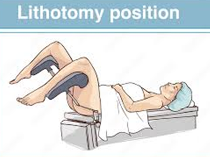
What is rumping?
Rumping described the moment during a breech birth where the buttocks descends through and emergs from the amternal pelvis. This is the equivilent of crowning in a cepahlic birth and signifies the point of no return, once rumping has occurred vaginal delivery is usually inevitable.
From this point sustained cord compression occurs and ideally the foetus will be birthed in the next 5-7 minutes.
What do you look for when assessing cord condition and what does it indicate?
The cord condition gives an indication of the perfusion of blood and oxygen to the foetus. Good colour and tone with a full umbilical cord indicated a well oxygenated foetus whereas a pale, floppy baby with a skinny white cord indicated foetal compromise.
Is the cord full and tumescent or coiled and blue/purple with great blood flow?
Is the cord pulsating or is it flat, empty and appears like a ribbon?
What is a transverse lie?
Where the baby is laying horizontally across the uterus instead of verticle.
What is an external cephalic version (ECV?)
External cephalic version is a procedure performed to turn a foetus from a breech or transverse lie into a cephalic (head-down) position by applying controlled external pressure to the abdomen. It is usually performed around 37 weeks gestation, must be a singleton pregnancy with intact membranes, normal amniotic fluid colume, no contraindications for a vaginal birth and a foetal CTG and ultrasound to confirm position before and after the procedure.
Procedure can cause pain and discomfort, short term changes to foetal heart rate that usually settles on its own, rare bleeding or placental abruption, rare rupture of membrane and in very rare cases an emergency caesarean may be needed.
What is meconium?
A newborn’s first stool. It is usually thick, sticky, greenish-black material made up of intestinal epithelial cells, fine hair, mucus, amniotic fluid, bile and water. It usually begins to form around 12 weeks gestation and accumulates in the fetal intestines until birth. Normally the neconium is passed within 24-48 hours of birth however if it is passed before birth it mixes with amniotic fluid creating a meconium-stained liquor that can indicate fetal distress or hypoxia. Meconium-sttained liquor is usually identified uring rupture of the membranes or delivery and can range from thin (lightly stained) to thick (viscous and particulate)
Meconium can be caused if the foetus is mature or post-term without indicating they are in distress or can occur due to redued oxygen supply stimulating vagal activity causing increased guy peristalsis and relaxation of the alan sphicter, umbilical cord compression causing a vagal response or maternal factors like hypertension, diabetes, smoking or infections.
MSL categorises labour and birth as high risk due to the link with foetal distress.