Anatomy and Physiology - Digestive System
 Call Kai
 Learn
 Practice Test
 Spaced Repetition
 Match
 Flashcards
 Knowt Play
1/34

There's no tags or description
Looks like no tags are added yet.
35 Terms

Accessory digestive organs
Assist in breakdown of food
Some produce secretions that empty into the GI tract (salivary glands, liver, pancreas)
Teeth and tongue: chewing and swallowing
Gallbladder: concentrates and stores liver secretions (bile) until release into the small intestine


Gastrointestinal (GI) tract
Form a continuous tube lined with a mucous membrane
Within the lumen (inner opening), food broken down into smaller components to be absorbed
Oral cavity → pharynx → esophagus → stomach → small intestine → large intestine
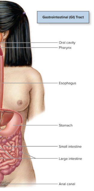
Functions of digestive system
Ingestion into the oral cavity: First step in process of digesting and absorbing nutrients
Motility: Voluntary and involuntary muscular contractions - mixing and moving materials through the GI tract
Secretion: Process of producing and releasing fluid products facilitating digestion (digestive enzymes, acid, bile)
Digestion: Breakdown of ingested food into smaller structures
Mechanical digestion: material physically broken down by chewing and mixing; no enzymes involved
Chemical digestion: involves enzymes to break chemical bonds and change large complex molecules into smaller molecules
Absorption: Transport of digested molecules, electrolytes, vitamins, water from GI tract into blood or lymph
Elimination: Expulsion of indigestible components that are not absorbed
Mucosa
Mucous membrane composed of an epithelium and lamina propria that lines the body tracts (e.g., digestive) that open to the external environment
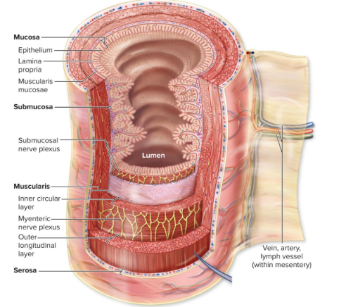
GI tract tunics
Inner to outer: mucosa → submucosa → muscularis (smooth muscle) → serosa
Serosa is connected to veins, arteries, and lymph vessels (within the mesentery, or the fold of peritoneum that attaches the small intestine to the posterior abdominal wall)

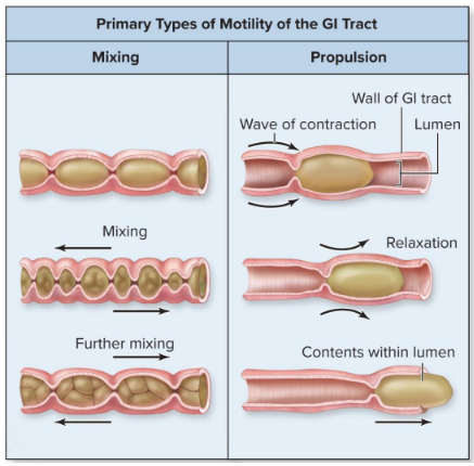
Primary types of mobility of the GI tract
Mixing: muscles contract in opposing directions, blending material (but not moving it)
Propulsion: alternative waves of contraction and relaxation pushes contents within lumen (ex: peristalsis)

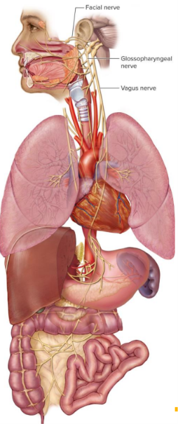
Nerves (regulation of the digestive system)
Facial nerve (CN VII): extends to the sublingual and submandibular glands to stimulate salivary secretions
Glossopharyngeal nerve (CN IX): extends to the parotid gland to stimulate salivary secretions
Vagus nerve (CN X): innervates most of the organs of the digestive system (e.g., stomach, small intestine, portions of the large intestine, pancreas, liver) and stimulates their digestive activities

Enteric nervous system/ENS (regulation of the digestive system)
500 million neurons; the “second brain”
Functions independently of the CNS and vagus control
Sensory and motor neurons within submucosal plexus (in the submucosa) and myenteric plexus (in the muscularis), extending from the esophagus to the anus
Innervates smooth muscle and glands of GI tract
Coordinates mixing and propulsion reflexes
Autonomic nervous system/ANS (regulation of the digestive system)
Parasympathetic innervation promotes GI tract activity
Sympathetic innervation opposes GI tract activity
Nerve reflexes (regulation of the digestive system)
By ENS and ANS; control GI tract wall
Receptors initiate reflexes
Baroreceptors detect stretch in GI tract wall
Chemoreceptors monitor chemical contents in lumen
Can be a short or long reflex
Short reflex (nerve reflexes - regulation of the digestive system)
Local reflex, only involves ENS; coordinates small segments of GI tract
Long reflexes (nerve reflexes - regulation of the digestive system)
Involves sensory input to CNS and autonomic motor output; coordinates GI tract motility, secretions, and accessory digestive organs
Hormonal control (regulation of the digestive system)
Several circulating (e.g., gastrin) and local (e.g. histamine) hormones participate in regulation of digestion

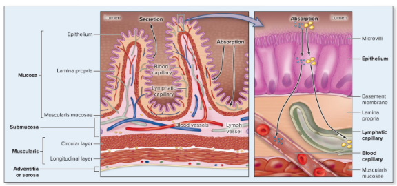
Absorption across epithelial lining
Digested substances are transported from the lumen of the GI tract thru the epithelium via membrane transport processes
Epithelium has microvilli protruding into the lumen
Lamina propria has blood and lymphatic capillaries
Most nutrients are absorbed into the blood capillaries; lipid and lipid-soluble vitamins are absorbed into the lymphatic capillaries
Upper GI tract organs & accessory structures
Oral cavity, salivary glands, pharynx, esophagus, and stomach
(Duodenum also considered part of upper GI tract)
Oral cavity (upper GI tract)
Mechanical digestion begins (along with chemical digestion)
Saliva secreted from salivary glands in response to food
Contains salivary amylase, enzyme initiating digestion of starch
Mixed with ingested materials to form bolus (mass of swallowed material)
Medications composed of small, nonpolar molecules (for example, nitroglycerin) can be placed under the tongue where they pass through the epithelium by simple diffusion; goes straight into the bloodstream for rapid action
Mastication (oral cavity - upper GI tract)
AKA chewing; uses the masseter muscle
Mechanically reduces bulk to facilitate swallowing
Increases surface area to facilitate exposure to digestive enzymes
Promotes salivation
Requires coordinated activities of teeth, lips, tongue, cheeks, jaws
Controlled by nuclei in medulla and pons (mastication center)
Salivary glands (oral cavity - upper GI tract)
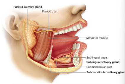
Exocrine glands
Saliva contains salivary amylase and drains into salivary ducts
Parotid: largest; located anterior and inferior to the ear; produces 25-30% of saliva
Submandibular: inferior to oral cavity floor, medial to mandible’s body; 60-70%
Sublingual: inferior to the tongue, medial & anterior to submandibular; only 3-5%

Pharynx (upper Gi tract)
Bolus moved to pharynx during swallowing
Mucus secreted to facilitate swallowing
Esophagus (upper GI tract)
Bolus transported from pharynx into stomach
Lubricated by mucus secretions

Phases of swallowing
Voluntary phase: bolus of food is pushed by the tongue against the hard palate in the oral cavity, moves towards the oropharynx, and reaches the epiglottis
Pharyngeal phase (involuntary): as the bolus moves into the oropharynx, the soft palate and uvula close off the nasopharynx, and the larynx elevates so the epiglottis closes over the laryngeal opening
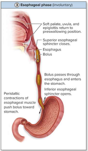
Esophageal phase (involuntary):
Soft palate, uvula, and epiglottis return to the preswallowing position
Superior esophageal sphincter closes
Bolus passes through the esophagus and enters the stomach
Inferior esophageal sphincter opens
Peristaltic contractions of esophageal muscle push bolus toward the stomach
Reflux esophagitis
Inflammation of esophagus due to acidic chyme refluxing into esophagus
Pain posterior to sternum called heartburn
Seen most frequently in overweight individuals, smokers, after large meals, and people with hiatal hernias (portion of stomach protruding through diaphragm)
Treatment: lifestyle changes, head elevation, limiting meal size
Gastroesophageal reflux disease (GERD)
Can result from chronic reflux esophagitis
Erodes esophageal tissue; scar tissue buildup over time → narrowed lumen
Barrett esophagus: esophageal epithelium changes from stratified squamous to columnar secretory epithelium in an attempt to protect from gastric secretions, but increases risk of cancerous growth

Stomach regions and esophageal-stomach border (upper GI tract)
Bolus mixed with gastric secretions by smooth muscle contractions → chyme formed
Cardia: small, narrow, superior entryway from esophagus to stomach lumen
Fundus: above the cardia; dome-shaped region
Body: largest region; below cardia and fundus, leads to the pylorus
Pylorus: narrow, funnel-shaped terminal region that opens into the duodenum; orifice opened/closed by pyloric sphincter
3 layers of smooth muscle comprise the muscularis tunic

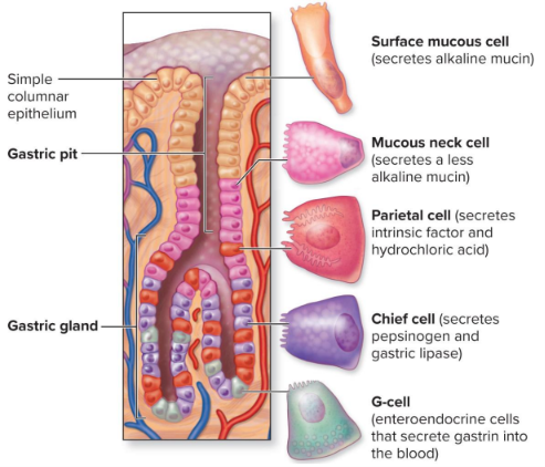
Gastric pits and glands
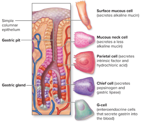
Gastric secretions produced by epithelial cells of stomach
Gastric pit
Surface mucous cell: secretes alkaline mucin (when hydrated = mucus); also makes up simple columnar epithelium
Mucous neck cell: secretes a less alkaline mucin
Gastric gland
Chief cell: secretes pepsinogen and gastric lipase
G-cell: enteroendocrine cells that secrete gastrin into the blood
Formation of HCl from parietal cells
Hydrogen ion (H+):
Carbonic anhydrate converts CO2 and water to H+ and bicarbonate
H+ is pumped into the lumen of the stomach by an H+/K+ pump
Chloride ion (Cl-):
An exchange occurs as bicarbonate is transported out of the parietal cell into the blood
Hydrochloric acid (HCl):
Pepsinogen activation and usage
Pepsinogen (inactive) + HCl → pepsin (active)
Proteins in stomach lumen + HCl → denatured proteins + pepsin → oligopeptides

Hepatic portal system
e
