Cestodes of the Intestinal Tract
 Call Kai
 Learn
Learn Practice Test
 Spaced Repetition
 Match
Match Flashcards
 Knowt Play
1/29

Earn XP
Description and Tags
Apart of the Helminth GI Infection Notes
Name | Mastery | Learn | Test | Matching | Spaced | Call with Kai | Chat |
|---|
No analytics yet
Send a link to your students to track their progress
30 Terms
What are the cestodes of the intestinal tract we went over?
Dibothriocephalus latus (Diphyllobothrium latum)
Dipylidium canium
Hymenolepis nana
Taenia spp.
Epidemiology of Dibothriocephalus latus/Diphyllobothrium latum
worldwide temperate regions (cool lake regions), in which freshwater fish are eaten raw or insufficiently cooked
in the early 1970s, ~9 million infected worldwide (5 million in Europe, 4 million in Asia and some in Americas)
Animal reservoirs: bears, walruses, members of the canine and feline families, and birds
Life Cycle of Diphyllobothriid
infection occurs upon consumption of plerocercoids larvae in undercooked fish (infective stage)
larvae development into adults in the small intestine
adults shed eggs in the stool (diagnostic stage)
eggs in the water develop into coracidia (exit through operculum) which are ingested by crustacean (first intermediate host)
coracidia develop into larvae crustacean is consumed by fish (second intermediate host)
larvae migrate to deeper tissue of fish

Infective stage and diagnostic stage of Diphyllothriid
infective stage: plerocercoids larvae in undercooked fish
diagnostic stage: eggs in the stool
First and second intermediate host for Diphyllothriid
first intermediate host: crustacean
second intermediate host: fish
Clinical Syndrome of Dibothriocephalus latus/Diphyllobothrium latum
mostly asymptomatic
abdominal pain, nausea, vomit, weight loss
40% with low serum levels of vitamin B12
Diagnosis of Dibothriocephalus latus/Diphyllobothrium latum
stool examination reveals the bile-stained operculated egg with its known at the bottom of the shell
typical proglottids with the rosette uterine structure may also be found in stool specimens
Treatment and prevention of Dibothriocephalus latus/Diphyllobothrium latum
praziquantel (does not guarantee the removal of the scolex - attached to the intestines)
vitamin B12 supplements, as needed
avoid consumption of insufficiently cooked fish or may freeze the fish (-20C for 7 days) prior to consumption
Epidemiology of Diphylidium canium
most common tapeworm of dogs and cats worldwide (usually asymptomatic)
no good evidence of prevalence, but the consensus is that it is high
occasionally found in humans, espcecially children
Life Cycle of Dipylidium
infection occurs upon consumption of cysticercoid larvae in flea (infective stage)
in the small intestine of the host, the cysticercoid develops into an adult
as proglottids mature, they detach from the adult and are shed in the stool (diagnostic stage)
in the environment, the proglottids disintegrate and release egg packets
the flea (intermediate host) ingests the egg packets
oncosphere within the eggs are released into the flea’s intestine which develops into a cysticercoid

Clinical Syndromes of Dipylidium canium
light infections are asymptomatic (rear end itching)
heavy infections produce abdominal discomfort and diarrhea
maybe self-limiting, with spontaneous cure
Diagnosis of Dipylidum canium
stool examination reveals colorless egg packets
proglottids may be in feces brought to physicians by patients
Infective stage and diagnostic stage for Dipylidum
infective stage: Consumption cysticercoid larvae in flea
diagnostic stage: proglottids
Intermediate host for Dipylidium
flea
Treatment and prevention of Dipylidium canium
praziquantel
dogs and cats
remove fleas
deworm
avoid getting licked by animals
Epidemiology of Hymenolepis nana
worldwide distribution (most common cestode infection)
affects millions of people, primarily children (50 to 75 million) (poor sanitation/personal hygiene)
highest in Sicily (46%), Argentina (34%), and Soviet Union (26%)
in the US, mostly found in Southeast (and among institutionalized children) or internationally adopted children
animal reservoir: rodents
Life Cycle of H. nana
infection occurs upon consumption of eggs or cysticercoid larvae in arthropod (infective stage) in contaminated water/food/hands
oncospheres are released in the intestines and develop into cysticercoid larvae
larvae develop into adults in the small intestine; adult produces eggs that are passed in the stool (diagnostic stage)
eggs in the environment are ingested by an arthropod (intermediate host - beetle or flea)
eggs develop into cysticercoid larvae
and alternative mode of infection - autoinfection (eggs hatch in the intestines) (can persist for years) (also means person to person transmission)

Clinical Syndromes of Hymenolepis nana
light infections are asymptomatic
heavy infections produce diarrhea, abdominal pain, headache, anal itching, anorexia
Diagnosis of Hymenolepis nana
stool examination reveals the characteristic H. nana egg with its six-hooked embryo and polar filaments (proglottids disintergrate)
Treatment and prevention of Hymenolepis nana
praziquantel (supportive care is usually sufficient)
education and improved sanitation
Infective stage and diagnostic stage of H. nana
infective: Consumption of egg or cysticercoid larvae in arthropod
diagnostic: eggs
Intermediate host of H. nana
beetle or flea
Name and differentiate the two tapeworms
Taenia solium (pork)
Taenia saginata (beef)
examination of the proglottids

Epidemiology of Taenia
worldwide distribution
~50 million people are infected
highest prevalence in central Asia, the near east, and central and eastern Africa
~1000 cases of cysticercosis annually in the US (most in immigrants from Latin America)
Human and animal reservoirs: T. solium - pig; T. saginata - cow
Life Cycle of Taenia
infection occurs upon consumption of raw/undercooked meat containing cysticercoid larvae (infective stage)
in the small intestines, the larvae develop into the adult
adult produces eggs that are passed in the stool (diagnostic stage) or proglottids detach from the adult and are passed in the stool
eggs/proglottids are in the environment are ingested by the intermediate host (either the cow - T. saginata or pig - T. solium)
oncospheres hatch from the eggs and migrate to the striated muscles where they develop into cysticercoid larvae
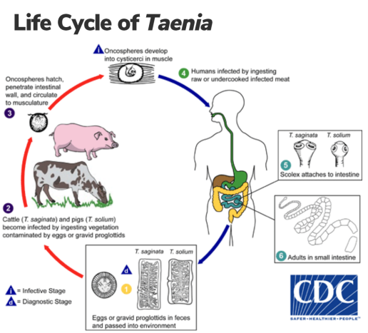
Infective stage and diagnostic stage of Taenia
infective stage: Consumption of meat containing cysticercoid
diagnostic stage: eggs or proglottids
Cysticercosis Transmission
humans are definitive hosts
consumption of water or vegetation contaminated with T. solium eggs from human feces
autoinfection may occur when eggs from a person infected with the adult worm are transferred from the perianal area to the mouth on contaminated fingers
Clinical Syndromes of Taenia
most are asymptomatic; abdominal discomfort, diarrhea and weight loss can occur
Cysticercosis (T. solium larvae) in muscle/connective tissue, neurocysticerosis (60%), lungs, and eyes (common in Mexico and Central America)
Diagnosis of Taenia
stool examination may reveal proglottids and eggs
eggs of T. saginata and T. solium are identical so eggs alone are not sufficient for species identification
examination of the proglottids to differentiate the 2 species
Treatment and prevention of Taenia
praziquantel (surgery may be needed for removal of cysts)
education and sanitation
cook meat to safe temperatures
pork: 145 F
beef 160 F
improved meat inspection