Membrane Transport Part 2
 Call Kai
 Learn
 Practice Test
 Spaced Repetition
 Match
 Flashcards
 Knowt Play
1/38

There's no tags or description
Looks like no tags are added yet.
39 Terms
Osmosis
movement of water across the cell membrane
helps maintain fluid and electrolytes balance
Fluid Balance
maintaining the proper amount and distribution of water in the body
Electrolyte Balance
maintaining appropriate concentrations of dissolved ions, such as Na⁺, K⁺, Cl⁻, and Ca²⁺, in body fluids.
Aquaporiins
allows the rapid passage of water, while blocking the passage of ions
How does aquaporin block out ions
The pores are too narrow for hydrated ions to enter the channel (.e. Na+ , K+, Cl- , Ca 2+)
Aquaporins are particularly impermeable to H+ as a hydronium ions (H3O+)
These ions diffuse through water fast
This relay mechanism requires Moving and breaking the bonds between adjacent water molecules
What distinguishes ion channels from aqueous pores
Ion selectivity
Ion channels are not continuously open
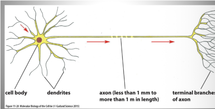
Structure of a neuron
membrane potential
A voltage difference across the plasma membrane
Resting membrane potential of a neuron
steady voltage difference across the neuron’s plasma membrane (usually -70mV)
Why is the cell more negative inside
because of negatively charged proteins inside the cell and greater permeability of the membrane to K+ (i.e. leaky K+ channels, K+ ions move dow the gradient out the cell)
Voltage gated Na+ channels
Tetramer of 4 identical polypeptide chains
Each domain has a voltage sensor (contains unusual transmembrane helix with many positively charged amino acids).
When membrane is polarized, charged helix exposed to negatively charged cytosolic side of membrane
Depolraization: helices experience electrostatic force. Helices expose charges to extracellular side of the membrane (opens channel)
Open Na+ channels
depolarization
Channels open in response to stimuli
A small amount fo Na+ enters the cell, down its electrochemical gradient
This influx of positive charge depolarizes the membrane even more, opening more Na+ channel (self amplification-> positive feedback)

Closed Na+ channels
A sufficient depolarization causes voltage gated NA+ channel
Na⁺ enters the cell down its electrochemical gradient.
The influx of positive charge depolarizes the membrane further, opening more Na⁺ channels through positive feedback.
This produces the rapid rising phase of the action potential.
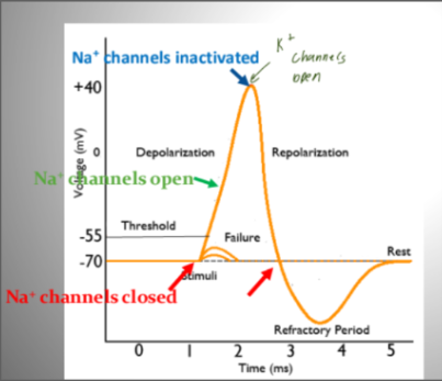
Inactivated Na+ channel
Unable to reopen, until after the membrane potential until after initial negative value
After opening briefly, Na⁺ channels automatically become inactivated, even if the membrane is still depolarized.
Na⁺ can no longer pass through the channels.
Meanwhile, voltage-gated K⁺ channels open, allowing K⁺ to leave the cell and repolarize the membrane.
The Na⁺ channels remain inactivated until the membrane potential returns sufficiently toward its resting, negative value. They can then return to the closed state and become available to open again.
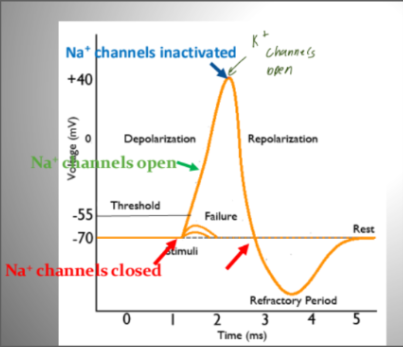
NA+ ion transport after depolarization
Voltage-gated Na+ channels open in response to a stimulus
A small amount of N+ enters the cell, down its electrochemical gradient
The influx of positive charge depolarizes the membrane further, and opens more Na+ channels (self-amplification, positive feedback), from -70 mV to +40 mV
At this point, net electrochemical driving force for Na+ is zero, cell would come to new resting state (with all Na+ channels open).
What saves cell from spasm?
Chat: voltage-gated Na⁺ channels become inactivated, and voltage-gated K⁺ channels open to repolarize the membrane.
Action Potental
Resting potential: The neuron is at about −70 mV; voltage-gated Na⁺ and K⁺ channels are closed.
Threshold: A stimulus depolarizes the membrane enough to trigger an action potential.
Depolarization: Voltage-gated Na⁺ channels open, and Na⁺ rushes into the cell, making the inside more positive.
Repolarization: Na⁺ channels inactivate, and voltage-gated K⁺ channels open. K⁺ exits, making the inside more negative.
Hyperpolarization: K⁺ channels close slowly, so extra K⁺ exits and the membrane briefly becomes more negative than resting potential.
Return to resting potential: K⁺ channels close, and the membrane returns to its resting state.
Na⁺ goes in → depolarization; K⁺ goes out → repolarization.

When can a membrane fire a second action potential?
only after the refractory period ends
Absolute Refractory Period
when all Na+ channels are in the inactivated state and can’t open, regardless of how strong the stimulus is.
From the beginning of depolarization until the early repolarization phase of the action potential
Relative refractory period
From late repolarization until hyperpolarization, until na+ channels have fully recovered and membrane conductance returns to resting levels
Some Na+ channels have recovered , but K+ channels remain open, making the membrane more negative than at rest (hyperpolarization
A stronger than normal stimulus is required to reach threshold
What is the purpose of K+ channels in action potential
To restore the negative membrane potential needed for another action potential
What causes hyperpolarization of the membrane
K+ channels are slow to close, leaky channel causes the cell to more negative than normal resting potential (hyperpolarization)
How does the membrane return to resting membrane potential
Voltage gated K+ channels close
Na+/K+ pum: restores sodium and potassium gradients
Unidirectional propagation
Propagates in one direction along the axon because the region of the membrane that just fired an action potential is in its refractory period
Depolarization: Na⁺ enters the axon, triggering an action potential.
Spread: Positive charge spreads to the adjacent region, bringing it to threshold and triggering another action potential.
Refractory period: Behind the action potential, Na⁺ channels are inactivated and K⁺ exits the cell.
Unidirectional propagation: The region behind cannot immediately fire again, so the action potential moves forward along the axon.

Channelrhodopsins
Photosensitive ion channel
When neurons are exposed to blue light, these channels open , depolarizing and activating cells
Optogenetics
Optogenetics
a technique that uses light-sensitive proteins to control and study the activity of specific cells, particularly neurons, with high temporal and spatial precision.
Structure of a CTFR Protein
Large glycoprotein found at cell membrane
Found in epithelial cells -> specialized cells that cover the internal and external body surfaces

What areas of the body are most affected by cystic fibrosis? Where is CTFR found
Skin, lungs, and digestive tract
Found in the digestive tract
What makes CFTR a “unique” ABC transporter?
uses the energy from ATP hydrolysis in the opening and closing of a channel
CFTR functions as an ATP-regulated Cl⁻ channel, allowing chloride ions to pass through the membrane down their electrochemical gradient.
Normal function of CTFR protein
Allows the release of Cl- from the cell → changes the regulation of water in epithelial cells → releases water and creates mucus
Lack of Cl movement causes lack of H20 movement
Helps establish ion concentration gradient that regulates water movement by osmosis.
Defective CTFR Protein
In the airways, defective CTFR reduces chloride secretion and can increase sodium absorption, decreasing water content on the airway surface
Why do CTFR mutations cause thick mucus
mutated protein doesn’t push cl- out fo cells → water isn’t removed from cell either → mucus becomes thicker → harder to breathe and for cilia to remove bacteria
What happens to mucus secretion in the pancreas and lung when the CTFR gene is mutated
Thick mucus secretions in the pancreas and lungs → digestive enzymes are not able to reach the small intestine
Symptoms of CF
Chromic pulmony (lung) disease
Thick secretion in airway passages
Breathing problems
Coughing/ wheezing
Pancreatic insufficiency
Excessive appetite but poor weight gain
Greasy blot stools
Saltier sweat
Meconium ileus
Meconium ileus
neonatal small‑bowel obstruction caused by abnormally thick, sticky meconium blocking the terminal ileum, and it is strongly associated with cystic fibrosis, occurring in most affected infants
How is cystic fibrosis diagnosed
Detection of meconium ileus at birth
Check blood samples for IRT (immunoreactive trypsinogen)
Sweat test
Genetics test
Why do we use a sweat test?
less salt is reabsorbed from sweat into sweat gland in person with Cystic Fibrosis
Treatments for Cystic Fibrosis
Antibiotics
Vibrating vests or bronchodilators to loosen mucus so it can be cleared]
Medications with digestive enzymes
In severe cases, you can do a lung transplant
Targeted Therapy based on CFTR gene mutation
What do CTFR drugs do
help CTFR protein reach the plasma membrane to function
Prevents both Cl- ions and water from leaving lung epithelial cells
mutation in the CFTR protein in lung epithelial cells